Jan 14, 2022
Protocol 3D Gait Analysis using Overground Approach - MUMC+
- Rachel Senden1,
- Rik Marcellis1,
- Paul Willems2,
- Jeroen Vermeulen3,
- Adhiambo Witlox4,
- Kenneth Meijer2
- 1Department of Physical Therapy, MUMC+, The Netherlands;
- 2Department of Nutrition and Movement Sciences, NUTRIM School of Nutrition and Translational Research in Metabolism, MUMC+, The Netherlands;
- 3Department of Neurology, MUMC+, The Netherlands;
- 4Department of Orthopaedic Surgery, MUMC+, The Netherlands
- MUMC


Protocol Citation: Rachel Senden, Rik Marcellis, Paul Willems, Jeroen Vermeulen, Adhiambo Witlox, Kenneth Meijer 2022. Protocol 3D Gait Analysis using Overground Approach - MUMC+. protocols.io https://dx.doi.org/10.17504/protocols.io.b2axqafn
License: This is an open access protocol distributed under the terms of the Creative Commons Attribution License, which permits unrestricted use, distribution, and reproduction in any medium, provided the original author and source are credited
Protocol status: Working
We use this protocol and it’s working
Created: November 23, 2021
Last Modified: January 14, 2022
Protocol Integer ID: 55351
Keywords: 3D gait analysis, Overground approach, Plug-in Gait, protocol 3d gait analysis, based 3d gait analaysi, overground approach, using overground approach, step, protocol
Abstract
This protocol describes the steps that are conducted for an overground based 3D gait analaysis at the MUMC+.
Materials
Overground 3D motion capture system:
A 9-m walkway with an instrumented force plate (1000 Hz, AMTI Biomechanics Force Platform model OR6-7)
Eight infrared cameras (100 Hz, Vicon T10S Motion System, Nexus 1.0, Oxford, UK) surrouding the walkway and motion capture software (Nexus 1.8.5)
Two 2D cameras
Twenty reflective markers: fixed according to the lower limb Plug-in Gait with four additional medial markers for functional calibration
The wireless TrignoTM EMG system (TrignoTM Research, Delsys Europe, UK), including 16 EMG sensors
Nexus 2.7 for data quality check and stepdetection
Matlab (R2016a, Mathworks, Natick, MA, USA) and custom made algorithms to calculate gait parameters
Description Set-up
The overground approach for 3D gait analysis consists of overground walking over a 9-m walkway with an instrumented force plate (1000 Hz, AMTI Biomechanics Force Platform model OR6-7) surrounded by 10 infrared cameras (100 Hz, Vicon T10S Motion System, Nexus 1.0, Oxford, UK) and two 2D cameras.
Nexus 1.8.5. is used to record the data.
Muscle activity during gait is measured using the wireless TrignoTM EMG system (TrignoTM Research, Delsys Europe, UK), comprising 16 EMG sensors.
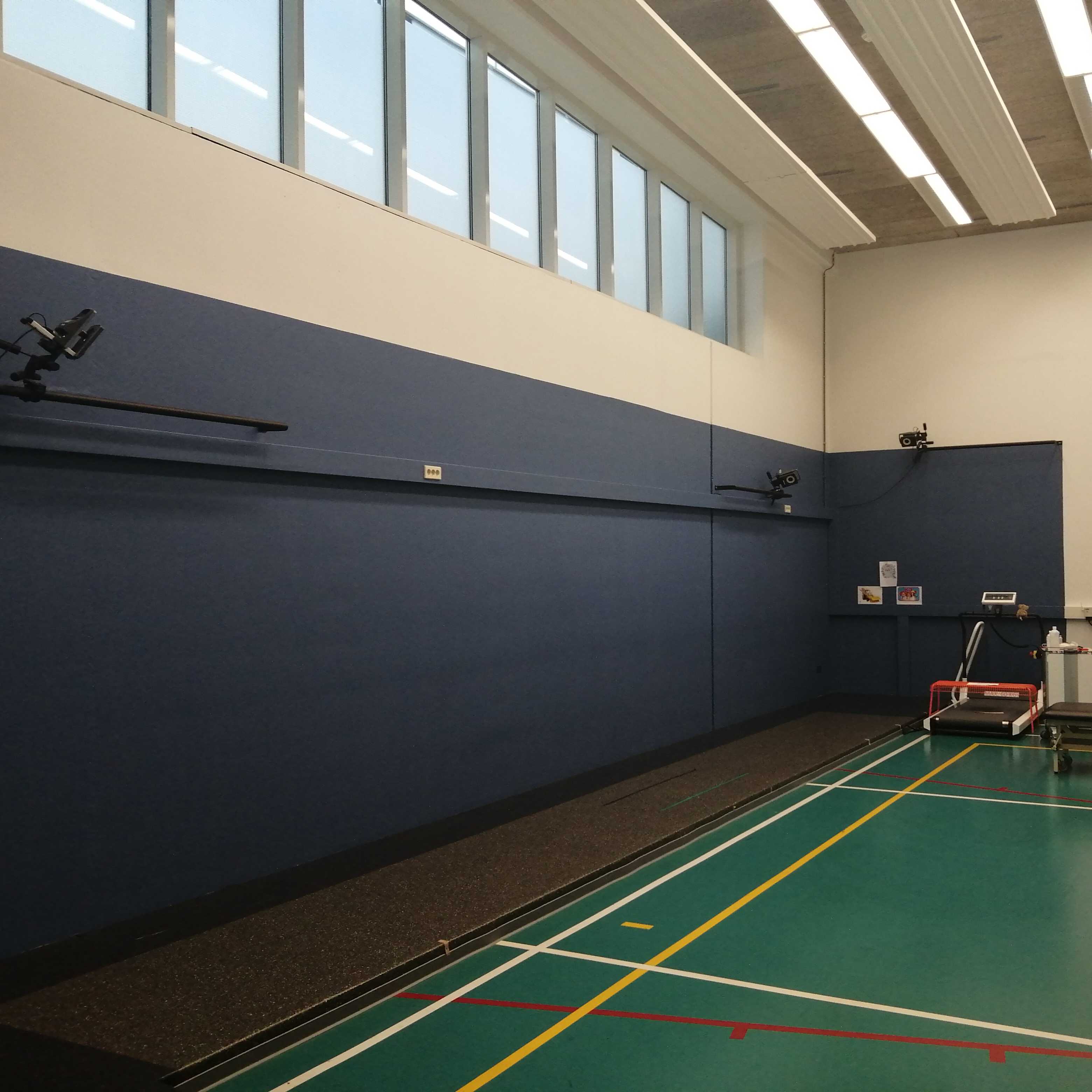
Figure: The 9 meter instrumented walkway surrounderd by 3D motion capture.
System preparation
Before the subject enters the lab, the Vicon system is set-up by the operator. This includes:
- the configuration of the Vicon system hardware
- the calibration of the vicon system
- zero level of the force plate
- the prepararation of a database in Nexus 1.8.5.
A detailed description of these steps can be found in the Vicon Nexus product guide (https://documentation.vicon.com/nexus/v2.2/Nexus1_8Guide.pdf, from p.33).
Subject enters the lab
After entering the lab, the operator introduces the lab to the subject and gives a brief explanation of the measurement that will be performed to the subject (and parents/guardians in case of children < 17 years).
Subject preparation
1. The subject is asked to change clothes (underwear is advised, but tight, non-reflective sportswear is also allowed)
2. Body weight (kg), body height (mm), knee width (mm) and ankle width (mm) are measured by the operator (RS, RM).
3. The subject is asked to ly on the treatment couch.
- Leg length (mm;) is measured supine, from SIAS to medial malleolus,
- The muscles described below, are palpated and marked by the operator. To palpate the muscles, the SENIAM recommendations for determination of sensor location are used (seniam.org).
The following muscles of right and left leg are included:
- m. rectus femoris
- m. vastus lateralis
- m. semitendinosus
- m. biceps femoris (lateral hamstrings)
- m. gastrocnemius medialis
- m. soleus
- m. tibia anterior
- m. peroneus longus
4. Sixteen EMG sensors are fixed to the muscles marked in step 4.3 using double sided adhesive tape specific for EMG sensors. Before sticking, the skin is shaved and cleaned with alcohol to ensure that the EMG sensors are properly fixed.
5. EMG signals (muscle activity) are checked in Nexus 1.8.5.
The subject is asked to perform movements like squatting, standing on tip toe.
6. A rest EMG measurement is done in Nexus 1.8.5., with the subject lying in supine position. The subject is asked not to move. 30seconds are recorded.
7. The subject gets standard gymnastic shoes from the researcher/operator.
9. Sixteen reflective markers are attached using double sided adhesive tape, directly onto the skin, at specific bony landmarks according to the Plug-In Gait (PiG) lower limb model as shown in the figure below.
More information about the marker placement is described in the following document: http://www.idmil.org/mocap/Plug-in-Gait+Marker+Placement.pdf (sacrum marker is not used).
Markers are placed by two experienced examiners (RS, RM).
Figure: Lower body Plug-in Gait marker set.
Reference figure: doi:10.1371/journal.pone.0102098.g001
5. Four additional markers are placed at the medial knee condyles and medial malleoli of both legs, required for the functional knee calibration.
6. 16 EMG sensors are attached to
Subject calibration
1. Subject characteristics such as body weight, body height, leg length, knee and ankle width are inserted into Nexus 1.8.5.
Static initialization of the model is done by recording an initialization pose. In addition, a functional knee joint calibration is performed. For all the calibration steps, the medial markers (knee and ankle) are used.
2. A static standing subject calibration is conducted with the subject standing still in T-pose on the force plate for 5 seconds: the feet are positioned at shoulder width, in parallel to the X-axis of the laboratory coordinate system and arms are spread at shoulder height. A recording of the static calibration is done in nexus 1.8.5.
A detailed description of the steps taken into Nexus: https://documentation.vicon.com/nexus/v2.2/Nexus1_8Gui
de.pdf, from p. 229)
3. A functional knee calibration is performed to determine the knee axis. The subject has to flex and extend the knee about five times through a range of approximately 0 to 45° of flexion. This calibration is done for each leg individually. The functional knee calibration is recorded in Nexus 1.8.5. and processed in Matlab .(R2016a, Mathworks, Natick, MA, USA).
This functional knee calibration procedure is based on the treadmill based approach for 3D gait analysis (see protocol titled 'Protocol 3D Gait Analysis using Treadmill Approach (CAREN) - MUMC+').
Measurement
Subjects are asked to walk at their self-chosen comfortable walking speed along the 9m walkway.
They are not instructed to target the force plates.
At least five correct hits on the force plate for both the left and right feet are collected, which has been shown to provide reliable kinematic data (CE Diss. Gait Posture 2001; 14(2): 98-103).
The comfortabel walking speed was measured using two movement detection portals placed 4 meter apart and centered with regard to the middle of the force plate during at least five randomly selected trials.
After completion of measurement
1. After competing the walking trials, the markers are removed by the operator
2. The subject gets dressed
3. The subject leaves the lab
Data recording and processing
Although data recording is done in Nexus 1.8.5., the quality check and stepdetection is done in Nexus 2.7.
Step detection (heel strikes and toe-offs) is based on combination of foot marker kinematics and force plate data (threshold of 50N; JA Zeni, JG Richards, JS Higginson. Gait Posture 2008; 27(4):710-4.).
The raw data (.c3d files) is then uploaded into Matlab (R2016a, Mathworks, Natick, MA, USA) where gait parameters are calculated using custom made algorithm which are based on published formulas and principles.
The produced gait parameters include:
- spatiotemporal parameters: e.g. walking speed (m/s), step length (cm), cadence (steps/min)
- 3D joint kinematics: joint angles in sagittal, frontal and transverse plane
- 3D joint kinetics: joint moments in sagittal, frontal, transversal plane and anterior-posterior, medio-lateral and vertical ground reaction forces.