Oct 24, 2025
Protein Extraction Protocol from the Salivary Glands of Hemipterans: Application in Mahanarva spectabilis
- Angelo José Rinaldi1,
- Monique da Silva Bonjour1,
- Humberto Josué de Oliveira Ramos1,
- Alexander Machado Auad2
- 1UFV;
- 2Embrapa
- Bioline
- Metabolomics Protocols & Workflows


Protocol Citation: Angelo José Rinaldi, Monique da Silva Bonjour, Humberto Josué de Oliveira Ramos, Alexander Machado Auad 2025. Protein Extraction Protocol from the Salivary Glands of Hemipterans: Application in Mahanarva spectabilis. protocols.io https://dx.doi.org/10.17504/protocols.io.14egnrr9ml5d/v1
License: This is an open access protocol distributed under the terms of the Creative Commons Attribution License, which permits unrestricted use, distribution, and reproduction in any medium, provided the original author and source are credited
Protocol status: Working
We use this protocol and it's working
Created: October 23, 2025
Last Modified: October 24, 2025
Protocol Integer ID: 230603
Keywords: salivary glands, protein extraction, proteomics, TCA, acetone, urea, CHAPS, PMSF, Mahanarva spectabilis, Hemiptera, protein extraction protocol from the salivary gland, protein extraction process from the salivary gland, protein extraction process, extraction of insoluble protein, protein extraction protocol, extraction of soluble protein, resolubilization for proteomic analysis, proteomic analysis, extraction, salivary gland, insoluble protein, protein precipitation with tca, protein precipitation, soluble protein, spittlebug mahanarva, hemipteran, hemiptera, application in mahanarva
Abstract
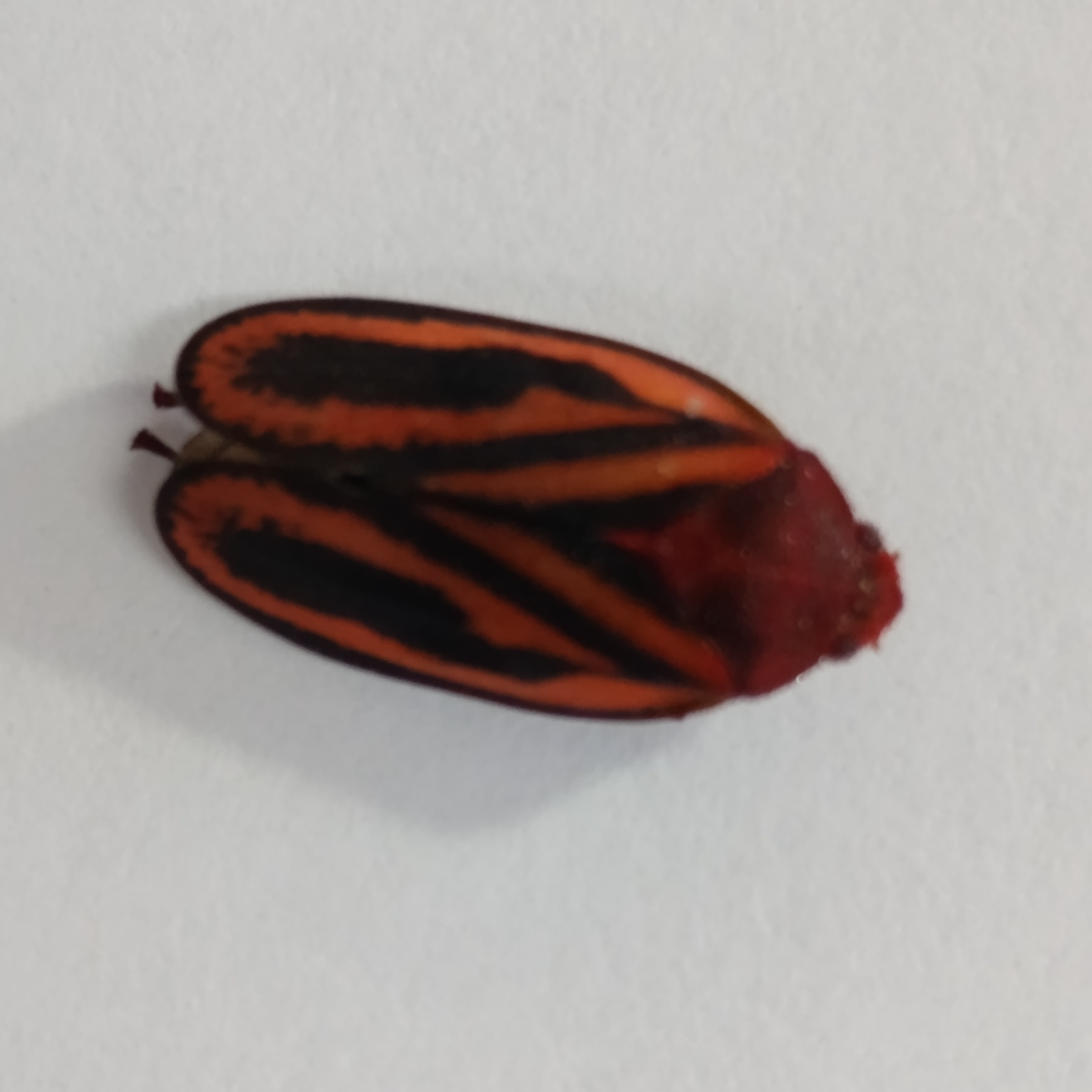
This protocol describes the collection and protein extraction process from the salivary glands of the spittlebug Mahanarva spectabilis (Hemiptera: Cercopidae). The method combines dissection under a stereomicroscope, mechanical and chemical lysis, centrifugation, protein precipitation with TCA/acetone, and resolubilization for proteomic analysis. It includes two workflows:
- Workflow A – Extraction of soluble proteins (supernatant)
- Workflow B – Extraction of insoluble proteins (pellet)
Guidelines
- Always perform the entire procedure on ice (0–4 °C) or in a refrigerated environment to prevent protein degradation.
- Prepare all buffers and reagents fresh or store them no longer than one week at 4 °C (without PMSF).
- Add protease inhibitors (PMSF or cocktail) immediately before use to ensure full protection of proteins.
- Label all tubes and reagents clearly to avoid cross-contamination between samples.
- Calibrate and pre-cool all instruments (centrifuge, sonicator, etc.) prior to use.
- Keep the work area clean and organized — dedicate one ice tray and one set of tools per sample batch.
- Use low-binding plasticware (e.g., microtubes) for protein samples to minimize sample loss.
- For reproducibility, note the number of glands processed per sample and the approximate weight of tissue.
- Check pH of buffers before use; ensure that Tris and PBS buffers are within ±0.1 pH unit of the target value.
- Perform all protein-handling steps using sterile, chilled pipette tips to avoid contamination.
Materials
- Stereomicroscope
- Fine forceps and scalpel
- Microtubes (1.5 mL, low protein-binding) and Falcon tubes (15/50 mL)
- Manual or mechanical tissue homogenizer
- Probe sonicator (with amplitude control)
- Refrigerated centrifuge
- Orbital shaker
- Ice bath and water bath (37 °C)
- Pipettes and sterile tips
- −80 °C ultrafreezer
- Spectrophotometer (Bradford or BCA assay)
- SDS-PAGE or LC-MS/MS system
Troubleshooting
Problem
Low protein yield, possible cause: PMSF degradation, insufficient tissue
Solution
Prepare PMSF fresh; increase number of glands
Problem
Protein degradation, possible cause: Delay or poor cooling
Solution
Keep on ice at all times; add PMSF promptly
Problem
Pellet difficult to dissolve, possible cause: Over-drying
Solution
Gradually rehydrate or increase buffer volume
Problem
Assay interference, possible cause: Detergent or urea incompatibility
Solution
Dilute sample or use compatible assay kits
Safety warnings
- Trichloroacetic acid (TCA) is corrosive — handle only under a fume hood, wearing gloves and eye protection.
- Acetone is highly flammable — keep away from open flames, sparks, and heating elements.
- Phenylmethylsulfonyl fluoride (PMSF) is toxic and unstable in aqueous solution — prepare fresh in isopropanol immediately before use, and dispose of properly after use.
- Urea decomposes slowly, producing cyanate — do not store buffers containing urea for more than 1 week.
- Use face protection and hearing protection during probe sonication to prevent aerosol formation and hearing damage.
- Always wear gloves, lab coat, and protective eyewear when handling biological material and reagents.
- Dispose of chemical and biological waste following your institution’s biosafety and hazardous waste protocols.
Before start
Work at 4 °C or on ice at all times.
- Use complete personal protective equipment (PPE): lab coat, nitrile gloves, safety goggles, face shield, and ear protection for sonication.
- Prepare all buffers and solutions fresh or store them at 4 °C for no more than one week (without PMSF).
- Add protease inhibitors (PMSF) immediately before use.
- Pre-chill acetone and TCA/acetone solution to −20 °C for at least 1 h before use.
- Ensure the centrifuge is pre-cooled to 4 °C.
Reagents and Preparation
Acidified Water (pH ≈ 3.0)
Use: Initial storage of dissected glands.
Preparation (100 mL):
- Add 8.3 µL of concentrated HCl (12 M) to 100 mL Milli-Q water.
- Mix well and check that pH ≈ 3.0.
- Store at 4 °C for up to one week.
Protein Extraction Buffer (Workflow A)
Final composition:
- 50 mM Tris-HCl (pH 7.5)
- 150 mM NaCl
- 1 mM EDTA
- 1 % Triton X-100
- 1 mM PMSF (add freshly before use)
Preparation (100 mL):
- Dissolve 0.605 g Tris base in 80 mL Milli-Q water.
- Add 0.876 g NaCl and 0.037 g EDTA·Na₂.
- Add 1 mL Triton X-100 (use wide-bore tip).
- Adjust pH to 7.5 with 1 M HCl.
- Bring to 100 mL with Milli-Q water.
- Filter-sterilize (0.22 µm).
- Immediately before use, add PMSF to 1 mM final from a 100 mM isopropanol stock.
- Keep on ice during use.
10 % (w/v) TCA in Acetone (Chilled)
Use: Protein precipitation.
Preparation (100 mL):
- Dissolve 10 g trichloroacetic acid (TCA) in 100 mL anhydrous acetone.
- Mix until fully dissolved (use fume hood).
- Store at −20 °C in an amber bottle for up to one month.
100 % Acetone (Chilled)
Use: Washing of protein pellets.
- Keep acetone at −20 °C prior to use.
- Always use chilled acetone for washes.
Solubilization Buffer (Workflow B)
Final composition:
- 8 M urea
- 2 % CHAPS
- 50 mM Tris-HCl (pH 8.0)
- 1 mM EDTA
- 1 mM PMSF (fresh)
Preparation (100 mL):
- Dissolve 48 g urea in 60 mL Milli-Q water (gentle stirring).
- Add 2 g CHAPS, mix until dissolved.
- Add 0.605 g Tris base and 0.037 g EDTA·Na₂.
- Adjust pH to 8.0 with 1 M HCl.
- Bring to 100 mL final volume.
- Store at 4 °C for up to 7 days.
- Add PMSF to 1 mM immediately before use.
PBS 1× (Phosphate-Buffered Saline)
Use: Washing step for pellets.
Final composition:
- 137 mM NaCl
- 2.7 mM KCl
- 10 mM Na₂HPO₄
- 1.8 mM KH₂PO₄
- pH 7.4
Preparation (1 L):
- Dissolve 8.0 g NaCl, 0.2 g KCl, 1.44 g Na₂HPO₄, and 0.24 g KH₂PO₄ in 800 mL Milli-Q water.
- Adjust pH to 7.4 with 1 M HCl.
- Bring to 1 L with water.
- Store at 4 °C for up to one month.
Safety Notes
- TCA is corrosive → handle in fume hood with gloves and goggles.
- Acetone is flammable → keep away from ignition sources.
- PMSF is toxic and unstable in water → prepare only under a fume hood and discard properly.
- Urea decomposes to cyanate → prepare fresh when possible.
- Dispose of all waste according to institutional chemical-safety regulations.
Workflow A — Soluble Protein Fraction (Supernatant)
Workflow A — Soluble Protein Fraction (Supernatant)Gland Collection
- Dissect salivary glands from adult M. spectabilis under a stereomicroscope.
- Place immediately into microtubes containing acidified water (pH 3).
Keep on ice or store at −80 °C until extraction.
Buffer Addition
- Transfer glands into 1.5 mL microcentrifuge tubes.
- Add 500 µL of extraction buffer. Keep on ice.
Homogenization and Lysis
- Homogenize manually or mechanically until uniform suspension forms.
- Sonicate (probe): 5 cycles of 30–60 s at 20–30 % amplitude, with 1 min on ice between cycles.
Cold Incubation
- Incubate the homogenate on ice for 30 min for optimal solubilization.
Centrifugation (Clarification)
- Spin at 12 000 × g for 10 min at 4 °C.
- Transfer the supernatant (soluble proteins) to a new tube.
- Store pellet at −80 °C for Workflow B.
Protein Precipitation
- Add an equal volume of chilled 10 % TCA/acetone (1:1).
- Mix gently and incubate on ice for 30 min.
Post-Precipitation Centrifugation
- Centrifuge at 12 000 × g for 10 min at 4 °C.
- Carefully discard the supernatant.
Pellet Washing
- Wash pellet three times with 1 mL chilled acetone (100 %).
- Each wash: mix gently, spin 12 000 × g for 5 min at 4 °C.
- Air-dry the pellet for 3–5 min (do not overdry).
Resuspension and Storage
- Resuspend pellet in a suitable buffer (e.g., 8 M urea + 2 % CHAPS).
- Vortex or pipette until fully dissolved.
- Store at −20 °C or −80 °C.
- Quantify protein concentration (Bradford or BCA) before SDS-PAGE or MS analysis.
Workflow B — Insoluble Protein Fraction (Pellet)
Pellet Washing
- Add 1 mL cold PBS 1×, vortex gently, and spin 12 000 × g for 5 min at 4 °C.
- Discard supernatant.
Pellet Solubilization
- Add 500 µL solubilization buffer (8 M urea + 2 % CHAPS + PMSF 1 mM).
- Homogenize by pipetting or gentle vortexing.
Incubation for Solubilization
- Shake at room temperature for 1–2 h or incubate at 37 °C for 30 min to assist solubilization.
Centrifugation
- Spin 12 000 × g for 10 min at 4 °C.
- Transfer the supernatant (solubilized proteins) to a new tube.
Preparation for Analysis
- Quantify protein concentration (Bradford or BCA).
- For SDS-PAGE: add sample buffer containing SDS + β-mercaptoethanol and heat to 95 °C for 5 min.
- For proteomics: perform enzymatic digestion (e.g., trypsin) followed by LC-MS/MS.
Storage
- Store samples at −20 °C or −80 °C in aliquots to avoid freeze–thaw cycles.
Protocol references
- Lomate & Bonning (2016). Distinct properties of proteases and nucleases in the gut, salivary gland and saliva of Nezara viridula. Scientific Reports 6:27587.
- Ramm et al. (2015). Morphology and Proteome Characterization of the Salivary Glands of Halyomorpha halys. Journal of Insect Physiology 76:29–37.
- Alhaddad et al. (2011). Comparative Behavioral and Protein Study of Salivary Glands of an Insect. Annals of the Entomological Society of America 104(3):543–551.
- He et al. (2024). Comparative Transcriptomic Analysis of Salivary Glands of Hemiptera. BMC Genomics 25(1):87.
- Ranganathan et al. (2019). Insight into Salivary Gland Proteomes of Two Polyphagous Hemipteran Bugs. Proteomics 19(10):e1800436.
Acknowledgements
Fundação de Amparo à Pesquisa do Estado de Minas Gerais (FAPEMIG)
Empresa Brasileira de Pesquisa Agropecuária (Embrapa) Gado de Leite JF/MG
Universidade Federal de Viçosa (UFV)