Oct 30, 2025
Version 1
Preparation of cryo- and paraffin sections of spheroids for histological and immunofluorescence analysis V.1
Peer-reviewed method
- Larisa Tratnjek1,
- Aleksandar Janev1,
- Nataša esnik1,
- Uroš erkvenik1,2,
- Mateja Erdani Kreft1
- 1University of Ljubljana, Faculty of Medicine, Institute of Cell Biology;
- 2University of Ljubljana, Biotechnical Faculty, Department of Biology
- PLOS ONE Lab ProtocolsTech. support email: [email protected]


External link: https://doi.org/10.1371/journal.pone.0342659
Protocol Citation: Larisa Tratnjek, Aleksandar Janev, Nataša esnik, Uroš erkvenik, Mateja Erdani Kreft 2025. Preparation of cryo- and paraffin sections of spheroids for histological and immunofluorescence analysis. protocols.io https://dx.doi.org/10.17504/protocols.io.ewov11oyovr2/v1
Manuscript citation:
Tratnjek L, Janev A, Resnik N, Cerkvenik U, Kreft ME (2026) Integrated light and electron microscopy workflow for morphological, molecular and ultrastructural analysis of spheroids. PLOS One 21(3). doi: 10.1371/journal.pone.0342659
License: This is an open access protocol distributed under the terms of the Creative Commons Attribution License, which permits unrestricted use, distribution, and reproduction in any medium, provided the original author and source are credited
Protocol status: Working
We use this protocol and it's working
Created: August 05, 2025
Last Modified: October 30, 2025
Protocol Integer ID: 224093
Keywords: spheroids, 3D in vitro model, histological staining, immunofluorescence, cryosectioning, paraffin embedding, bladder cancer, SV-HUC-1, T24, light microscopy., spheroid samples for histological staining, paraffin sections of spheroid, spheroids from other cell type, immunofluorescence labelling on both cryosection, using spheroid, human urothelial cell line, preparing spheroid sample, small size of spheroid, spheroid, recapitulate tumour microenvironment, immunofluorescence analysis, immunofluorescence labelling, dimensional cell culture, solid tumour, dimensional models such as spheroid, paraffin sections for light, paraffin section, cell connectivity, microscopy analysis, differences in cell, other cell type, eosin staining, tissue origin, cell
Funders Acknowledgements:
Slovenian Research and Innovation Agency ARIS
Grant ID: P3-0108
Slovenian Research and Innovation Agency ARIS
Grant ID: J7-2594
Slovenian Research and Innovation Agency ARIS
Grant ID: MRIC UL IP-0510
Abstract
Traditional two-dimensional cell cultures inadequately model cancer progression, prompting increased use of three-dimensional models such as spheroids that better recapitulate tumour microenvironments found in solid tumours, including features such as hypoxia and acidic pH. The small size of spheroids presents challenges for handling and processing using traditional methods.
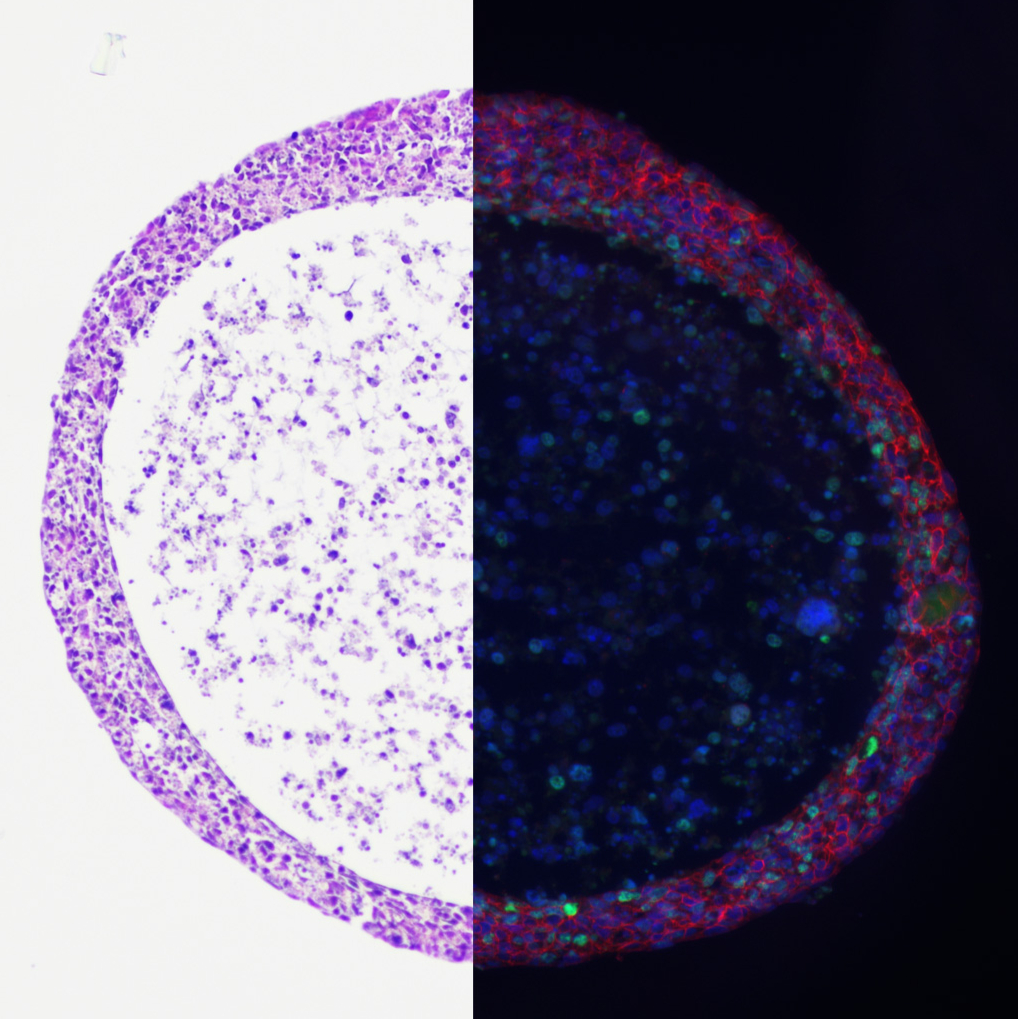
This protocol describes a detailed procedure for preparing spheroid samples for histological staining and immunofluorescence labelling on both cryosections and paraffin sections for light and fluorescence microscopy analysis. The workflow is demonstrated using spheroids derived from normal (SV-HUC-1) and cancerous (T24) human urothelial cell lines but can be readily adapted to spheroids from other cell types and tissue origins. It enables comprehensive morphological and molecular characterization of two-dimensional sections using haematoxylin-eosin staining and immunofluorescence, revealing features such as necrotic cores and differences in cell-cell connectivity.
Materials
The following materials and reagents were used:
- 1 mL and 200 µL micropipette tips – Brand, Germany (Cat. No.: 732012 and 732028)
- Anti-E-cadherin antibody – BD Biosciences, USA (Cat. No.: 610182)
- Anti-Ki-67 antibody – Abcam, United Kingdom (Cat. No.: 16667)
- Anti-N-cadherin antibody – Cell Signaling Technology, USA (Cat. No.: 13116)
- Bovine Serum Albumin (BSA) – Sigma-Aldrich, USA (Cat. No.: A2153)
- Microcentrifuge tubes (0.5 mL and 1.5 mL) – LLG Labware, United Kingdom (Cat. No.: 9409026 and 6.490 852)
- Cover glasses – Brand, Germany (Cat. No.: 470060)
- Distilled water – Institute of Cell Biology, ULMF, Slovenia
- Entellan new mounting medium – Merck (Sigma-Aldrich), Germany (Cat. No.: 107961)
- Eosin – Merck (Sigma-Aldrich), Germany (Cat. No.: 1.09844)
- Ethanol (100%) – Carlo Erba Reagents, France (Cat. No.: 4146072)
- Goat anti-mouse Alexa Fluor 488 and 555 – Molecular Probes, Thermo Fisher Scientific, USA (Cat. No.: A11001 and A21422)
- Goat anti-rabbit Alexa Fluor 488 and 555 – Molecular Probes, Thermo Fisher Scientific, USA (Cat. No.: A11008 and A21428)
- Hydrophobic barrier pen – Invitrogen, USA (Cat. No.: R3777)
- Mayer’s Haematoxylin solution – Merck, Sigma-Aldrich, Germany (Cat. No.: 54275)
- Microscope slides – Epredia, Menzel-Thermo Scientific, Germany (Cat. No.: AA00000112E01 MNZ10)
- Microscope slide racks and jars – Merck Brand, Sigma-Aldrich, USA
- Mounting medium with DAPI (Aqueous Fluoroshield) – Abcam, United Kingdom (Cat. No.: AB104139)
- Paraffin (Histosec pastilles) – Merck, Germany (Cat. No.: 115161)
- Paraformaldehyde – Sigma-Aldrich, Germany (Cat. No.: 158127)
- Pasteur pipette (3 mL) – Brand, Germany (Cat. No.: 747765)
- Phosphate-buffered saline (PBS) – prepared from:
Potassium chloride – Merck (Sigma-Aldrich), Germany (Cat. No.: 104936)
Disodium hydrogen phosphate – Merck (Sigma-Aldrich), Germany (Cat. No.: 106580)
Potassium dihydrogen phosphate – Merck (Sigma-Aldrich), Germany (Cat. No.: 104873)
Sodium chloride – Chem-Lab, Belgium (Cat. No.: CL00.1429)
- Sodium citrate dihydrate – Merck, Germany (Cat. No.: 106447)
- Spatulas – Sigma-Aldrich, USA
- Sterile 96-well ultra-low attachment U-shaped bottom microplates – Corning, USA (Cat. No.: 7007)
- Sterile surgical blades (BB510) – B. Braun (Cat. No.: 4512201760)
- Sucrose – Sigma-Aldrich, Germany (Cat. No.: S9378)
- Tissue freezing medium – Leica, Germany (Cat. No.: 14020108926)
- Vial stopper – Neolab, Germany
- Watch glasses – Sigma-Aldrich, USA
- Xylene – Carlo Erba Reagents, France (Cat. No.: 492301)
The following equipment was used:
- Bench-mounted fume cupboard System Delta 30 – Wesemann, Germany
- Centrifuge MyFuge Mini – Benchmark Scientific, USA
- Cryostat CM1950 – Leica, Germany
- Fluorescence microscope AxioImager.Z1 equipped with ApoTome – Carl Zeiss, Germany
- Laminar air-flow cabinet M182 (II) – Iskra Pio, Slovenia
- Light microscope Eclipse Ci – Nikon Instruments Inc., Japan
- Microtome RM 2135 – Leica, Germany
- Microwave oven – GoldStar, LG Electronics, South Korea
- Orbital shaker KS 260 – IKA, Germany
- Scale AE 163 – Mettler Toledo, Switzerland
- Thermoblock – Marijan Krokter s.p., Slovenia
- Thermostat – Kambič, Slovenia
- Thermostat (oven) – Instrumentaria, Croatia
Protocol for the formation of normal and cancer urothelial spheroids
1w
SV-HUC-1 and T24 spheroids were grown in 96-well ultra-low attachment U-shaped bottom microplates (Corning, New York, NY, USA) and incubated in a humidified incubator at 5% CO2 and 37°C. Seeding densities of 100,000 cells per well (in 200 µL of culture medium) were used to generate spheroids. Spheroids were grown for 7 days prior to their preparation for histological and immunofluorescence analysis.
Note
During seeding, ensure that pipette tips do not touch the bottom or sides of the wells to avoid damaging the surface coating of the ultra-low attachment U-shaped bottom microplates.
Note
Spheroid loss during washing, solution exchanges, and procedures such as paraffin embedding is expected and correlates with operator experience level and handling technique precision. Always process 20–30% excess spheroids beyond experimental requirements to compensate for potential losses.
1w
Protocol for preparation of cryosections
5h
Spheroid transfer and collection
2.1 Use a 1 mL micropipette fitted with a tip or a 3 mL plastic Pasteur pipette to transfer the spheroids from the 96-well ultra-low attachment U-bottom cell culture microplate to a 1.5 mL microcentrifuge tube filled with cold 4% (w/v) formaldehyde in PBS (pH 7.2-7.4).
Note
To prevent spheroids from adhering to the pipette wall and to ensure maximal transfer, pre-coat the micropipette tips or Pasteur pipette with 1% BSA in PBS by dipping their full length of the tip in 1% BSA in PBS.
Note
Cold fixative (4°C) was used in the experiments, which improves preservation of phosphorylated proteins and other sensitive antigens in tissue for immunolabelling, compared to warm fixation (protocol adapted from Lerch et al.,2020, Scientific Reports;10(1):2147).
2.2 Quickly aspirate the culture medium and spheroid with the pipette. You should be able to see the spheroid inside the tip. If it is not there, return the medium to the well and aspirate again until you can visually confirm the presence of the spheroid.
2.3 Allow the spheroid to settle at the bottom of the 1 ml tip or Pasteur pipette, then pipette it into the microcentrifuge tube containing fixative.
30m
Fixation
3.1 Fix the spheroids in cold 4% (w/v) formaldehyde in PBS (pH 7.2-7.4) for 15 minutes at room temperature, ensuring that the spheroids are completely submerged in the fixative.
Note
If there is weak or insufficient staining after immunolabelling of cryosections, test different fixation times (e.g., 5, 10, and 20 minutes) to determine the optimal time for spheroid preservation without over-fixation as this can affect antibody penetration.
3.2 Wash the spheroids three times in PBS for 5 minutes by gently aspirating the solution and gentle adding fresh PBS with a pipette.
Note
To change the solution in which the spheroid is submerged in a microcentrifuge tube, gently aspirate as much of the solution as possible with a 200 µL/1 mL micropipette fitted with a tip without disturbing the spheroid and add the appropriate volume of the new solution. However, to prevent accidental spheroid loss, maintain 10-20 μL of residual liquid at the container bottom during solution exchanges.
Note
When performing solution exchanges, first allow spheroids to settle at container bottom. For accelerated settling: tubes may be gently spun at 2000×g (6000 rpm) for 5 seconds to ensure complete spheroid sedimentation. Visually confirm the presence of spheroids after each step.
30m
Cryoprotection
4.1 After removing the PBS, submerge the spheroids in 30% sucrose in PBS for at least 2 hours at room
temperature or overnight at 4°C.
2h
Fig 1. Embedding a spheroid in
the freezing medium for cryosectioning. A:[CU1] [ME2] A small amount of
freezing medium [CU3] was applied to the
specimen disk of the cryostat and placed in the cryostat for 1-2 minutes to
partially harden. B-C: Using a scalpel, the spheroids were transferred
onto the freezing medium before it is completely hardened. The red arrow
indicates the spheroid. D: The embedded spheroids were left in the
cryostat to complete hardening. Then, 5 µm sections were cut in the cryostat.
5.1 Transfer and immerse the spheroids for 5 minutes into tissue freezing medium (OCT) applied on a watch glass using a 1 mL micropipette with a tip or 3 mL plastic Pasteur pipette.
5.2 Transfer spheroid into fresh OCT on a watch glass for another 5 minutes at room temperature.
5.3 Apply a small portion of the freezing medium onto the specimen disk of the cryostat to form a small pile and place it in the cryostat for 1-2 minutes until the freezing medium turns from clear to turbid (Fig 1A).
5.4 Before the freezing medium completely hardens (usually takes about 1-2 minutes), transfer 1-2 spheroids on top of the pile using a scalpel razor tip or small spatula (with the specimen disk still in the cryostat) from the freezing medium in a watch glass (Fig 1B, C). Leave the embedded spheroids in the cryostat for 15 minutes to finish hardening the freezing medium (Fig 1D).
Note
This ensures that the spheroids are not submerged in the freezing medium but rather placed on top of it (Fig 1C).
Note
Carefully remove any air bubbles from the freezing medium during embedding to prevent cutting problems while sectioning.
Fig 1. Embedding a spheroid in the freezing medium for cryosectioning. A: A small amount of freezing medium is applied to the specimen disk of the cryostat and placed in the cryostat for 1-2 minutes to partially harden. B-C: Before the freezing medium is completely hardened, spheroids were transferred onto the freezing medium using a scalpel. The red arrow indicates the spheroid. D: The embedded spheroids were left in the cryostat to complete hardening. Then, 5 µm sections were cut in the cryostat.
30m
Sectioning and storage
6.1 Cut the samples embedded in freezing medium into 5 - 10 µm sections using a cryostat at -20 °C
ambient cryostat (as well blade stage and specimen holder) temperature.
6.2 Mount the sections onto microscopic slides and leave them to dry at room temperature for 1 hour.
6.3 The slides with sections can be stored at -20°C or -80°C for several months and up to a year,
respectively.
Note
Alternatively, embedded spheroids (the frozen blocks with spheroids) can be stored at -20°C or -80°C on a specimen disk or wrapped in parafilm for months and then cut in a cryostat at a later point. In both cases, the sample should never be thawed.
1h 30m
Protocol for preparation of paraffin sections
5h 20m
Spheroid transfer and collection
7.1 Use 1 mL micropipette fitted with a tip or a 3 mL plastic Pasteur pipette to transfer the spheroids from the 96-well ultra-low attachment U-bottom cell culture microplate to a microcentrifuge tube filled with cold 4% (w/v) formaldehyde in PBS (pH 7.2 – 7.4).
Note
To prevent spheroids from adhering to the pipette wall and to ensure maximal transfer, pre-coat the micropipette tips or Pasteur pipette with 1% BSA in PBS by dipping their full length of the tip in 1% BSA in PBS.
Note
Cold fixative (4 °C) was used in the experiments, which improves preservation of phosphorylated proteins and other sensitive antigens in tissue for immunolabelling, compared to warm fixation (protocol adapted from Lerch et al.,2020, Scientific Reports;10(1):2147).
7.2 Quickly aspirate the culture medium and spheroid with the pipette. You should be able to see the spheroid inside the tip. If it is not there, return the medium to the well and aspirate again until you can visually confirm the presence of the spheroid.
7.3 Allow the spheroid to settle at the bottom of the 1 ml tip or Pasteur pipette, then pipette it into the microcentrifuge tube containing fixative.
Note
You can transfer several spheroids (3-5) into a single microcentrifuge tube or work with individual spheroids.
30m
Fixation
8.1 Incubate the spheroids in cold 4% (w/v) formaldehyde in PBS (pH 7.2 – 7.4) for 30 minutes at room temperature, ensuring that the spheroids are completely submerged in the fixative.
Note
If there is weak or insufficient staining after immunolabelling of paraffin sections, test different fixation times (e.g., 20- and 40-minute, 1 hour) to determine the optimal time for spheroid preservation without over-fixation, as this can affect antibody penetration.
8.2 Wash with PBS four times for 5 minutes at room temperature.
Note
To change the solution in which the spheroid is submerged in a microcentrifuge tube, gently aspirate as much of the solution as possible with a 200 µL/1 mL micropipette fitted with a tip without disturbing the spheroid and add the appropriate volume of the new solution. However, to prevent accidental spheroid loss, maintain 10-20μL of residual liquid at the container bottom during solution exchanges.
Note
When performing solution exchanges, first allow spheroids to settle at container bottom. For accelerated settling: tubes may be gently spun at 2000×g (6000 rpm) for 5 seconds to ensure complete spheroid sedimentation. Visually confirm the presence of spheroids after each step.
45m
Dehydration and clearing
Perform all steps at room temperature with an orbital shaker set to 100 rpm, (0.056 × g, orbital diameter 10 mm).
9.1 Submerge in 50% ethanol for 5 minutes.
9.2 Submerge in 70% ethanol for 5 minutes.
9.3 Submerge in 90% ethanol for 5 minutes.
9.4 Submerge in 100% ethanol for 5 minutes.
9.5 Transfer spheroids to glass vials.
9.6 Mix 2/3 100% ethanol with 1/3 xylene and submerge spheroids for 5 minutes.
9.7 Mix 1/2 100% ethanol with 1/2 xylene and submerge spheroids for 5 minutes.
9.8 Submerge in 100% xylene for 5 minutes.
35m
10.1 Using a 1 mL micropipette fitted with a tip, transfer the spheroids into a rubber vial stopper (Fig 2) or a sample embedding mould of a similar size/volume.
10.2 Remove excess xylene from the vial stopper on a warm plate by pipetting and add liquid paraffin (pre-warmed to 60 °C). Immediately transfer the spheroids into a thermostat to embed with paraffin at 58 °C for 2 hours. Then allow the paraffin to solidify at room temperature or shift the vial stopper to the cold plate.
Note
Paraffin-embedded spheroid blocks (Fig 2) can be stored long term at room temperature.
10.3 Remove the paraffin block from the vial stopper and mount it onto a wooden block of appropriate size (Fig 2) by melting the point of contact between the paraffin and wooden block to firmly fix the paraffin to the wooden block.
10.4 Cut the paraffin embedded samples with a microtome into 6-7 μm sections.
Note
Blocks of paraffin embedded spheroids can be cooled in the refrigerator before sectioning, which makes sectioning easier.
10.5 Collect the sections on microscope slides after being flattened in a water heated thermoblock at 60 °C.
10.6 Place the slides in an oven at 65 °C for 10–20 minutes until the paraffin starts to melt, which mounts the sections to the slides. Slides can be stored long term at room temperature.
Fig 2. Paraffin embedding of spheroids. The spheroids were transferred into a rubber vial stopper (A, B left, grey coloured). Liquid paraffin was poured in and left for 2 hours at 58 °C to penetrate throughout the entire spheroid. The paraffin-embedded spheroids were then placed at room temperature to solidify the paraffin and mounted onto wooden blocks (A, B right). A: top view. B: side view.
3h 30m
Protocol for histological staining of cryo- and paraffin sections of spheroids with haematoxylin-eosin
40m
Pretreatment of cryosections
11.1 Transfer slides with cryosections of spheroids (from the freezer) into racks and/or containers.
11.2 Warm cryosections to room temperature and rinse with PBS for 5 minutes
5m
Pretreatment of paraffin sections
12.1 Transfer slides with paraffin sections of spheroids into racks and/or containers.
12.2 Deparaffinize the paraffin sections in xylene for 5 minutes at room temperature.
12.3 Submerge the paraffin sections in 100% ethanol for 5 minutes at room temperature.
12.4 Submerge the paraffin sections in 70% ethanol for 5 minutes at room temperature.
12.5 Rinse (submerge) the paraffin sections twice with distilled water for 5 minutes each at room
temperature.
20m
Histological staining process of cryosections and paraffin sections
Perform all steps at room temperature.
13.1 Incubate the cryosections and paraffin sections for 15 seconds in Mayer's haematoxylin solution.
13.2 Wash the sections with distilled water (submerge).
13.3 Rinse the sections under running water for 1 minute.
13.4 Incubate the sections in eosin for 2 minutes (submerge).
5m
Dehydration, clearing and mounting
Perform all steps at room temperature.
14.1 Submerge the sections in 90% ethanol twice for 1 minute each.
14.2 Submerge the sections in 100% ethanol twice for 1 minute each.
14.3 Submerge the sections in xylene for 5 minutes.
14.4 Add a drop of mounting medium (Entellan new inclusion medium) to the samples and cover with coverslips.
Note
Samples can be stored long term at room temperature.
10m
Protocol for immunofluorescence labelling of spheroids’ paraffin and cryosections
21h 30m
Immunofluorescence labelling can be performed on cryosections and paraffin sections of spheroids. The protocol described below includes immunofluorescence labelling of E-cadherin, N-cadherin and Ki-67. Samples were examined under a fluorescence microscope.
Note
The use of blocking solutions and dilution should be adjusted and optimized depending on the
primary antibodies.
Pretreatment of cryosections
16.1 Transfer slides with cryosections of spheroids into racks and/or containers.
16.2 Warm cryosections to room temperature and rinse with PBS for 5 minutes.
5m
Pretreatment of paraffin sections
17.1 Transfer microscopic slides with paraffin sections of spheroids into racks and/or containers at room temperature.
17.2 Deparaffinize paraffin sections as described in the "Histological Analysis" section.
17.3 For antigen retrieval of paraffin sections, we recommend using a microwave and a 10 mM sodium citrate buffer with a pH of 6.0. Heat slides until boiling is initiated, then maintain at a sub-boiling temperature for 10 minutes. This typically requires heating slides at full power for 2.5-3 minutes (600 W), then adjusting to 20-30% power. After microwaving, cool the slides on the bench top for 30 minutes and then rinse them with PBS for 5 minutes.
Note
Avoid boiling the citrate buffer, as bubbling can damage the sections and cause them to detach.
8m
Immunofluorescence labelling of E-cadherin, N-cadherin and Ki-67 on cryosections and paraffin sections
18.1 Block the samples with 1% BSA/PBS at room temperature for 1 hour (submerge the sections).
18.2 Incubate the samples with mouse anti-E-cadherin antibody (diluted 1:100) and rabbit anti-N cadherin antibody (diluted 1:100) or rabbit anti-Ki-67 antibody (diluted 1:100) in 1% BSA/PBS overnight at 4°C (using a hydrophobic pen add 100-200 µL of the antibody solution per slide). For the negative control, omit the primary antibodies and incubate the samples in 1% BSA in PBS at 4°C overnight.
18.3 Submerge the sections in PBS at least 3 times for 5 minutes each.
18.4 Add a mixture of goat anti-rabbit and anti-mouse secondary antibodies (Alexa Fluor 488 and 555; Invitrogen) diluted 1:400 in 1% BSA/PBS for 90 minutes at room temperature (using a hydrophobic pen add 100-200 µL of the antibody solution per slide).
18.5 Wash the samples by submerging them in PBS at least 3 times for 5 minutes each (3-5 times).
18.6 Mount the samples in mounting medium containing with 4′,6-diamidino-2-phenylindole (DAPI) for DNA labelling.
21h 17m
Protocol references
Lerch, M. L., Kenerson, H. L., Chafin, D., Westerhoff, M., Theiss, A., Otter, M., Yeung, R. S., & Baird, G. S. (2020). Effect of immediate cold formalin fixation on phosphoprotein IHC tumor biomarker signal in liver tumors using a cold transport device. Scientific reports, 10(1), 2147. https://doi.org/10.1038/s41598-020-58257-3
Acknowledgements
We thank Sanja Čebraja, Nada Pavlica Dubarič, Marko Radanović and Sabina Železnik for their technical support and expertise in preparing spheroids and samples for electron microscopy. We also thank Urška Dragin Jerman, PhD, for her expertise and assistance with confocal microscopy. The research was done using the Research infrastructure ELIXIR-SI (https://elixir-slovenia.org), funded by the European Regional Development Fund, the Ministry of Science, Education and Sports and by the Slovenian Research and Innovation Agency.