Oct 31, 2025
Preparation and gold sputter coating of whole-mount spheroids for scanning electron microscopy analysis
Peer-reviewed method
- Larisa Tratnjek1,
- Aleksandar Janev1,
- Nataša esnik1,
- Uroš erkvenik1,2,
- Mateja Erdani Kreft1
- 1Institute of Cell Biology, Medical Faculty, University of Ljubljana;
- 2Department of Biology, Biotechnical Faculty, University of Ljubljana
- PLOS ONE Lab ProtocolsTech. support email: [email protected]


External link: https://doi.org/10.1371/journal.pone.0342659
Protocol Citation: Larisa Tratnjek, Aleksandar Janev, Nataša esnik, Uroš erkvenik, Mateja Erdani Kreft 2025. Preparation and gold sputter coating of whole-mount spheroids for scanning electron microscopy analysis. protocols.io https://dx.doi.org/10.17504/protocols.io.x54v95yoql3e/v1
Manuscript citation:
Tratnjek L, Janev A, Resnik N, Cerkvenik U, Kreft ME (2026) Integrated light and electron microscopy workflow for morphological, molecular and ultrastructural analysis of spheroids. PLOS One 21(3). doi: 10.1371/journal.pone.0342659
License: This is an open access protocol distributed under the terms of the Creative Commons Attribution License, which permits unrestricted use, distribution, and reproduction in any medium, provided the original author and source are credited
Protocol status: Working
We use this protocol and it's working
Created: August 01, 2025
Last Modified: October 31, 2025
Protocol Integer ID: 223870
Keywords: Spheroids, three-dimensional in vitro model, Scanning electron microscopy (SEM), Sputter coating, Surface ultrastructure, SV-HUC-1, T24, bladder cancer, high-resolution imaging, whole-mount, resolution imaging of spheroid surface ultrastructure, spheroid surface ultrastructure, using spheroid, mount spheroid, spheroid, fragility of spheroid, dimensional spheroid model, human urothelial cell line, studying tumour heterogeneity, features of the tumour microenvironment, dimensional cell culture, electron microscopy analysis, microscopy analysis, electron microscopy, cancer stem cell dynamic, scanning electron microscopy, cancer research, tumour microenvironment, preparation for ultrastructural analysis, gold sputter coating, tumour heterogeneity, ultrastructural analysis, tissue origin, relevant platform for cancer research, surface analysis
Funders Acknowledgements:
Slovenian Research and Innovation Agency ARIS
Grant ID: P3-0108
Slovenian Research and Innovation Agency
Grant ID: J7-2594
Slovenian Research and Innovation Agency ARIS
Grant ID: MRIC UL IP-0510
Abstract
Three-dimensional spheroid models provide a more physiologically relevant platform for cancer research compared to conventional two-dimensional cell cultures, as they closely mimic features of the tumour microenvironment, including hypoxia and acidic pH. These models are essential for studying tumour heterogeneity, cancer stem cell dynamics, and therapy resistance. However, the small size and fragility of spheroids make sample handling and preparation for ultrastructural analysis challenging.
This protocol provides a detailed, step-by-step workflow for the preparation and gold sputter coating of whole-mount spheroids, optimized for scanning electron microscopy (SEM) surface analysis. The method is demonstrated using spheroids derived from normal (SV-HUC-1) and cancerous (T24) human urothelial cell lines but can be readily adapted to spheroids generated from other cell types and tissue origins. Following this protocol enables reproducible, high-resolution imaging of spheroid surface ultrastructure.
Materials
Materials
- 1 mL and 200 μL micropipette tips (Brand, Germany, Cat. No.: 732012 and 732028)
- 10 mL glass vials (SGD Pharma, Germany)
- Bovine Serum Albumin (BSA) (Sigma-Aldrich, USA, Cat. No.: A2153)
- Cacodylic acid sodium salt trihydrate (Serva, Germany, Cat. No.:1554002)
- Microcentrifuge tubes 0.5 mL and 1.5 mL (LLG Labware, United Kingdom, Cat. No.: 9409026 and 6.490 852)
- Distilled water (Institute of cell biology, ULMF, Slovenia)
- Ethanol (100 %) (Carlo Erba Reagents, France, Cat. No.: 4146072)
- Glutaraldehyde (Serva, Germany, Cat. No.: 23114.01)
- Hexamethyldisilazane (HMDS) (Sigma-Aldrich, USA, Cat. No.: 440191)
- Metal stubs (12.7 diameter) (Mikrolux, Croatia)
-Osmium tetraoxide (Roth, Germany, Cat. No.:8371.3)
- Paraformaldehyde (Sigma-Aldrich, Germany, Cat. No.: 158127)
- Pasteur pipette (3 mL) (Brand, Germany, Cat. No.:747765)
- Phosphate buffer saline (PBS):
- Potassium chloride (Merck (Sigma-Aldrich); Germany, Cat. No.:104936)
- Disodium hydrogen phosphate (Merck (Sigma-Aldrich); Germany, Cat. No.:106580)
- Potassium dihydrogen phosphate (Merck (Sigma-Aldrich); Germany, Cat. No.:104873)
- Sodium chloride (Chem-Lab, Belgium, Cat. No.:CL00.1429)
- Sterile 96-well ultra-low attachment U-shaped bottom microplates (Corning, USA, Cat. No.: 7007)
- Ultrasmooth carbon discs (SPI supplies, USA, Cat. No.:04967-B1)
Equipment
- Bench-mounted fume cupboard System Delta 30 (Wesemann, Germany)
- Centrifuge MyFuge Mini (Benchmark Scientific, USA)
- Laminar air-flow cabinet M182 (II) (Iskra Pio, Slovenia)
- Scanning electron microscope Vega 3 (Tescan, Brno, Czech Republic)
- Sputtering coating machine SCD 040 (Balzers Union, Liechtenstein)
Protocol for the formation of normal and cancer urothelial spheroids
1w
SV-HUC-1 and T24 spheroids were grown in 96-well ultra-low attachment U-shaped bottom microplates (Corning, New York, NY, USA) and incubated in a humidified incubator at 5% CO2 and 37°C. Seeding densities of 100,000 cells per well (in 200 μL of culture medium) were used to generate spheroids. Spheroids were grown for 7 days prior to their preparation for scanning electron microscopy analysis.
Note
During seeding, ensure that pipette tips do not touch the bottom or sides of the wells to avoid damaging the surface coating of the ultra-low attachment U-shaped bottom microplates.
Note
Spheroid loss during washing, solution exchanges, and procedures such as dehydration is expected and correlates with operator experience level and handling technique precision. Always process 20–30% excess spheroids beyond experimental requirements to compensate for potential losses.
1w
Spheroid transfer and collection
30m
Room temperature
2.1 Use a 1 mL micropipette fitted with a tip or a 3 mL plastic Pasteur pipette to transfer the spheroids from the 96-well ultra-low attachment U-bottom cell culture microplate to a 1.5 mL microcentrifuge tube filled with cold 2% formaldehyde (w/v) and 2% glutaraldehyde (v/v) in 0.2 M cacodylate buffer (pH 7.4).
Note
To prevent spheroids from adhering to the pipette wall and to ensure maximal transfer, pre-coat the micropipette tips or Pasteur pipettes with 1% BSA in PBS by dipping their full length of the tip in 1% BSA in PBS.
2.2 Quickly aspirate the culture medium and spheroid with the pipette. You should be able to see the spheroid inside the tip. If it is not there, return the medium to the well and aspirate again until you can visually confirm the presence of the spheroid inside the tip of the pipette.
2.3 Allow the spheroid to settle at the bottom of the tip, then pipette it into the microcentrifuge tube containing fixative.
Note
You can transfer several spheroids (3-5) into a single microcentrifuge tube or work with individual spheroids.
30m
Fixation
1h 30m
3.1 Fix the spheroids with cold 2% formaldehyde (w/v) and 2% glutaraldehyde (v/v) in 0.2 M cacodylate buffer (pH 7.4) for 1 hour at 4 °C ensuring that the spheroids are completely submerged in the fixative.
3.2 Rinse the samples with 0.2 M cacodylate buffer three times for 10 minutes each at Room temperature .
Note
To change the solution in which the spheroid is submerged in a microcentrifuge tube, gently aspirate as much of the solution as possible with a 200 μL/1 mL micropipette fitted with a tip without disturbing the spheroid and add the appropriate volume of the new solution. However, to prevent accidental spheroid loss, maintain 10-20μL of residual liquid at the container bottom during solution exchanges.
Note
When performing solution exchanges, first allow spheroids to settle at the container bottom. For accelerated settling, tubes may be gently spun at 2000×g (6000 rpm) for 5 seconds to ensure complete spheroid sedimentation. Visually confirm the presence of spheroids after each step.
3.3 Transfer the samples to glass vials and post-fix the samples in 1% (w/v) osmium tetroxide in 0.2 M cacodylate buffer for 1 hour at Room temperature .
3.4 Rinse samples in 0.2 M cacodylate buffer for three times for 10 minutes each at Room temperature .
1h 30m
Dehydration
25m
Room temperature
4.1 Submerge the samples in 30% ethanol for 5 minutes.
4.2 Submerge the samples in 50% ethanol for 5 minutes.
4.3 Submerge the samples in 70% ethanol for 5 minutes.
4.4 Submerge the samples in 90% ethanol for 5 minutes.
4.5 Submerge the samples in 100% ethanol twice for 5 minutes each.
25m
Drying
1d
Room temperature
5.1 Submerge the samples in acetone twice for 10 minutes each.
5.2 Submerge the samples in hexamethyldisilazane (HMDS) with a 1:1 ratio of acetone:HMDS for 10 minutes.
5.3 Submerge the samples in HMDS twice for 10 minutes each.
5.4 Aspirate the HMDS and air-dry the samples overnight at room temperature (open the vial/microcentrifuge tube with the spheroids immersed into remaining HMDS and leave it in the fume hood overnight).
1d
Mounting
1h
Room temperature
Mount spheroids on metal stubs (Fig 1) using a sticky carbon disc (apply double-sided conductive tape to the stub and remove the protective layer) with tweezers (do not squeeze the spheroids to avoid damaging them), or simply by dropping spheroids from glass vials onto the sticky carbon disc (by turning the vial upside down so that the spheroids fall onto the conductive tape).
Note
Several spheroids can be mounted on individual metal stub (Fig 1A).
1h
Sputter Coating
1h
Sputter-coat the samples with gold according to the manufacturer's instructions (Fig 1B).
Note
The SCD 040 sputter coating machine (Balzers Union) with Baltec gold sputter target (50 mm) was used in our experiments to apply a gold layer approximately 16 nm thick, operating at 30 mA for 1 minute.
Note
Samples should be stored in an air-tight container at Room temperature .
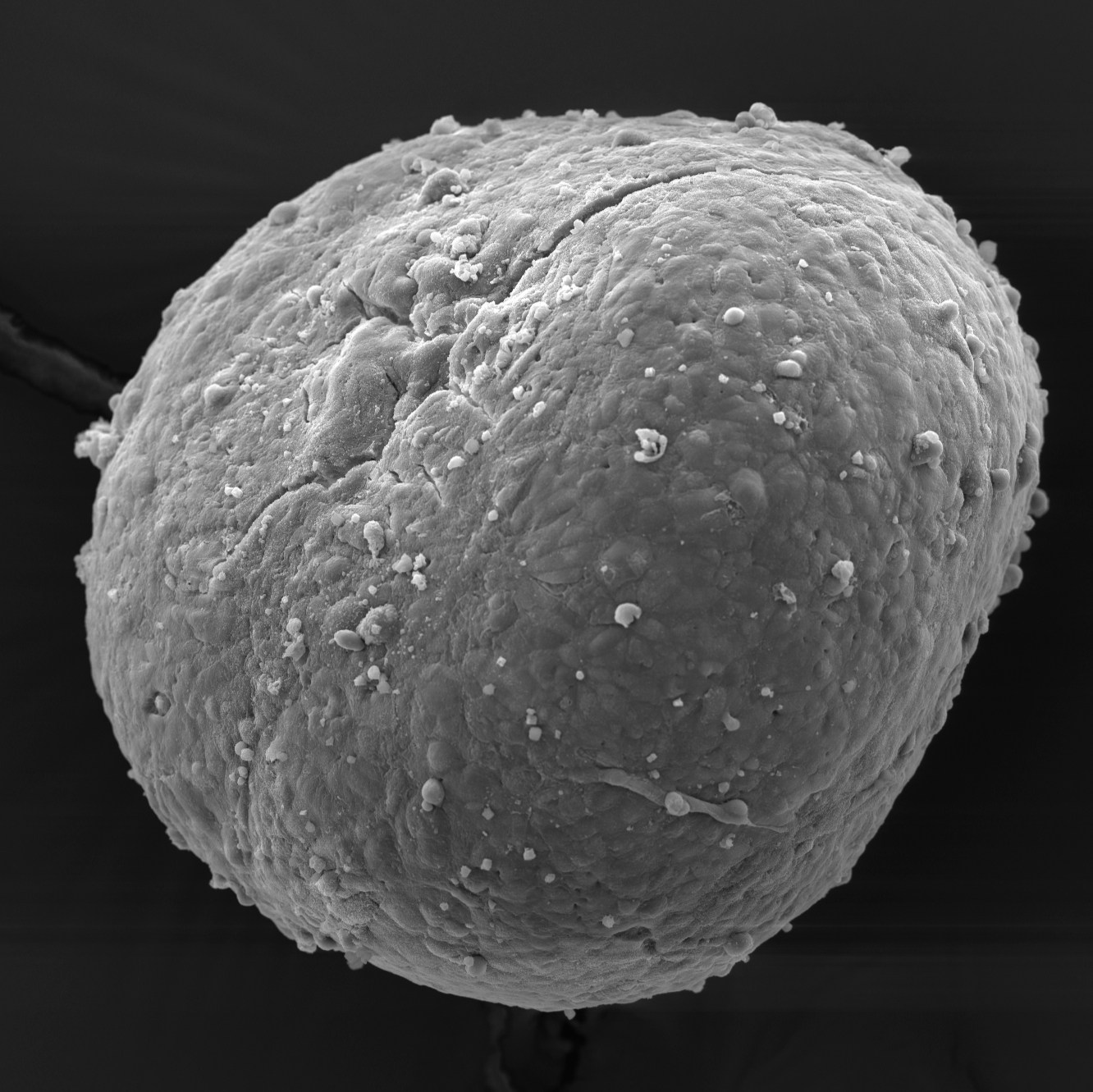
Fig 1. SV-HUC-1 spheroids mounted on metal stubs
and sputter-coated with gold. A: side view. B: top view.
1h
Acknowledgements
We thank Sanja Čebraja, Nada Pavlica Dubarič, Marko Radanović and Sabina Železnik for their technical support and expertise in preparing spheroids and samples for electron microscopy. We also thank Urška Dragin Jerman, PhD, for her expertise and assistance with confocal microscopy. The research was done using the Research infrastructure ELIXIR-SI (https://elixir-slovenia.org), funded by the European Regional Development Fund, the Ministry of Science, Education and Sports and by the Slovenian Research and Innovation Agency.