Oct 07, 2025
Plaque assay T7 bacteriophage
Forked from a private protocol
- 1iGEM TU Delft, Snaccine 2025
- Duncan Whyte: Improved and adjusted protocol


Protocol Citation: Duncan Whyte 2025. Plaque assay T7 bacteriophage. protocols.io https://dx.doi.org/10.17504/protocols.io.rm7vz91p4gx1/v1
Manuscript citation:
Improved and adjusted
Original
Plaque assay
- iGEM Wageningen
License: This is an open access protocol distributed under the terms of the Creative Commons Attribution License, which permits unrestricted use, distribution, and reproduction in any medium, provided the original author and source are credited
Protocol status: Working
We use this protocol and it's working
Created: October 06, 2025
Last Modified: October 07, 2025
Protocol Integer ID: 229174
Keywords: Plaque Assay, phage, lambda, T7, plaque assay, plaque, counting plaque, assay, plaque assay t7 bacteriophage, plaque forming unit, t7, assay, pfu
Abstract
This is a protocol for the quantification of T7 in a sample in Plaque Forming Units (PFU) per mL.
Image Attribution
Protocol image: Marijn Ceelen, iGEM Wageningen; from original protocol.
Phage safety work area image: Duncan Harry Whyte, iGEM TU Delft 2025
Materials
MATERIALS
Liquid LB medium
LB agar
Prepare a safe phage work area.
To avoid contaminating other lab work with phages, take the required measures. Always make sure to wear gloves when working with phages. Designate a certain lab bench area for phage work. Use a special phage incubator. Prepare and maintain Virkon to decontaminate the phage area after all work. Use filter tips for pipettes, and designate pipette(s) (required range: 11 µL - 111 µL ) to remain phage-work pipettes. We recommend using fluorescent tape to mark pipettes and other phage-contaminated equipment.
To avoid contaminating other lab work with phages, take the required measures. Always make sure to wear gloves when working with phages. Designate a certain lab bench area for phage work. Use a special phage incubator. Prepare and maintain Virkon to decontaminate the phage area after all work. Use filter tips for pipettes, and designate pipette(s) (required range: 11 µL - 111 µL ) to remain phage-work pipettes. We recommend using fluorescent tape to mark pipettes and other phage-contaminated equipment.
Here is an example of the phage area we used.
Create an E. coli culture
Streak LB plate with E. coli, e.g. BL21(DE3) and incubate overnight at 37 °C
Pick a colony from this plate and use it to inoculate 3 mL of LB. Incubate this culture at 37 °C until the OD reaches 2-3. We recommend an overnight culture.
Prepare LBa and LBa plates
Prepare enough plates with LB and agar, one for every dilution sample and one for the control. For a first assay, we recommend 6 samples (100 to 10-4 and a control). For assays where the rough PFU is known, you might be able to leave out some of these dilutions.
Preheat the plates to 37 °C . Consider labelling the plates in advance.
We recommend preparing molten soft (0.5%) agar LB at 50 °C , but you can also make it before it is needed. You'll need 3 mL per dilution sample, but we recommend using a bottle with more than enough and reusing whatever is left.
Make a range dilutions, by a factor 10 each, and a control
Make a range of dilutions . Mix 100 µL of phage dilution with 200 µL of E. coli culture. Then incubate for 10 minutes at room temperature. After 00:10:00 add 3 mL of liquid soft LB agar (LB agar with 0.7% agar) (50 °C ).
Prepare the required number of tubes. We recommend using 15 mL Falcon tubes, generically called Screw-top centrifuge tubes. Label these with (recommended) a combination of coloured stickers (per different T7 sample) and a marker (for different dilutions). Label the dilution samples with 0, -1, -2, etc.
Add 100 µL of ddH2O (MilliQ) to the control. Add 100 µL of ddH2O (MilliQ) to all dilution samples except for the first dilution, labelled 0. Do not add any ddH2O to this sample.
Enter the phage area. Using a pipette tip with filter, pipette 111 µL of the desired T7 phage sample into the sample labelled 0.
With filter tips, pipette 11 µL from the sample labelled 0 to the next sample, -1. Mix the sample, or gently vortex it.
Now repeat this previous step for every dilution. That is, take 11 µL from the previous dilution sample and add it to the next one. Then mix or gently vortex.
Finally, discard 11 µL from the final sample (or leave it in and adjust the calculation at the end). Now every sample should contain 100 µL of T7, reduced in concentration by a factor of ten compared to the previous sample.
Add the E. coli and pour the plates
25m
Now add 200 µL of the E. coli culture to every dilution sample and to the control. Use filter tips. Gently mix. Wait 00:10:00 . Do not wait more than a phage replication time (rather conservatively, 00:20:00 at room temperature).
10m
Add 3 mL of liquid soft LB agar (LB with 0.5% agar) (50 °C ). Mix and quickly pour the resulting mixture on the LB plates (preheated at 37 °C ). Note: this means the plates will have two layers, the first without any E. coli or T7 and the second with. Incubate for at least 00:15:00 or more at Room temperature , to let the second layer harden. Turn the plates upside-down.
15m
Incubate Overnight in a specialised phage work area incubator at 37 °C .
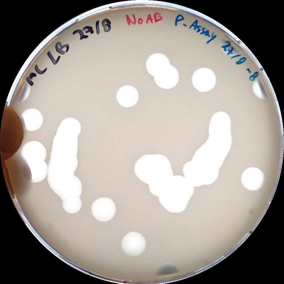
Find the plate with the most easily identifiable number of plaques, e.g. 10-100. Calculate the Plaque Forming Units (PFU)/mL by the following formula: PFU/mL = N × 1/Vdiluted = 𝑁 × 1/DF × 1/V0.
N is the number of plaques of lysis counted on the plate (expressed as PFU).
Vdiluted is the total volume of the original phage sample added to this plate (equal to DF × V0)
DF is the dilution factor of this plate (i.e. 10x, where x is the label 0, -1, -2, etc.)
V0 is the volume of phage dilution of the first plate, i.e. 100 µL .
When comparing PFU/mL, remember to take into account the strain of E. coli used.