Aug 20, 2025
Picrosirius red staining for histology
- 1Charles Perkins Centre Histology Facility, The University of Sydney, NSW, 2006, Australia
- The University of Sydney


Protocol Citation: Samson Dowland 2025. Picrosirius red staining for histology. protocols.io https://dx.doi.org/10.17504/protocols.io.ewov11oxpvr2/v1
License: This is an open access protocol distributed under the terms of the Creative Commons Attribution License, which permits unrestricted use, distribution, and reproduction in any medium, provided the original author and source are credited
Protocol status: Working
We use this protocol and it's working
Created: August 10, 2025
Last Modified: August 20, 2025
Protocol Integer ID: 224421
Keywords: histology, histochemistry, collagen, microscopy, pathology, histopathology, polarised, psr dye stains collagen fibre, histology picrosirius red, collagen in tissue section, iii collagen fibre, using polarised light microscopy, collagen, polarised light microscopy, histochemical technique for the identification, tissue component, picrosirius red, tissue section, fibrosi, tissue remodelling, histochemical technique, immunohistochemistry
Abstract
Picrosirius red (PSR, aka Sirius red) staining is a histochemical technique for the identification of collagen and its specificity makes it suitable for robust quantitative assessments of collagen in tissue sections. Such digital image analysis is particularly valuable for the evaluation of fibrosis and tissue remodelling.
The PSR dye stains collagen fibres red, while also making them birefringent. There is some non-specific red staining of tissue components, which may include nuclei, keratohyalin granules and mucus, however these are not rendered birefringent so do not impact quantification using polarised light microscopy. The non-specific nuclear staining can be prevented by completing the optional heteropolyacid pre-treatment step in this protocol.
Some authors use PSR to distinguish between type I and type III collagen fibres, but recent analysis has shown that this is not always reliable, with immunohistochemistry providing a more definitive distinction.
Protocol materials
Phosphotungstic acid hydrateMerck MilliporeSigma (Sigma-Aldrich)Catalog #P4006
EthanolPOCD ScientificCatalog #ETHABS20M
XylenePOCD ScientificCatalog #XYL5P
DPX Mountant for histologyMerck MilliporeSigma (Sigma-Aldrich)Catalog #06522
Picric acidMay & Baker LTD
Sirius red F3BABDH Chemicals Ltd Poole EnglandCatalog #34149
Iron (III) chloride hexahydrateMerck MilliporeSigma (Sigma-Aldrich)Catalog #F2877
Hydrochloric AcidBanksia Scientific Company (AJAX FineChem)Catalog #AJA256
HaematoxylinFronine Laboratory SuppliesCatalog #JL555S
EthanolHurst ScientificCatalog #EIMS95-5L
EthanolPOCD ScientificCatalog #ETHABS70WV5
Glacial acetic acidAjax Finechem (ThermoFisher Scientific)Catalog #AJA1-500ML
Deparaffinisation and rehydration
32m
Heteropolyacid pre-treatment (optional)
3m 30s
Incubate in 0.2% phosphotungstic acid (PTA) solution
PTA solution: 0.2 g PTA in 100 ml distilled water
Phosphotungstic acid hydrateMerck MilliporeSigma (Sigma-Aldrich)Catalog #P4006
Note
This PTA pre-treatment prevents non-specific nuclear staining in PSR, thereby enhancing the distinction between collagen and cellular staining. Elimination of the non-specific staining is particularly beneficial if performing automated digital image analysis, improving rigour of the method.
Citation
LINK
2m
Rinse with distilled water
30s
Rinse with distilled water
30s
Rinse with distilled water
30s
Nuclear staining (optional)
20m
Stain in Weigert’s Haematoxylin (combine solution A & B immediately before use)
Solution A: 1 g Haematoxylin in 100 ml of 95% Ethanol
Solution B: 1.6 g Ferric chloride hexahydrate (Iron III chloride; FeCl3.6H2O) + 95 ml distilled water + 1 ml concentrated HCl
HaematoxylinFronine Laboratory SuppliesCatalog #JL555S EthanolHurst ScientificCatalog #EIMS95-5L
Iron (III) chloride hexahydrateMerck MilliporeSigma (Sigma-Aldrich)Catalog #F2877
Hydrochloric AcidBanksia Scientific Company (AJAX FineChem)Catalog #AJA256
10m
Wash with running tap water to remove excess stain
5m
Differentiate with acid alcohol (5-10 dips) to clear the slides
[1% Hydrochloric Acid (HCl) in 70% Ethanol]
Hydrochloric AcidBanksia Scientific Company (AJAX FineChem)Catalog #AJA256 EthanolPOCD ScientificCatalog #ETHABS70WV5
Wash with running tap water
Note
It is important to ensure that the slide is completely cleared of excess Weigert's Haematoxylin at this point. If there is carryover into the PSR solution, it becomes contaminated with the ferric chloride, resulting in non-specific red background staining.
5m
Collagen staining
Stain with picrosirius red solution
Solution: 0.1 g Sirius Red F3BA + 100 ml saturated aqueous picric acid 1.3 Mass Percent in distilled water
Sirius red F3BABDH Chemicals Ltd Poole EnglandCatalog #34149 CI: 35780
Picric acidMay & Baker LTD
Sirius red is also known as "Direct Red 80"
Solution can be re-used many times. Lasts 20 years.
Safety information
Picric acid is an unstable explosive when dry, where it becomes highly sensitive to shock, heat and friction. Ensure it remains hydrated at all times.
Picric acid reacts with metals to form unstable picrate salts which are more explosive. Do not use metal containers, lids or racks.
Picric acid is toxic if swallowed, inhaled or absorbed through the skin.
Consult the MSDS and follow all recommended safety precautions.
1h
Differentiation of PSR
30s
Differentiate the PSR staining with 2 changes of a 0.5% acetic acid solution, 5-10 dips each.
The timing of this differentiation (or number of dips) should be optimised for each specific tissue. Use a microscope to check that collagen is stained red and cells are stained yellow.
Solution: 0.5 ml glacial acetic acid + 99.5 ml distilled water
Glacial acetic acidAjax Finechem (ThermoFisher Scientific)Catalog #AJA1-500ML
30s
Drain acetic acid solution from the slides as much as possible
Dehydration, clearing and mounting
11m 30s
100% ethanol
Note: If you want to maintain the yellow cytoplasmic staining, ensure the dehydration is quick.
10 dips per ethanol step is usually sufficient.
EthanolPOCD ScientificCatalog #ETHABS20M
30s
100% ethanol
EthanolPOCD ScientificCatalog #ETHABS20M
30s
100% ethanol
Note: ensure whole slide and slide rack is fully immersed in ethanol to remove all traces of water, preventing water contamination of the xylene
EthanolPOCD ScientificCatalog #ETHABS20M
30s
100% Xylene
XylenePOCD ScientificCatalog #XYL5P
5m
100% Xylene
XylenePOCD ScientificCatalog #XYL5P
5m
Coverslip with a resinous mounting medium, such as DPX
DPX Mountant for histologyMerck MilliporeSigma (Sigma-Aldrich)Catalog #06522
Results
Expected result
Collagen: Red
Bone: Red
Muscle: Yellow
Epithelium: Yellow
Erythrocytes: Yellow
Nuclei: Brown - Black
Note - collagen and bone stained specifically with Sirius Red are birefringent, enabling identification and quantification under polarised light microscopy.
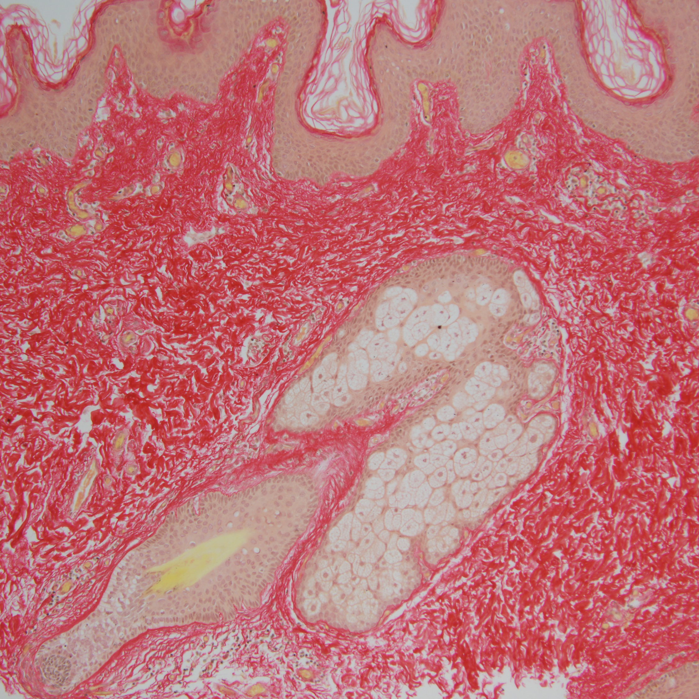
In the below images, the optional heteropolyacid pre-treatment steps were not used.
Skin tissue stained with PSR, without nuclear counterstain. Imaged with regular brightfield microscopy.
Skin tissue stained with PSR, without haematoxylin nuclear counterstain. Imaged with polarised light microscopy. Note that while keratin stains red with PSR, it does not exhibit birefringence as it is not collagenous.
Skin tissue stained with PSR, with Weigert's haematoxylin nuclear counterstain. Imaged with regular brightfield microscopy.
Skin tissue stained with PSR, with Weigert's haematoxylin nuclear counterstain. Imaged with polarised light microscopy. Note that while keratin stains red with PSR, it does not exhibit birefringence as it is not collagenous.
Protocol references
López De Padilla, C. M., Coenen, M. J., Tovar, A., De la Vega, R. E., Evans, C. H., & Müller, S. A. (2021). Picrosirius Red Staining: Revisiting Its Application to the Qualitative and Quantitative Assessment of Collagen Type I and Type III in Tendon. The journal of histochemistry and cytochemistry : official journal of the Histochemistry Society, 69(10), 633–643. https://doi.org/10.1369/00221554211046777
Citations
Step 2
Mukade Y, Kobayashi S, Nishijima Y, Kimura K, Watanabe A, Ikota H, Shirabe K, Yokoo H, Saio M. Phosphotungstic Acid-treated Picrosirius Red Staining Improves Whole-slide Quantitative Analysis of Collagen in Histological Specimens.
https://doi.org/10.1369/00221554221141140