Feb 10, 2026
Optimized SYBR Green qRT-PCR Workflow for Pharmacologic Perturbation in Head and Neck Squamous Cell Carcinoma (HNSCC) Models (TR146 and SCC58)
- 1University of Minnesota;
- 2Department of Diagnostic and Biological Sciences;
- 3School of Dentistry


Protocol Citation: Boluwatife OLU Afolabi 2026. Optimized SYBR Green qRT-PCR Workflow for Pharmacologic Perturbation in Head and Neck Squamous Cell Carcinoma (HNSCC) Models (TR146 and SCC58). protocols.io https://dx.doi.org/10.17504/protocols.io.ewov1k7bkgr2/v1
License: This is an open access protocol distributed under the terms of the Creative Commons Attribution License, which permits unrestricted use, distribution, and reproduction in any medium, provided the original author and source are credited
Protocol status: Working
We use this protocol and it's working
Created: February 09, 2026
Last Modified: February 10, 2026
Protocol Integer ID: 242945
Keywords: Head and Neck Squamous Cell Carcinoma (H..., Aryl Hydrocarbon Receptor (AHR), 4-nitroquinoline 1-oxide (4NQO), Benzo[a]pyrene (BaP), qRT-PCR, Gene Expression Analysis, Oral Squamous Cell Carcinoma (OSCC), pcr workflow for pharmacologic perturbation, general qpcr method, optimized sybr green qrt, ahr pathway activation, assessing transcriptional response, neck squamous cell carcinoma, transcriptional responses to pharmacologic perturbation, pcr workflow, standardized sybr green, relative gene expression change, squamous cell carcinoma, based qrt
Abstract
This protocol details a standardized SYBR Green-based qRT-PCR workflow for assessing transcriptional responses to pharmacologic perturbations (e.g., 4NQO, BaP) in head and neck squamous cell carcinoma (HNSCC) lines (TR146 and SCC58). Unlike general qPCR methods, this workflow is optimized for comparative toxicology, emphasizing strict matched-vehicle controls, light-sensitive handling for polycyclic aromatic hydrocarbons (PAHs), and plate-level normalization. It utilizes the ΔΔCt method with GAPDH normalization to quantify relative gene expression changes following DNA damage or AHR pathway activation.
Image Attribution
Created by the author (Boluwatife OLU Afolabi) using SciDraw
Guidelines
Suitable for:
- Dose-response and time-course analysis of environmental toxins.
- Determining transcriptional signatures.
- Evaluating AHR antagonist efficacy (e.g., CH223191).
Not suitable for:
- Absolute quantification (copy number).
- Clinical diagnostic tissue samples without modification.
Materials
Cell Models
- TR146 Cells: Oral squamous carcinoma.
- SCC58 Cells: Oral squamous carcinoma.
- Culture Media: (e.g., DMEM/F12 or user-specific formulation) supplemented with FBS; PBS
Chemical Treatments
- 4-nitroquinoline 1-oxide (4NQO): (Stock dissolved in DMSO; Store at -20°C).
- Benzo[a]pyrene (BaP): (Stock dissolved in DMSO; Light Sensitive; Store at -20°C).
- CH223191 (AHR Antagonist): (Stock dissolved in DMSO; Light Sensitive; Store at -20°C).
- Vehicle Control: Dimethyl sulfoxide (DMSO), PCR grade.
Molecular Biology Reagents
- RNA Extraction: Zymo Quick-RNA Kit (or equivalent).
- cDNA Synthesis: Bio-Rad cDNA Synthesis Kit.
- qPCR Master Mix: SYBR Green qPCR Master Mix (e.g., Bio-Rad SsoAdvanced).
- Primers: Gene-specific forward/reverse primers (validated for ~60°C Tm).
- Plates: 96-well 0.2 mL PCR plates (e.g., Bio-Rad Hard-Shell) and optical seals.
Safety warnings
- Carcinogen Hazard: 4NQO and BaP are potent mutagens and carcinogens. Handle only in a certified biosafety cabinet while wearing appropriate PPE (double gloves, safety glasses).
- Waste Disposal: All tips, tubes, and media contacting these chemicals must be disposed of as hazardous chemical waste, not biohazard trash.
- Light Sensitivity: BaP and CH223191 are photo-reactive or prone to degradation. Perform treatments in low light and wrap tubes/plates in foil.
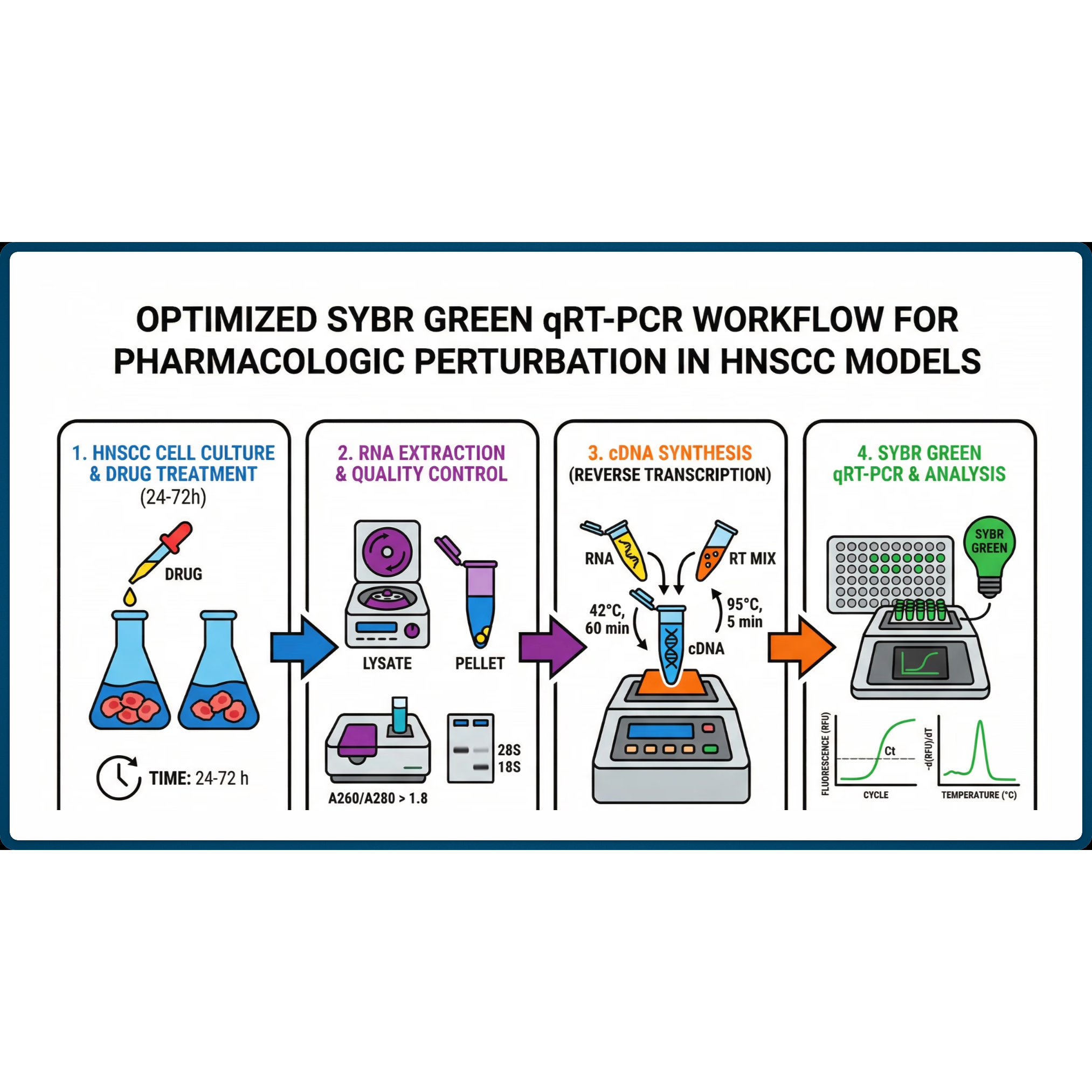
Step 1: Cell Seeding and Treatment
Seeding: Seed TR146 or SCC58 cells into 6-well plates (approx. 2–5×10⁵ cells/well) to reach ~70-80% confluency at the time of treatment.
Stock Preparation: Prepare 1000x stock solutions of 4NQO, BaP, or CH223191 in sterile DMSO.
Treatment Media: Dilute stocks 1:1000 into fresh, pre-warmed media.
- Critical: Ensure the final DMSO concentration is ≤0.1% in all wells, including the Vehicle Control.
Exposure: Aspirate old media, wash with PBS and apply treatment media.
- For BaP/CH223191: Immediately wrap plates in aluminum foil to protect from light.
Incubation: Incubate at 37°C / 5% CO2 for the designated time course (e.g., 6h, 12h, 24h).
Step 2: RNA Extraction & cDNA Synthesis
Harvest: Aspirate media and wash cells once with PBS.
Lysis: Add lysis buffer directly to the well (per Zymo kit instructions) and scrape cells.
- QC Check: Assess cell morphology prior to lysis. If significant cell death is visible (>20%), data may be confounded by apoptosis rather than specific signaling.
Extraction: Proceed with Zymo Quick-RNA protocol including the in-column DNase I treatment step to remove genomic DNA.
Quantification: Measure RNA concentration (Nanodrop).
A260/280 ratio should be > 2.0.
cDNA Synthesis: Normalize all samples to 500 ng total RNA input. Synthesize cDNA using the Bio-Rad iScript/cDNA kit (20 µL reaction volume).
- Store cDNA at -20°C.
Step 3: qRT-PCR Plate Setup
Primer Mix: Prepare a working stock of Forward/Reverse primers (final reaction concentration usually 300–500 nM).
Reaction Mix (per well):
- SYBR Green Master Mix: 5.0 µL
- Forward Primer: 1.0 µL
- Reverse Primer: 1.0 µL
- Nuclease-Free Water: 1.0 µL
- cDNA Template: 2.0 µL (diluted 1:5 to 1:10 if necessary)
- Total Volume: 10.0 µL
Plating: Pipette into 96-well plate. Run technical duplicates or triplicates.
Sealing: Apply optical seal and briefly centrifuge (1 min at 1000 x g) to remove bubbles.
Step 4: Thermal Cycling Conditions
| Step | Temperature | Time | Note | |
| Activation | 95.0°C | 3:00 | Polymerase activation | |
| Denature | 95.0°C | 0:15 | ||
| Anneal/Extend | 55.0°C | 0:30 | ||
| Repeat | GOTO Step 2 | 39x | Total 40 cycles | |
| Melt Curve | 65.0°C – 95.0°C | 0.5°C inc | 0:05 per step |
Optimization Note: 55°C is used here for specific primer compatibility. Standard Bio-Rad cycling often uses 60°C for Step 3. Ensure your primers are validated for 55°C.
Step 5: Data Analysis (Microsoft Excel / GraphPad Prism)
Export: Export quantization data (Ct values) to Excel.
QC Filter: Flag any replicates where Ct variance > 0.5.
Normalize (ΔCt): Calculate ΔCt=Ct(Target)−Ct(GAPDH) for each sample.
Normalize to Control (ΔΔCt): Calculate ΔΔCt=ΔCt(Treated)−ΔCt(Vehicle Control)
Fold Change: Calculate relative expression using the formula:
Fold Change=2−ΔΔCt
Statistics: Perform one-way ANOVA (for dose-response) or t-tests (paired by plate) in GraphPad Prism.
Protocol references
The Methodological Standard (The "Pfaffl" or Livak Method)
- Livak, K. J., & Schmittgen, T. D. (2001). Analysis of relative gene expression data using real-time quantitative PCR and the 2^−ΔΔCT method. Methods, 25(4), 402–408.
Characterization of the TR146 Cell Line
- Rupniak, H. T., Rowlatt, C., Lane, E. B., et al. (1985). Characteristics of four new human cell lines derived from squamous cell carcinomas of the head and neck. Journal of the National Cancer Institute, 75(4), 621–635.
4NQO as an Oral Cancer Model
- Kanojia, D., & Vaidya, M. M. (2006). 4-Nitroquinoline-1-oxide induced experimental oral carcinogenesis. Oral Oncology, 42(7), 655–667.
Acknowledgements
Special acknowledgement to Drs. Mark Herzberg, Karen Johnstone, and Chong Wang (University of Minnesota School of Dentistry) for their constant guidance.