Aug 24, 2022
Version 2
Optimized protocol for translatome analysis of mouse brain endothelial cells V.2
Peer-reviewed method
- Won-Jong OH1,
- Namsuk Kim1,
- Mi-Hee Jun1,
- Jin-Young Jeong1
- 1Korea Brain Research Institute
- PLOS ONE Lab ProtocolsTech. support email: [email protected]


External link: https://doi.org/10.1371/journal.pone.0275036
Protocol Citation: Won-Jong OH, Namsuk Kim, Mi-Hee Jun, Jin-Young Jeong 2022. Optimized protocol for translatome analysis of mouse brain endothelial cells. protocols.io https://dx.doi.org/10.17504/protocols.io.8epv59or6g1b/v2Version created by Neurovascularlab KBRI
Manuscript citation:
Kim N, Jun MH, Jeong JY, Oh WJ (2022)Optimized protocol for translatome analysis of mouse brain endothelial cells. PLOS ONE 17(9): e0275036. https://doi.org/10.1371/journal.pone.0275036
License: This is an open access protocol distributed under the terms of the Creative Commons Attribution License, which permits unrestricted use, distribution, and reproduction in any medium, provided the original author and source are credited
Protocol status: Working
We use this protocol and it's working
Created: August 23, 2022
Last Modified: August 24, 2022
Protocol Integer ID: 69048
Keywords: Brain endothelial cells, RiboTag, Translatome, RNA sequencing, pure mrnas from bec, many transcriptome analyses for bec, mouse brain endothelial cells brain endothelial cell, specificity of the mrna pool, hemagglutinin epitope under cre recombinase activation, molecular analysis of bec, mrna, becs at the molecular level, mrna level, optimized protocol for translatome analysis, mrna pool, translated protein, diverse neurodegenerative disease, many transcriptome analysis, molecular pathogenesis of these neurological disease, cre recombinase activation, low yield of mrna, pure mrna, bulk gene expression analysis, gene expression, protein, impairment of bec, parenchyma cells from toxin, quality cdna library for rna, translatome analysis, rna, using ribotag mice, molecular pathogenesi, ribotag mice, brain homeostasi, hemagglutinin epitope, ribosomal component
Abstract
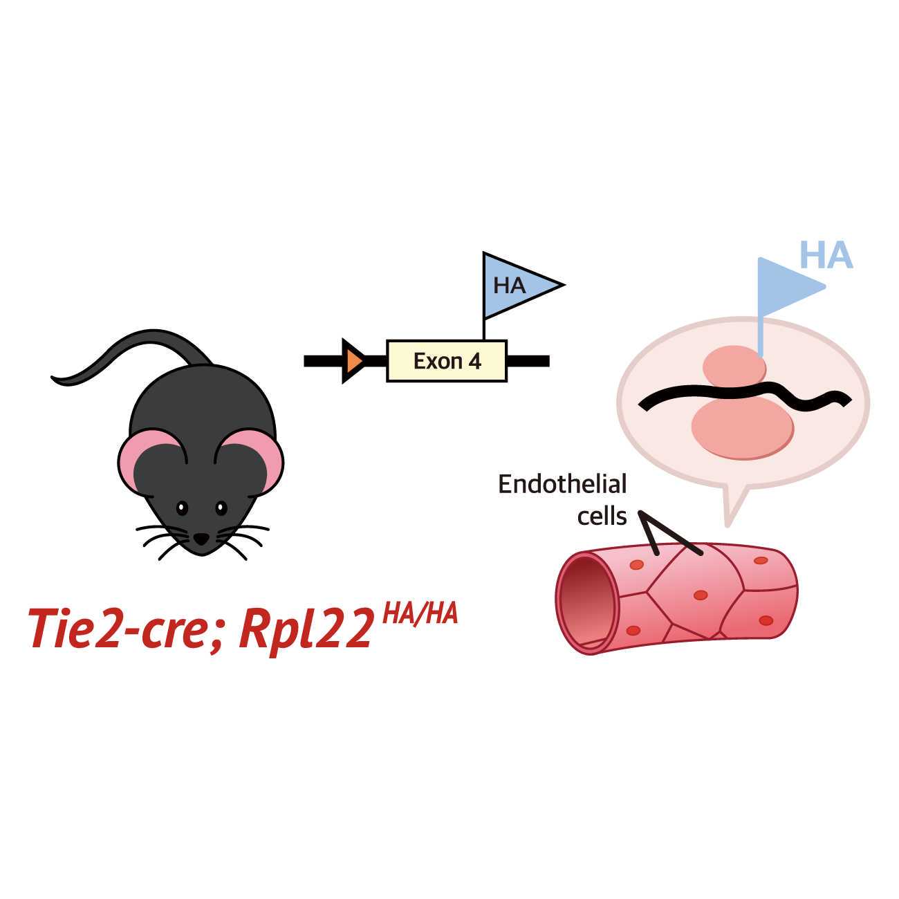
Brain endothelial cells (BECs) are important conduits that deliver oxygen and nutrients, protect parenchyma cells from toxins, and drain wastes to maintain brain homeostasis. Impairment of BECs has been implicated in diverse neurodegenerative diseases, including Alzheimer’s disease and Parkinson’s disease. Therefore, molecular analysis of BECs is important for understanding the molecular pathogenesis of these neurological diseases. Even though many transcriptome analyses for BECs have been developed, mRNA levels do not necessarily correlate with the levels of actively translated proteins. Translatome analysis using RiboTag mice, in which Rpl22, a ribosomal component, is tagged by the hemagglutinin epitope under Cre recombinase activation, could serve as an excellent tool that overcomes these caveats.However, implementation of this technique is limited by high noise-to-signal ratios as well as the low yield of mRNAs from BECs, which limits bulk gene expression analysis. In this study, we established a protocol to isolate highly pure mRNAs from BECs in the cortex of eight- to twelve-week-old male Tie2-Cre; Rpl22HA/HA mice by using a cell strainer to trap blood vessels prior to immunoprecipitation. According to the results of RT–PCR, the specificity of the mRNA pools isolated by our protocol was much higher than that of the pools isolated by the standard protocol. We were also able to generate a high-quality cDNA library for RNA-seq with the small amount of mRNA isolated with our protocol. Thus, this optimized method will be useful for future studies of BECs at the molecular level.
Guidelines
1. An RNase-free environment is essential. Use barrier pipet tips to avoid RNase contamination. Wipe down the surface of an experimental table and all equipment including surgical tools, pipets, etc., with RNase Zap.
2. Homogenization buffer and high-salt buffer should be freshly prepared.
3. Washes should be done in cold conditions.
4. Tissue samples should be processed fresh directly to RNA yield.
5. The average amount of BEC mRNA => whole cortex ( 8-12 weeks): 7.3 ng, visual cortex ( 8-12 weeks): 1.05 ng, visual cortex (2 weeks): 0.25 ng per mouse.
Materials
| A | B | C | |
| REAGENT or RESOURCE | SOURCE | IDNETIFIER | |
| Antibodies | |||
| Mouse anti-HA | Millipore | Cat# 05-904 RRID: AB_417380 | |
| Chemicals, peptides, and kits | |||
| TRIzolTM Reagent | Thermo | Cat# 15596026 | |
| Cycloheximide | Sigma-Aldrich | Cat# 1810 | |
| Magnesium chloride | Sigma-Aldrich | Cat# M8266 | |
| Potassium chloride | Sigma-Aldrich | Cat# P9333 | |
| DNase1 | Invitrogen | Cat# 18068015 | |
| Pierce™ Protein A/G Magnetic Beads | Thermo | Cat# 88803 | |
| Chloroform | Sigma | Cat# C2432 | |
| 20X TE Buffer (pH 7.5) | Promega | Cat# A2651 | |
| Ethyl alcohol, Pure | Sigma | Cat# E7023 | |
| Glycogen, Molecular Biology Grade | Roche | Cat# 10901393001 | |
| RNasin® Ribonuclease Inhibitor | Promega | Cat# N2115 | |
| Pierce™ Protein A/G Magnetic Beads | Thermo Fisher Scientific | Cat# 88803 | |
| Halt™ Protease and Phosphatase Inhibitor Cocktail | Thermo Fisher Scientific | Cat# 78444 | |
| NEBNext® Single Cell/Low Input RNA Library Prep Kit for Illumina® | NEB | Cat# E6420L | |
| NEBNext® Multiplex Oligos for Illumina® | NEB | Cat# E7600S | |
| High Sensitivity D5000 Screen tape | Agilent | Cat# 5067-5592 | |
| High Sensitivity D1000 Screen tape | Agilent | Cat# 5067-5584 | |
| High Sensitivity RNA Screentape | Agilent | Cat# 5067-5579 | |
| High Sensitivity D5000 Screen tape Reagent | Agilent | Cat# 5067-5593 | |
| High Sensitivity D1000 Screen tape Reagent | Agilent | Cat# 5067-5585 | |
| High Sensitivity RNA Screen tape Reagent | Agilent | Cat# 5067-5580 | |
| High Sensitivity D5000 Screen tape ladder | Agilent | Cat# 5067-5594 | |
| High Sensitivity D1000 Screen tape ladder | Agilent | Cat# 5067-5587 | |
| High Sensitivity RNA Screen tape ladder | Agilent | Cat# 5067-5581 | |
| Glass homogenizer | WHEATON | Cat# 357542 | |
| Disposable scalpel | Bard-Parker | Cat# 371611 | |
| Experimental models: Organisms/strains | |||
| Mouse: Tie2-Cre | The Jackson Laboratory | Stock# 008863 | |
| Mouse: Ai9 | The Jackson Laboratory | Stock# 007909 | |
| Mouse:RiboTag mice (Rpl22HA/HA) | The Jackson Laboratory | Stock# 011029 | |
Safety warnings
TRIzol is a highly corrosive and toxic chemical that can cause burns on contact with the skin as well as systemic poisoning.
Chloroform can cause a person to become unconscious and even be fatal at high doses.
Before start
1. An RNase-free environment is essential. Use barrier pipet tips to avoid RNase contamination. Wipe down the surface of an experimental table and all equipment including surgical tools, pipets, etc., with RNase Zap.
2. Homogenization buffer and high-salt buffer should be freshly prepared.
Vessel isolation
2h
The whole mouse cortex of a Tie2-Cre; Rpl22HA/HA mouse is isolated in the chilled DMEM. Then, tissues are dissociated by using a glass homogenizer (WHEATON, 357542) in 10 mL of chilled DMEM.
10m
1000 x g, 4°C, 00:10:00
10m
After discarding the supernatants, the pellets are resuspended in 15 mL of 20 % BSA-DMEM to avoid myelin contamination.
5m
2500 x g, 4°C, 00:15:00
15m
This process is repeated three times.
40m
After discarding the supernatants, the pellets are resuspended in 5 mL of chilled PBS.
5m
PBS containing blood vessels is passed through a 40-micrometer cell strainer.
5m
Immunoprecipitation
1d 1h
The strainer mesh containing vessels is then cut with a disposable scalpel (Bard-Parker, 371611) and transferred into a microcentrifuge tube for lysis in 600 µL of homogenization buffer containing 1 % (v/v) NP-40, 100 millimolar (mM) KCl, 50 millimolar (mM) Tris (7.4 ), 12 millimolar (mM) MgCl2, cycloheximide (100 mg/mL ), heparin (1 mg/mL ), Halt Protease and Phosphatase Inhibitor Cocktail (Thermo Fisher Scientific, 78444), RNA inhibitor (5 units/ml, Promega, N2615), and 1 millimolar (mM) DTT).
5m
The lysates are incubated On ice for 00:05:00 .
5m
12000 x g, 4°C, 00:10:00
10m
After being transferred to a new 1.5 ml microcentrifuge tube, the supernatants are incubated with a mouse monoclonal antibody against the HA epitope tag (1:200, Millipore, 05-904) for 04:00:00 at 4 °C with rotation by using a multimixer (NanoEnTek, 4519).
.
4h
Protein A/G magnetic beads (Thermo Fisher Scientific, 88803) equilibrated in homogenization buffer for 30 min are added to the antibody-lysate solution and incubated Overnight at 4 °C with gentle rotation.
16h
The next day, after a brief spin-dwon, the magnetic beads are washed five times with 1 mL high salt buffer ( 1% NP-40, 300 millimolar (mM) KCL, 50 millimolar (mM) Tris (7.4 ), 12 millimolar (mM) MgCl2, cycloheximide (100 mg/mL ), and 0.5 millimolar (mM) DTT).
30m
mRNA isolation
5h 55m
After the last wash, all supernatants are removed and 1 mL of TRIzol reagent (Invitrogen, 15596026) is added to the bead-antibody-tissue homogenate, followed by 200 µL of chloroform (Sigma-Aldrich, C2432).
Safety information
TRIzol is a highly corrosive and toxic chemical that can cause burns on contact with the skin as well as systemic poisoning.
Chloroform can cause a person to become unconscious and even be fatal at high doses.
10m
12000 x g, 4°C, 00:10:00
10m
The upper aqueous layer (approximately 600 µL ) is transferred into a new 15 ml conical tube, and 60 µL of 4 Molarity (M) LiCl, 120 µL of 20 X TE (0.2 Molarity (M) Tris-HCl, 20 millimolar (mM) EDTA, 7.5 , Promega, A2651), 1.8 mL of 100% ethyl alcohol (Sigma, E7023), and 3 mL of glycogen (Roche, 10901393001) are added for RNA precipitation.
15m
The mRNA mixture is incubated Overnight at -20 °C
16h
The following day, samples are centrifuged 12000 x g, 4°C, 00:10:00 . After the supernatants are discarded, 1 mL of 75% ethyl alcohol is added to the pellets for washing.
10m
After centrifugation7500 x g, 4°C, 00:05:00 and subsequent supernatant removal, the samples are air-dried for 00:05:00 at Room temperature . Do not overdry the beads.
10m
The dried pellets are then resuspended in 16 µL of RNase-free water.
5m
2 µL of DNase I and 2 µL of 10X DNase I Reaction Buffer (Invitrogen, 18068-015) are added to the reaction mixture, which is then incubated for 00:15:00 at Room temperature .
15m
DNaseI is inactivated by adding 25 millimolar (mM) of EDTA and heating at 65 °C for 00:10:00 .
10m
For RNA precipitation, 2.2 µL of 4 Molarity (M) of LiCl, 4.8 µL of 20 X TE (0.2 Molarity (M) Tris-HCl, 20 millimolar (mM) EDTA, 7.5 (Promega, A2651), 66 µL of 100% ethyl alcohol (Sigma-Aldrich, E7023), and 1 µL of glycogen (Roche, 10901393001) are added to the RNA mixture, followed by Overnight incubation at -20 °C .
16h
The next day, the RNA mixture is centrifuged 12000 x g, 4°C, 00:10:00
10m
After removing the supernatants, 1 mL of 75% ethyl alcohol is added to the pellets for washing.
5m
After centrifugation 7500 x g, 4°C, 00:05:00 , the supernatants are discarded.
5m
The pellets are then air-dried and finally resuspended in 10 µL of RNase-free water.
5m
Generation of cDNA library
2h 25m
The amount of isolated mRNA is measured by using High Sensitivity RNA ScreenTape (Agilent, 5067-5579), High Sensitivity RNA ScreenTape Reagent(Agilent, 5067-5580), and a High Sensitivity RNA ScreenTape ladder(Agilent, 5067-5581) from the Agilent 4200 TapeStation System according to the manufacturer's instructions.
One nanogram of mRNA obtained from RiboTag immunoprecipitation is reverse-transcribed into cDNA using the NEBNext Single Cell/Low Input RNA Library Prep Kit for Illumina (NEB, E6420L) according to the manufacturer’s protocol.
One nanogram of mRNA is added to the mixture containing 1 µL of NEBNext Single Cell RT (Reverse Transcription) Primer Mix. The 9 µL of the final volume is achieved by adding nuclease-free water.
The mixture is incubated at 70 °C for 00:05:00 with the heated lid set to 105 °C for annealing and then held at 4 °C .
5m
The RT mixture is prepared in a separate tube as follows On ice ; 5 µL of NEBNext Single Cell RT buffer, 1 µL of NEBNext Template Switching Oligo, 2 µL of NEBNext Single Cell RT Enzyme Mix, 3 µL of nuclease-free water. It is important to vortex the NEBNext Single Cell RT buffer prior to use for optimal performance.
The RT mixture (11 µL ) is combined with the annealed sample (9 µL ). Mix well by pipetting up and down at least 10 times.
The reaction is incubated in a thermocycler with the following steps: the heated lid is set to 105 °C , followed by 01:30:00 at 42 °C and 00:10:00 at 70 °C , and then held at 4 °C
1h 40m
The cDNA amplification mix is prepared as follows: 50 µL of NEBNext Single Cell cDNA PCR Master Mix, 2 µL of NEBNext Single Cell cDNA PCR Primer, and 28 µL of nuclease-free water.
80 µL of cDNA amplification mix are added to 20 µL of the sample with pipetting.
The reaction is performed in a thermocycler with the following PCR cycling conditions.
| Cycle step | Temperature | Time | Cycles | |
| Initial Denaturation | 98 °C | 45 sec | 1 | |
| Denaturation | 98 °C | 10 sec | 32 | |
| Annealing | 62 °C | 15 sec | ||
| Extension | 72 °C | 3 min | ||
| Final Extension | 72 °C | 5 min | 1 | |
| Hold | 4 °C |
For the next step, the NEBNext Bead Reconstitution Buffer and the SPRI (Solid Phase Reversible Immobilization) beads should be warmed to Room temperature for at least 00:30:00 before use.
30m
60 µL SPRI beads are added to the PCR. (mix well by pipetting up and down at least 10 times).
The samples are incubated for at least 00:05:00 at Room temperature .
5m
The samples are placed on the magnetic stand (Promega, Z5342) to separate the beads from the supernatant.
After 00:05:00 , the supernatant is removed. then, 200 µL of 80% freshly prepared ethanol is added for washing. The samples are Incubated at Room temperature for 00:00:30 ,, and then the supernatant is carefully removed and discarded.
5m 30s
. This process is repeated twice. The samples are air-dried for 00:05:00 at Room temperature . Do not overdry the beads.
5m
50 µL of 0.1X TE (diluted from 1X TE buffer) is added to the samples to elute the cDNA from the beads.
The samples are mixed well and incubateed for at least 00:02:00 at Room temperature .
2m
Next, 45 µL of NEBNext Bead Reconstitution Buffer is added to the cDNA-Bead mixture. Mix well by pipetting up and down at least 10 times and incubate for at least 00:05:00 at Room temperature .
5m
The samples are placed on a magnetic stand to separate the beads.
After 00:05:00 , the supernatant is carefully removed.
5m
Then, 200 µL of 80% freshly prepared ethanol is added to the tube to wash the beads. After 00:00:30 of incubation at Room temperature , . This process is repeated twice.
30s
The beads containing cDNA are air-dried for 00:05:00 at Room temperature . Do not overdry the beads.
5m
cDNA is eluted from the beads by adding 33 µL of 1X TE. Mix well by pipetting up and down at least 10 times. The sample is incubated for at least 00:02:00 at Room temperature .
2m
The sample is placed on the magnetic stand. After 00:05:00 of incubation at Room temperature , 30 µL of the solution is transferred to a new tube.
5m
The cDNA quality and quantity can be assessed by using High Sensitivity D5000 ScreenTape (Agilent, 5067-5592), High Sensitivity D5000 ScreenTape Reagent (Agilent, 5067-5593), and a High Sensitivity D5000 ScreenTape ladder (Agilent, 5067-5594) in the Agilent 4200 TapeStation System.
40 ng of cDNA is used for Illumina NGS (Next Generation Sequencing) library preparation.
40 ng of cDNA in 1X TE is mixed with 7 µL of NEBNext Ultra II FS Reaction Buffer and 2 µL of NEBNext Ultra II FS Enzyme Mix in a PCR tube. The final volume of the mixture is brought to 35 µL , and the sample is vortexed for 00:00:05 .
5s
In a thermocycler, with the heated lid set to 75 °C , the following program is performed: 00:25:00 at 37 °C and 00:30:00 at 65 °C .
55m
While the PCR is running, prepare the solution for the next step. NEBNext Adaptor for Illumina is diluted by 25-fold in the NEBNext Adaptor Dilution Buffer.
The following components should be added directly to the above sample (35 µL ). The adaptor should be added separately to each sample (DO NOT premix with ligation master mix and enhancer).
| Component | Volume | |
| FS Reaction Mixture | 35 μl | |
| NEBNExt Ultra II Ligation Master Mix | 30 μl | |
| NEBNext Ligation Enhancer | 1 μl | |
| NEBNext Adaptor for Illumina (dilluted 1:25) | 2.5 μl |
The samples are mixed well by using pipetting the entire volume up and down at least 10 times. The ligation mixture is incubated at 20 °C for 00:15:00 in a thermocycler without the heated lid.
15m
3 µL of USER Enzyme, a mixture of uracil DNA glycosylase (UDG) and the DNA glycosylase-lyase endonuclease VIII, is added to the ligation mixture and mixed well. Incubate at 37 °C for 00:15:00 .
15m
For the next step, the NEBNext Bead Reconstitution Buffer and the SPRI beads should be warm to Room temperature for at least 00:30:00 before use.
30m
57 µL of SPRI beads are added to the PCR reaction. The sample is incubated for at least 00:05:00 at Room temperature .
5m
The sample is placed on a magnetic stand.
After 00:05:00 incubation, the supernatant is removed. Then, 200 µL of 80% freshly prepared ethanol is added to the tube. After incubation at Room temperature for 00:00:30 , the supernatant is removed. . This process is repeated twice.
5m 30s
The beads containing cDNA are air-dried for 00:05:00 at Room temperature . Do not overdry the beads.
5m
17 µL of 0.1X TE is added to resuspend the beads. The cDNA-bead mixture is incubated for at least 00:02:00 at Room temperature .
2m
The sample is placed on a magnetic stand. After 00:05:00 , 15 µL of the cleared solution is transferred to a new PCR tube.
5m
The following components are combined into a new PCR tube.
| Component | Volume | |
| Adaptor Ligated DNA Fragments | 15 μl | |
| NEBNext Ultra II Q5 Master Mix | 25 μl | |
| Index Primer / i7 | 5 μl | |
| Index Primer / i5 | 5 μl |
Labelling with dual barcodes is performed by using the following PCR cycling conditions.
| Cycle step | Temperature | Time | Cycles | |
| Initial Denaturation | 98 °C | 30 sec | 1 | |
| Denaturation | 98 °C | 10 sec | 8 | |
| Annealing | 65°C | 75 sec | ||
| Final Extension | 65 °C | 5 min | ||
| Hold | 4 °C | � |
For the next step, the NEBNext Bead Reconstitution Buffer and the SPRI beads should be warmed to Room temperature for at least 00:30:00 before use.
30m
The PCR mixture is resuspended in 45 µL of SPRI beads. The sample is incubated for at least 00:05:00 at Room temperature .
5m
The cDNA-bead mixture is placed on a magnetic stand to separate the beads from the supernatant.
After 00:05:00 , the supernatant is removed and discarded.
5m
200 µL of 80% freshly prepared ethanol are added to the tube in the magnetic stand. . This process is repeated twice.
The beads containing cDNA are air-dried on a magnetic stand for 00:05:00 at Room temperature .
5m
The cDNA library is eluted by adding 33 µL of 0.1X TE. Mix well by pipetting up and down 10 times.
The sample is placed on a magnetic stand. After 00:05:00 , 30 µL of the sample containing the cDNA library is transferred to a new tube. Libraries can be stored at -20 °C .
5m
Before NGS, the quality of the final cDNA libraries is checked by using High Sensitivity D1000 ScreenTape (Agilent, 5067-5584), High Sensitivity D1000 ScreenTape Reagent(Agilent, 5067-5585), and a High Sensitivity D1000 ScreenTape ladder (Agilent, 5067-5587) in the Agilent 4200 TapeStation System.