Jan 23, 2026
Opsonization-assisted phagocytosis assay using particles conjugated to synthetic peptides via glutaraldehyde
- 1University of the Philippines Manila


Protocol Citation: Brian Andrich Pollo 2026. Opsonization-assisted phagocytosis assay using particles conjugated to synthetic peptides via glutaraldehyde. protocols.io https://dx.doi.org/10.17504/protocols.io.81wgbozz1lpk/v1
Manuscript citation:
King RAN, Climacosa FMM, Santos BMM, Caoili SEC. A Human Erythrocyte-based Haemolysis Assay for the Evaluation of Human Complement Activity: Https://DoiOrg/101177/0261192920953170 2020;48:127–35. https://doi.org/10.1177/0261192920953170.
License: This is an open access protocol distributed under the terms of the Creative Commons Attribution License, which permits unrestricted use, distribution, and reproduction in any medium, provided the original author and source are credited
Protocol status: Working
We use this protocol and it's working
Created: January 23, 2026
Last Modified: January 23, 2026
Protocol Integer ID: 239209
Keywords: opsonization, phagocytosis, glutaraldehyde, synthetic peptides, human leukocytes, phagocytosis assay, assisted phagocytosi, synthetic peptides via glutaraldehyde, using human leukocyte, opsonization with human plasma, wbc interaction, human leukocyte, leukocyte, phagocytic index, inactivated plasma, synthetic peptide, wbc, glutaraldehyde, human plasma, glutaraldehyde crosslinking, using particle
Funders Acknowledgements:
DOST-PCHRD
Grant ID: MD PhD Dissertation Grant
Disclaimer
DISCLAIMER – FOR INFORMATIONAL PURPOSES ONLY; USE AT YOUR OWN RISK
The protocol content here is for informational purposes only and does not constitute legal, medical, clinical, or safety advice, or otherwise; content added to protocols.io is not peer reviewed and may not have undergone a formal approval of any kind. Information presented in this protocol should not substitute for independent professional judgment, advice, diagnosis, or treatment. Any action you take or refrain from taking using or relying upon the information presented here is strictly at your own risk. You agree that neither the Company nor any of the authors, contributors, administrators, or anyone else associated with protocols.io, can be held responsible for your use of the information contained in or linked to this protocol or any of our Sites/Apps and Services.
Abstract
This protocol describes the opsonization and phagocytosis assay using human leukocytes and synthetic-peptide-decorated particles: L. floteri_, RBCs, and yeast. Particle conjugation is achieved through glutaraldehyde crosslinking. Following opsonization with human plasma (positive control) or heat-inactivated plasma (negative control), particle–WBC interactions are measured via microscopy, and the phagocytic index is determined.
Attachments

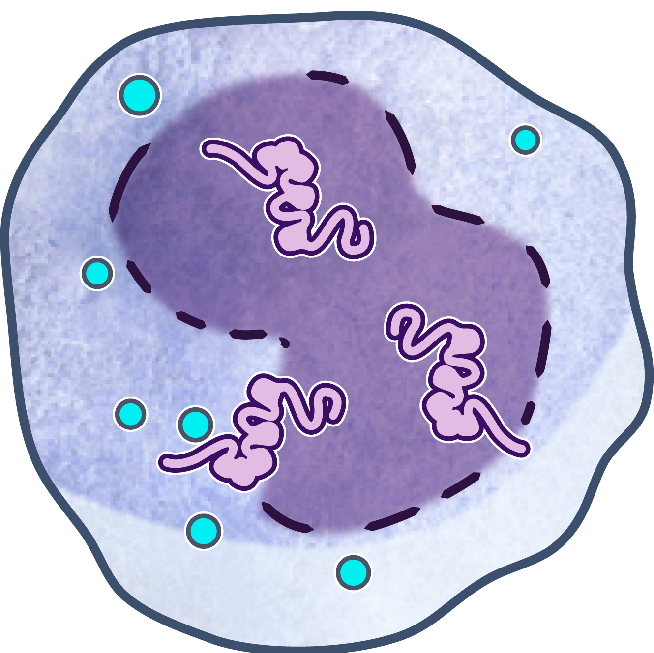
Image Attribution
Courtesy of NIAIDRyan Kissinger, Public domain, via Wikimedia Commons
Materials
A. Reagent Preparation
A.1 Prepare 10× RBC lysis buffer (100 ml)
- Ammonium chloride
- Sodium bicarbonate
- Disodium EDTA
- Distilled water
A.2 Prepare 2× HBSS (500 ml)
- NaCl
- KCl
- Na2HPO4
- KH2PO4
- Glucose
- CaCl2
- MgSO4
- NaHCO3
- Distilled water
A.3 Prepare Wright stain solution (100 ml)
- Wright stain powder
- Methanol
B. Particle–Peptide Conjugation
B.1 Prepare particle suspensions
- Lactobacillus reuteri in PBS
- RBCs in PBS
- Yeast in PBS
B.2 Conjugate synthetic peptides
- Glutaraldehyde
- Synthetic peptide
- Glycine
C. WBC Isolation
- Whole blood in CPDA-1 tubes
- 1× RBC lysis buffer
D. Cell Viability and Counting
- WBC suspension
- Trypan blue
E. Plasma Treatment
- Pooled human plasma
F. Opsonization of Particles
A. Reagent Preparation (Estimated time: 30–40 min, excluding storage)
Prepare 10× RBC lysis buffer (100 ml)
Weigh 0.82 g ammonium chloride.
Weigh 0.84 g sodium bicarbonate.
Weigh 0.37 g disodium EDTA.
Add all solids to a 100 ml volumetric flask.
Add distilled water to reach 100 ml total volume.
Mix until completely dissolved.
Store at 4 °C for up to 6 months.
Prepare 2× HBSS (500 ml)
Weigh: 8 g NaCl; 400 mg KCl; 48 mg Na2HPO4; 60 mg KH2PO4; 1 g glucose; 140 mg CaCl2 (anhydrous); 98 mg MgSO4 (anhydrous); 350 mg NaHCO3.
Add all solids to a 500 ml volumetric flask.
Add distilled water to 500 ml total volume.
Mix until dissolved.
Store at 4 °C for up to 1 month.
Prepare Wright stain solution (100 ml)
Weigh 300 mg Wright stain powder.
Add to 100 ml methanol.
Mix until dissolved.
Store at room temperature in a dark bottle.
B. Particle–Peptide Conjugation via Glutaraldehyde (Estimated time: ~1.5 h)
Prepare particle suspensions
Suspend Lactobacillus reuteri in PBS at ~1 × 10^8 cells/ml.
Suspend RBCs in PBS at ~1 × 10^8 cells/ml.
Suspend yeast in PBS at ~1 × 10^8 cells/ml.
Conjugate synthetic peptides
Add glutaraldehyde (stock: 25%w/v or 2.5M) to each suspension to a final concentration of 0.1% (v/v) (10.6mM).
Incubate 30 min at room temperature.
Add synthetic peptide to each suspension to a final concentration of 40 µg/ml (stock: 400 µg/ml).
Incubate 30 min at room temperature.
Quench with glycine to 100 mM final concentration (stock: 1M).
Incubate 10 min at room temperature.
Wash and resuspend
Centrifuge at 300 × g for 5 min.
Discard supernatant.
Resuspend pellet in PBS.
Repeat wash twice more.
Resuspend final pellet in HBSS or assay buffer.
C. WBC Isolation (Estimated time: 40–50 min)
Lysis of RBCs
Collect whole blood in CPDA-1 tubes.
Centrifuge at 300 × g for 5 min to separate plasma.
Transfer plasma to a fresh tube and place on ice.
Transfer 1 ml RBC pack with buffy coat to a new tube.
Add 10 ml of 1× RBC lysis buffer.
Incubate 10 min at room temperature with gentle rocking.
Centrifuge 5 min at 300 g.
Discard supernatant.
Repeat lysis if RBCs remain visible.
Washing and resuspension
Wash WBC pellet once with HBSS.
Centrifuge 5 min at 300 g.
Resuspend WBCs in HBSS or assay buffer.
D. Cell Viability and Counting (Estimated time: 10–15 min)
Mix 100 µl WBC suspension with 100 µl 0.4% trypan blue.
Incubate for 3 min at room temperature.
Load 100 µl onto hemocytometer.
Count viable (unstained) and non-viable (blue-stained) cells.
E. Plasma Treatment (Estimated time: 40 min, including incubation)
Thaw pooled human plasma at 37 °C for 10 min.
Keep plasma on ice until use.
For negative (complement-inactivated) control, heat-inactivate plasma at 56 °C for 30 min in a heat block.
F. Opsonization of Particles (Estimated time: 50 min)
Prepare particle suspensions at ~1 × 10^8 particles/ml.
Add 300 µl particle suspension to 1.2 ml plasma.
Incubate 30 min at 37 °C with shaking.
Centrifuge 15 min at 1,600 g.
Discard supernatant.
Resuspend pellet in 300 µl HBSS.
G. Phagocytosis Assay (Estimated time: 50–60 min)
Prepare WBC suspension at 5 × 10^6 cells/ml in HBSS.
Mix 200 µl WBC suspension with 200 µl opsonized particle suspension.
Incubate 20 min at 37 °C with rotation at 8 RPM.
Centrifuge 8 min at 250 g.
Discard supernatant.
Wash pellet with HBSS.
Repeat wash twice more.
Resuspend final pellet in ice-cold HBSS.
Place 100 µl suspension onto microscope slide.
Air-dry in a laminar flow hood.
Stain with Wright stain according to manufacturer’s instructions.
Air-dry again.
H. Microscopy and Data Collection (Estimated time: varies; ~15–20 min per sample)
Examine slides under 1000× magnification.
Count 50 neutrophils per condition.
Record the number of neutrophils containing ≥1 particle.
Record the number of particles per positive cell.
Calculate the phagocytic index: Phagocytic Index = (% positive neutrophils) × (mean particles per positive cell)
Protocol references
The Drevets, Canono, and Campbell (2015) protocol describes macrophage ingestion and killing assays, including distinguishing bound vs. internalized bacteria using fluorescence microscopy. Boero et al. (2021) use flow cytometry to evaluate neutrophil phagocytosis of S. aureus_, employing opsonization and fluorescence readouts for higher throughput.
Drevets DA, Canono BP, Campbell PA. Measurement of Bacterial Ingestion and Killing by Macrophages. Curr Protoc Immunol 2015;109:14.6.1-14.6.17. https://doi.org/10.1002/0471142735.IM1406S109.