Apr 09, 2026
Negative staining of macromolecules, viruses and isolated organelles using uranyl acetate for transmission electron microscopy analysis
- 1Ramaciotti Centre for Cryo EM, Monash University, Melbourne Australia


Protocol Citation: Jillian Danne, Georg Ramm, Denis Korneev 2026. Negative staining of macromolecules, viruses and isolated organelles using uranyl acetate for transmission electron microscopy analysis. protocols.io https://dx.doi.org/10.17504/protocols.io.261ge5zn7g47/v1
License: This is an open access protocol distributed under the terms of the Creative Commons Attribution License, which permits unrestricted use, distribution, and reproduction in any medium, provided the original author and source are credited
Protocol status: Working
We use this protocol and it's working
Created: October 03, 2024
Last Modified: April 09, 2026
Protocol Integer ID: 108978
Keywords: Negative staining, Uranyl Acetate, Proteins, Extracellular vesicles, Bacteriophages, Glow discharge, Transmission electron microscope, Viruses, Organelles, uranyl acetate for transmission electron microscopy analysis, negative staining of macromolecule, transmission electron microscopy analysis, transmission electron microscopy, microscopy, such as uranyl ion, negative staining, transmission electron microscope, uranyl ion, such as isolated organelle, visualisation of biological structure, isolated organelle, including individual protein, macromolecule, protein, protein complex, individual protein, biological structure, using uranyl acetate
Abstract
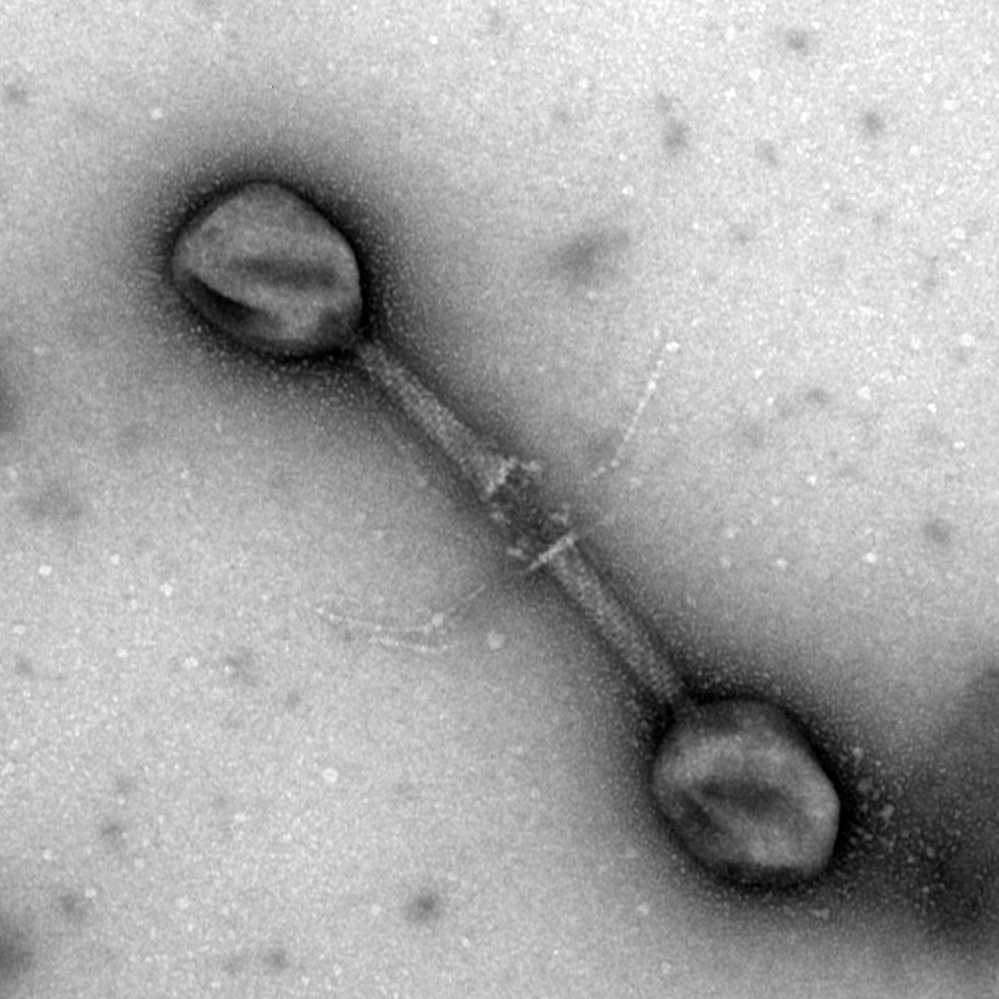
Negative staining is a rapid and simple technique that allows for the visualisation of biological structures in the nanometre size range, such as isolated organelles, viruses and macromolecules, including individual proteins and protein complexes. A sample suspended in solution is absorbed onto a glow discharged continuous carbon support film grid and electron dense heavy metals, such as uranyl ions are deposited around the sample providing external structural information in which the object of interest appears clear and the background dark when imaged using a transmission electron microscope.
Materials
MilliQ water
Uranyl acetateElectron Microscopy SciencesCatalog #22400
Equipment
Parafilm M
NAME
Bemis
BRAND
IA041
SKU
Equipment
Plain glass slides 76mm x 39mm x 1.0-1.2mm
NAME
Thermo Scientific
BRAND
AGL4222A
SKU
Equipment
Carbon Support Films, Copper 300 mesh
NAME
QUANTIFOIL
BRAND
N1-C73nCu30-01
SKU
Equipment
PELCO easiGlow™ Glow Discharge Cleaning System
NAME
PELCO
BRAND
91000
SKU
Equipment
Glass board
NAME
N/A
BRAND
N/A
SKU
Equipment
Fine Forceps
NAME
Forceps
TYPE
Dumont
BRAND
11251-10
SKU
LINK
Equipment
10ml Syringe, Luer lock tip
NAME
Terumo
BRAND
19046TE
SKU
Equipment
Acrodisc syringe filter 25mm, 0.2um pore size
NAME
Pall Corporation
BRAND
Z259969
SKU
Equipment
Falcon® Centrifuge Tubes
NAME
Polypropylene, Sterile, 15 mL
TYPE
Corning®
BRAND
352096
SKU
Equipment
Foil
NAME
N/A
BRAND
N/A
SKU
Equipment
Gilson Pipetman classic P20
NAME
Gilson
BRAND
F144056M
SKU
Equipment
Gilson Pipetman P200
NAME
Gilson
BRAND
F144058M
SKU
Equipment
Filter paper, grade 1, 12.5cm
NAME
Whatman
BRAND
1001-125
SKU
Equipment
Glass petri dish, 100mm
NAME
BRAND
BRAND
BR455751
SKU
Equipment
Grid storage box, 100 grid capacity
NAME
Gilder
BRAND
HL065
SKU
Equipment
JEOL JEM-1400 Plus 120keV Transmission electron microscope
NAME
JEOL
BRAND
N/A
SKU
Safety warnings
Uranyl acetate is radioactive and extremely toxic if ingested, inhaled or in contact with abraded or cut skin. This chemical should be handled with care using the appropriate personal protective equipment (PPE).
Negative staining
Wrap a pieces of parafilm around a glass slide and place a QUANTIFOIL carbon support film grid or custom made carbon coated formvar grid on the parafilm, carbon side up.
Glow discharge the grid on the slide for 30 seconds at 30mA.
Example: PELCO easiGlowTM Glow Discharge Cleaning System.
A glow discharge instrument uses air to clean transmission electron microscope (TEM) carbon support films by removing hydrocarbons. The film becomes negatively charged and hydrophilic, allowing an aqueous sample solution to spread onto the surface of the grid easily.
Place a sheet of parafilm on a clean flat bench, glass board or large glass petri dish lid using a small amount of water underneath to keep the parafilm flat.
Pipette 5-10 μl of sample solution onto the clean parafilm surface.
Pipette 10-50μl of 0.5-2% aqueous uranyl acetate onto the same clean parafilm surface.
Uranyl acetate is toxic and radioactive and must be handled with care using the appropriate personal protective equipment (PPE).
The uranyl acetate stock solution should be aqueous and not made up in phosphate buffered saline or phosphate buffer. Uranyl ions can react with phosphate ions to produce electron dense precipitates. Uranyl acetate can also precipitate on exposure to UV light, so the solution should be filtered prior to use, wrapped in foil and stored in a dark place.
Float the grid in the sample solution drop carbon side down for 1-3 minutes.
Sample purification is critically important for clear visualisation of a homogeneous negatively stained sample.
Place a sheet of parafilm underneath a piece of filter paper to prevent solutions contaminating the bench surface. Pick up the grid using fine forceps, tilt the grid at a 45-60 degree angle and bring the grid to the piece of filter paper to blot away the excess solution. Take care not to damage the carbon support film.
Using fine forceps, transfer the grid to the uranyl acetate drop sample side down and incubate for approximately 30-60 seconds. Uranyl acetate incubation times may require optimisation depending on the sample that is being negatively stained.
Uranyl ions primarily interact with proteins and lipids. In negative staining, these heavy metal ions surround the specimen rather than binding to it and scatter electrons, providing negative contrast during TEM imaging.
Blot away the excess uranyl acetate solution as described in Step 7. If staining is insufficient (Step 8), consider increasing the uranyl acetate incubation time or repeat Steps 5 & 8-9.
Place the grid on a sheet of filter paper sample side up, cover with a glass petri dish and dry for 10 minutes. Store grids in a grid storage box.
Place a grid in a transmission electron microscope grid holder for high resolution imaging using a transmission electron microscope.
Example: JEOL JEM-1400-Plus TEM at 80 keV equipped with a high sensitivity bottom mount CMOS 'Flash' camera.
Protocol references
Scarff, Charlotte A., et al. "Variations on negative stain electron microscopy methods: tools for tackling challenging systems." JoVE (Journal of Visualized Experiments) 132 (2018): e57199.
Théry, Clotilde, et al. "Isolation and characterization of exosomes from cell culture supernatants and biological fluids." Current protocols in cell biology 30.1 (2006): 3-22.
Bonilla, Natasha, et al. "Phage on tap–a quick and efficient protocol for the preparation of bacteriophage laboratory stocks." PeerJ 4 (2016): e2261.
Acknowledgements
We would like to thank Associate Professor Jeremy Barr, School of Biological Science, Monash University for providing the T4 bacteriophages that were negatively stained and imaged for this protocol.