Nov 07, 2023
MRI Imaging of the Gut
- 1Department of Neurologial Surgery, Weill Cornell Medicine


Protocol Citation: Santiago Unda, Rachel Retik, Michael G. Kaplitt 2023. MRI Imaging of the Gut . protocols.io https://dx.doi.org/10.17504/protocols.io.x54v9pwd4g3e/v1
License: This is an open access protocol distributed under the terms of the Creative Commons Attribution License, which permits unrestricted use, distribution, and reproduction in any medium, provided the original author and source are credited
Protocol status: Working
We use this protocol and it's working
Created: November 07, 2023
Last Modified: September 23, 2024
Protocol Integer ID: 90570
Keywords: ASAPCRN, MRI, gut, measure peristaltic activity in the upper portion, gut, peristaltic activity, test, upper portion
Funders Acknowledgements:
ASAP
Grant ID: ASAP-020608
Abstract
This test is used measure peristaltic activity in the upper portion of the gut.
Materials
- DietGel Recovery, Cat#72-06-5022 ClearH2O, ME, USA
- Gadolinium [Gd-DTPA powder] Cat#381667, Sigma Aldrich, St. Louis, USA
- 7T rodent MRI BioSpec 70/30; Bruker Instruments, Billerica, USA
Protocol
Habituate mice to eat Diet gel (Cat#DietGel Recovery, ClearH2O, ME, USA) for at least a week.
Note
Habituation takes ~7 days of training until mice consume voluntarily approximately 1g of diet gel.
Fast mice for 12 to 18 hours.
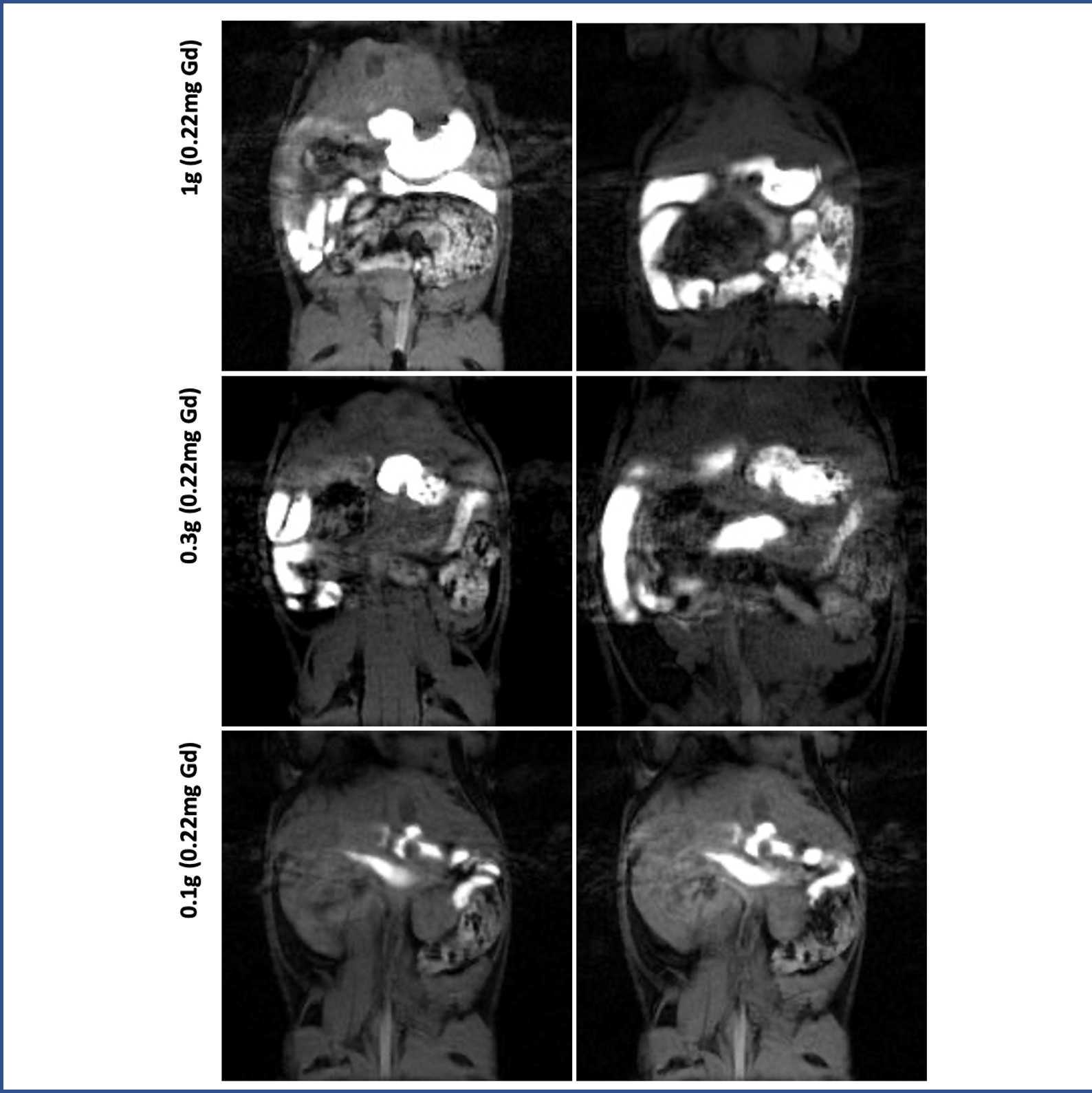
Prepare 10g of Diet gel mixed with 22.4mg of Gadolinium [Gd-DTPA powder] (Cat#381667, Sigma Aldrich, St. Louis, USA).
Note
Boil the diet gel in warm water until its composition is completely liquid, let it cool down for 2-3 min and add the gadolinium powder, mix it well and mixed solution will return to it semi-solid gel state.
Give 0.3g of Diet Gel-Gadolinium mix to each mouse.
Note
0.3g of the Diet Gel-Gadolinium per mouse might vary depending on mouse weight. For a 30g mouse this amount will be enough to fill immediately the upper GI tract, however in our experience an amount of 1g is enough to get a visible contrast in the GI lumen.
Note
Following 12 to 18 hours of food restriction, mice should eat the gel within 2 to 5 min.
Using a 7T rodent MRI (BioSpec 70/30; Bruker Instruments, Billerica, USA), position the animal in prone and monitor respiratory activity patterns.
Note
Respiratory gating is highly recommended for image quality.
Localize the longitudinal axis of the stomach.
Perform series of alternating volumetric and fast scans.
Note
Volumetric scans are performed using a FLASH sequence, parameters should be troubleshooted for each investigator, however reference parameters for repetition time, echo time, angles, and thickness can be taken from Lu et al, 2018.
Sequences commonly take 2 min, repeat for as long as your experimental setting requires.
Protocol references
Lu KH, Cao J, Oleson S, Ward MP, Phillips RJ, Powley TL, Liu Z. Vagus nerve stimulation promotes gastric emptying by increasing pyloric opening measured with magnetic resonance imaging. Neurogastroenterol Motil. 2018 Oct;30(10):e13380. doi: 10.1111/nmo.13380. Epub 2018 May 24. PMID: 29797377; PMCID: PMC6160317.