Aug 23, 2022
Mouse lung MAIT cell expansion and purification protocol
- 1University of California, San Diego;
- 2La Jolla Institute for Immunology


External link: https://www.lji.org/labs/kronenberg
Protocol Citation: Gabriel A Ascui, Eleni Phung, Alba Mendis, Mitchell Kronenberg 2022. Mouse lung MAIT cell expansion and purification protocol. protocols.io https://dx.doi.org/10.17504/protocols.io.x54v9yrpqg3e/v1
License: This is an open access protocol distributed under the terms of the Creative Commons Attribution License, which permits unrestricted use, distribution, and reproduction in any medium, provided the original author and source are credited
Protocol status: Working
We use this protocol and it's working
Created: March 29, 2022
Last Modified: June 05, 2023
Protocol Integer ID: 60070
Keywords: MAIT cells, in vitro, lung, mouse, role of mait cell, mait cell, lung immune responses against bacterial infection, expansion of mait cell, mouse lung mait cell expansion, immune response, immune system, cultured salmonella typhimurium brd509 vaccine strain, cytokine, antigen, microbial infection, cell, infection, mhc class, bacterial infection, flow cytometry, mait, infecting mice retro, salmonella, lung
Funders Acknowledgements:
NIH
Grant ID: 5R01AI137230-04
Disclaimer
The Authors declare no competing interests.
Abstract
Mucosal Associated Invariant T (MAIT) cells are unconventional T cells present abundantly in human tissues. MAIT cells interact with antigens presented by MR1, an MHC class I protein, to fight microbial infections. They also reportedly activate through cytokines to combat viruses. While MAIT cells exhibit the innate-like characteristic of rapid responses to infections, they also display adaptive-like qualities such as an effector-memory phenotype. Defining the role of MAIT cells in the immune system is essential to understand lung immune responses against bacterial infections. The following protocol outlines the expansion of MAIT cells via a cultured Salmonella Typhimurium BRD509 vaccine strain. Six days after infecting mice retro-pharyngeally with BRD509, MAIT cells were extracted from the lungs. The cells were then enriched and labeled with fluorescence for sorting. Once sorted, MAIT cells were plated and activated with anti-CD3/CD28. The cells were then analyzed with flow cytometry.
Protocol materials
Red Blood Cell Lysis Buffer Hybri-MaxMerck MilliporeSigma (Sigma-Aldrich)Catalog #R7757
Cell strainer 70um filterFalconCatalog #352350
gentleMACS C-Tube Miltenyi BiotecCatalog #130-093-334
Spleen Dissociation Medium 10 x 4 mL
STEMCELL Technologies Inc.Catalog #7915
LIVE/DEAD™ Fixable Yellow Dead Cell Stain Kit, for 405 nm excitationThermo FisherCatalog #L34967
EasySep™ Mouse Streptavidin RapidSpheres™ Isolation Kit For processing 1 x 10^9 cells
STEMCELL Technologies Inc.Catalog #19860
The Big Easy EasySep™ Magnet For isolating 1 x 10^9
STEMCELL Technologies Inc.Catalog #18001
MAIT cell in vivo expansion with BRD509
Salmonella enterica serotype Typhimurium BRD509 vaccine strain culture
Safety information
Work in BSL2 biosafety conditions.
6d
Day 0: Overnight culture of Salmonella Typhimurium BRD509 strain.
- Prepare 5 mL of LB media with 100 µg/ml of Streptomycin.
- Pick -80 °C BRD509 stock and inoculate tube.
- Incubate Overnight in agitation225 rpm, 37°C .
10m
Day 1: Prepare 4h culture of BRD509.
- Take 1 mL of overnight BRD509 culture and dilute with LB media until 5 mL .
- Culture for an additional 04:00:00 to 06:00:00 .
- Measure OD600 on spectrophotometer.
- Calculate the amount of culture that will need to be diluted in 900 µL of sterile PBS to get 10^6 CFU per mouse based on the following conversion table (input OD600):
| A | B | C | D | E | F | |
| A600 | 1 | A600 of culture | 0.6 | |||
| CFU/mL | 600000000 | CFU/mL | 360000000 | |||
| Desired CFU/mouse | 2.7777777777777777 | uL per mouse | ||||
| Resuspend in 900uL (30uL per mouse) | 83.33333333333333 | uL for 30 mice | ||||
A600 BRD509 spreadsheet
5h
Day 1: Infection Mice
1. Anesthetize the mice with isoflurane gas.
2. Administer retro-pharyngeal injections of 30 µL of diluted BRD509 per mouse.
3. Prepare dilutions of the original dose at 10^4 and 10^5 in sterile PBS.
4. Plate dilutions on LB agar plate with Streptomycin. Count colonies next day to calculate actual dose.
1h
MAIT cell sorting
2h
Prepare Lungs MAIT cells
Safety information
Work in BSL2 biosafety conditions.
Day 6: Collect lungs
1. Collect lungs from infected mice and process ingentleMACS C-Tube Miltenyi BiotecCatalog #130-093-334wwith 2 mL of Spleen Dissociation Medium 10 x 4 mL
STEMCELL Technologies Inc.Catalog #7915 using program 37C_m_LDK_1. This should take about 00:30:00 .
2. Filter single cell suspension through Cell strainer 70um filterFalconCatalog #352350 on 50 mL conical tube. Mash any remaining bits of tissue through the strainer with a syringe plunger. Wash strainer with HBSS with 10% FBS until 25 mL .
3. Centrifuge at 1500 rpm, 4°C, 00:05:00 .
4. Lyse red blood cells by adding 1 mL of Red Blood Cell Lysis Buffer Hybri-MaxMerck MilliporeSigma (Sigma-Aldrich)Catalog #R7757 . Incubate at Room temperature for 00:05:00 .
5. Add 14 mL of HBSS 10%FBS and centrifuge at 1500 rpm, 4°C, 00:05:00 .
6. Re-suspend cells in 2 mL of MACS buffer.
45m
MAIT cell enrichment
- Add 50 ul of Rat Serum to sample.
- Add the following biotin-conjugated antibodies:
| A | B | C | |
| Antigen | Stock [ ] (mg/ml) | V in 2 ml (ul) | |
| CD11b | 0.5 | 4 | |
| CD11c | 0.5 | 4 | |
| Ter119 | 0.5 | 4 | |
| F4/80 | 0.5 | 4 | |
| B220 | 0.5 | 4 | |
| Ly6G | 0.5 | 4 | |
Biotin-conjugated antibodies
3. Incubate atRoom temperature for 00:10:00 .
4. Vortex EasySep™ Mouse Streptavidin RapidSpheres™ Isolation Kit For processing 1 x 10^9 cells
STEMCELL Technologies Inc.Catalog #19860 for 00:00:30 . Add 50 µL of RapidSpheres to sample.
5. Mix and incubate for 00:05:00 at Room temperature .
6. Add 2 mL MACS Buffer.
7. Place tube into The Big Easy EasySep™ Magnet For isolating 1 x 10^9
STEMCELL Technologies Inc.Catalog #18001 . Incubate for 00:05:00 at Room temperature .
8. Gently pour supernatant in a new 5 ml tube.
20m 30s
Fluorescent labelling
- Stain cells with PE-conjugated or mouse MR1 tetramer loaded with 5-OP-RU at 1:300 dilution for 00:45:00 at Room temperature . Wash with PBS 2% FBS and centrifuge at 1500 rpm, 4°C, 00:05:00 .
Note
Mouse MR1 tetramers were provided by the NIH Tetramer Core Facility:
- Stain cells with LIVE/DEAD™ Fixable Yellow Dead Cell Stain Kit, for 405 nm excitationThermo FisherCatalog #L34967 at 1:500 dilution, FcBlock (2G4) at 1:500 dilution and 1:500 dilution of 1 mg/mL Free Streptavidin in PBS.
Note
NOTE: Make sure the PBS has no other protein content.
3. Incubate at 4 °C for 00:15:00 . Wash with PBS 2% FBS.
4. Stain cells with the following antibodies:
| A | B | C | D | E | F | G | |
| # | Marker | Population | Channel | Host | Clone | Dilution | |
| 2 | gd TCR | DUMP | PerCP-Cy5.5 | Mouse | GL3 | 1:400 | |
| 3 | IgD | DUMP | PerCP-Cy5.5 | Rat | 11-26c.2a | 1:300 | |
| 4 | CD11b | DUMP | PerCP-Cy5.5 | Rat | M1/70 | 1:300 | |
| 5 | CD11c | DUMP | PerCP-Cy5.5 | Rat | N418 | 1:300 | |
| 6 | B220 | DUMP | PerCP-Cy5.5 | Rat | RA36B2 | 1:300 | |
| 7 | 5-OP-RU | MAIT cells | PE | - | - | 1:300 | |
| 8 | TCR beta | T cells | APC-eF780 | Mouse | H57-597 | 1:300 | |
| 9 | Live/Dead | Yellow | BV570 | Rat | MEL14 | 1:200 | |
| 10 | CD45 | Lymphocytes | BV785 | Rat | 30.F11 | 1:300 |
MAIT cell Sorting Panel
4. Incubate at4 °C for 00:30:00 .
5. Wash cells with PBS 2% FBS. Resuspend in 4 mL .
1h 35m
Cell Sorting
- Sort MAIT cells with 85 nm nozzle on low pressure. Sort into 500 µL of sterile FBS in 5 ml FACS tubes.
- Gate samples as following: Lymphocytes/Singlets/Live/DUMP-CD45+/Tetramer+TCRbint
Expected result
You should obtain a 20-30% frequency of MAIT cells from the DUMP-CD45+ gate. Total number of MAIT cells should be between 20,000-50,000 per lung.
In vitro culture
2h
Day 1: MAIT cell activation with anti-CD3/CD28
Coat 48-well plate
While MAIT cells are been sorted, prepare anti-CD3e + anti-CD28 coated plates.
1. Dilute anti-CD3e (2C11) to 5 µg/ml and anti-CD28 to 2 µg/ml in sterile PBS.
2. Add 100 µL to each well of a 48-well plate.
3. Incubate at 37 °C for 02:00:00 .
4. Wash plate with 200 µL of PBS, twice.
5. remove all liquid from every well.
2h
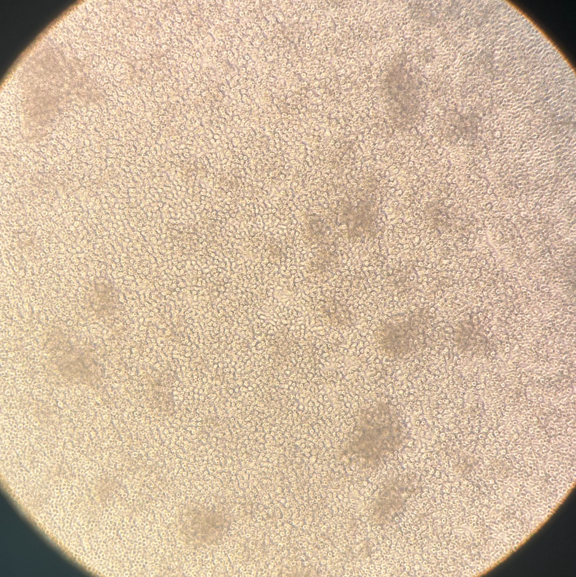
Plate MAIT cells
Note
- MAIT cell culture media: RPMI 10% FBS 1X Pen/Strep 1X Pyruvate 1X HEPES 55 uM β-mercaptoethanol.
1. Wash sorted cells with MAIT cell culture media.
2. Resuspend cells in MAIT cell at 0.5 x 10^6 cells/mL in MAIT cell culture media supplemented with the following cytokines:
| A | B | C | D | |
| Cytokine | Final [ ] | Stock [ ] | Dilution | |
| IL-2 | 10 ng/ml | 20 ug/ml | 1:2000 | |
| IL-7 | 20 ng/ml | 20 ug/ml | 1:1000 | |
| IL-1b | 10 ng/ml | 100 ug/ml | 1:1000 | |
| IL-23 | 10 ng/ml | 10 ug/ml | 1:1000 |
Cytokines
3. Plate 500,000 cells per well.
10m
Day 2: Put in fresh plate.
5m
Analyze MAIT cells through flow
- Stain cells with PE-conjugated or mouse MR1 tetramer loaded with 5-OP-RU at 1:300 dilution for 00:45:00 at Room temperature . Wash with PBS 2% FBS and centrifuge at 1500 rpm, 4°C, 00:05:00 .
- Stain cells with LIVE/DEAD™ Fixable Yellow Dead Cell Stain Kit, for 405 nm excitationThermo FisherCatalog #L34967 at 1:500 dilution, FcBlock (2G4) at 1:500 dilution and 1:500 dilution of 1 mg/mL Free Streptavidin in PBS.
Note
NOTE: Make sure the PBS has no other protein content.
3. Incubate at 4 °C for 00:15:00 . Wash with PBS 2% FBS.
4. Stain cells with the following antibodies:
| A | B | C | D | E | F | G | H | I | J | |
| # | Marker | Population | Channel | Host | Clone | Dilution | ||||
| 2 | gd TCR | DUMP | PerCP-Cy5.5 | Mouse | GL3 | 1:400 | ||||
| 3 | IgD | DUMP | PerCP-Cy5.5 | Rat | 11-26c.2a | 1:300 | ||||
| 4 | CD11b | DUMP | PerCP-Cy5.5 | Rat | M1/70 | 1:300 | ||||
| 5 | CD11c | DUMP | PerCP-Cy5.5 | Rat | N418 | 1:300 | ||||
| 6 | B220 | DUMP | PerCP-Cy5.5 | Rat | RA36B2 | 1:300 | ||||
| 7 | 5-OP-RU | MAIT cells | PE | - | - | 1:300 | ||||
| 8 | TCR beta | T cells | APC-eF780 | Mouse | H57-597 | 1:300 | ||||
| 9 | Live/Dead | Yellow | BV570 | Rat | MEL14 | 1:200 | ||||
| 10 | CD45 | Lymphocytes | BV785 | Rat | 30.F11 | 1:300 |
4. Incubate at4 °C for 00:30:00 .
5. Wash cells with PBS 2% FBS. Resuspend in 4 mL .
6. Analyze cells through flow.
1h 35m