Jun 04, 2025
Mouse Liver Periportal Assembloids Method
- Anna M. Dowbaj1,
- Aleksandra Sljukic1,
- Meritxell Huch1
- 1Max Planck Institute of Molecular Cell Biology and Genetics, Dresden, Germany
- Anna M. Dowbaj: equal contribution
- Aleksandra Sljukic: equal contribution
- Meritxell Huch: corresponding author



Protocol Citation: Anna M. Dowbaj, Aleksandra Sljukic, Meritxell Huch 2025. Mouse Liver Periportal Assembloids Method. protocols.io https://dx.doi.org/10.17504/protocols.io.36wgqqk6xgk5/v1
License: This is an open access protocol distributed under the terms of the Creative Commons Attribution License, which permits unrestricted use, distribution, and reproduction in any medium, provided the original author and source are credited
Protocol status: Working
We use this protocol and it's working
Created: May 08, 2025
Last Modified: June 04, 2025
Protocol Integer ID: 217966
Keywords: hepatocyte organoids, ductal organoids, portal fibroblasts, liver, homeostasis, fibrosis, biliary fibrosis, periportal assembloids, mouse liver periportal assembloids method, hepatocyte organoids with cholangiocyte, mouse liver periportal assembloids method the study, cellular contributions to cholestatic liver disease, periportal liver assembloid platform, cholangiocyte, bile into ductal structure, single cells of cholangiocyte, portal mesenchymal cell, combining hepatocyte organoid, hepatocyte organoid, primary adult hepatocyte, bile canaliculi network, key features of the liver, bile drainage dynamic, bile canaliculi formation, portal fibroblast, cholestatic liver disease, organoids to single cells step, biliary fibrosi, ductal cell, liver disease progression, mesenchymal cell, draining bile, liver, recapitulate tissue architecture, modeling of cholestatic injury, modulation of mesenchymal cell abundance, mesenchymal cell abundance, organoid, multicellular interaction, cell, vitro system, mouse assembloid protoco
Funders Acknowledgements:
Max Planck Gessellschaft
Grant ID: core grant
Abstract
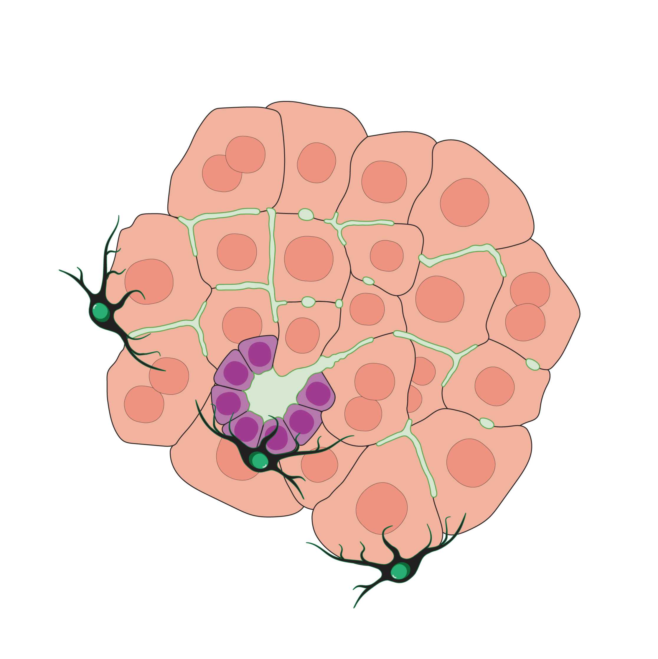
The study of liver disease progression requires in vitro systems that faithfully recapitulate tissue architecture and multicellular interactions. Here, we describe a periportal liver assembloid platform composed of primary adult hepatocytes, cholangiocytes, and portal mesenchymal cells. This system reconstructs key features of the liver’s periportal region and enables modeling of cholestatic injury and biliary fibrosis in vitro. By combining hepatocyte organoids with cholangiocytes and portal fibroblasts, we generate organized, functional assembloids that consistently form bile canaliculi networks capable of draining bile into ductal structures. Importantly, modulation of mesenchymal cell abundance within the assembloids is sufficient to trigger fibrotic-like responses, independent of an immune compartment. The system is compatible with genetic perturbations, including gene knockdown and creation of chimeric assembloids of mutant and wild-type cells, allowing the investigation of cell-autonomous mechanisms in a controlled environment. This protocol provides a robust and scalable approach to study bile canaliculi formation, bile drainage dynamics, and cellular contributions to cholestatic liver disease and fibrosis within a single integrated mouse model.
___________________________________________________________
Table of content for the Mouse assembloid protocol below:
To make assembloids we prepare single cells of cholangiocytes and portal fibroblasts and hepatocyte organoids released from Matrigel. Then, they are either “assembled” on the rocking platform, or in the Aggrewells. After assembly, the formed assembloids are seeded in Matrigel, and left for 6-7 days.
Where possible, we provide several alternative versions of the protocol, each producing periportal assembloids with similar efficiency.
PART 1: PREPARATION OF THE STARTING MATERIAL
Step 2. Processing of Cholangiocyte / Ductal cell (DC) Organoids to Single Cells
Step 3. Processing of Mesenchymal cells (Portal fibroblasts) to Single Cells
Step 4. Processing of Hepatocyte Organoids (HepOrgs) - Version 1
Step 5. Processing of Hepatocyte Organoids (HepOrgs) - Version 2
PART 2: ASSEMBLY METHODS
To make homeoststic-like assembloids, we combine 10 HepOrgs, 1000 ductal cells and 250 mesenchymal cells
To make fibrotic-like assembloids, we combine 10 HepOrgs, 1000 ductal cells and 2500 mesenchymal cells
Step 6. Assembly Method 1: ROCKING PLATFORM
Step 7. Assembly Method 2: AGGREWELL
PART 3: COLLECTION and SEEDING OF ASSEMBLOIDS
Step 8-9. Assembloid collection from rocking platform, and seeding (Version 1 and 2)
Step 10. Assembloid collection from Aggrewell, and seeding
Guidelines
Since for the generation of periportal assembloids it is required to prepare three cell types, here is general advice on preparing them:
Choice of cells:
Prepare cholangiocytes, portal fibroblasts and HepOrgs with different endogenous fluorescence so you can distinguish between cell types you are adding; this also helps with the validation if the method produced periportal assembloids.
- since HepOrgs are fully formed, larger structure (150-300
- for portal fibroblasts, it is helpful to use the endogenously labelled PDGFRα-H2B-GFP [B6.129S4-Pdgfratm11 (EGFP)Sor/J] mice, because the portal mesenchyme is already GFP+. You can find the original paper describing their isolation outlined in the section "Materials"
- for cholangiocytes, mTmG [Gt(ROSA)26Sortm4(ACTBtdTomato,-EGFP)Luo/J] are most frequently used.
_____________________________________________
Preparation of cells and organoids for assembly:
Steps 1-3 in the steps section do not need to be done sequentially. It is even better if several experimenters can prepare cells simultaneously.
However, if a person is working alone, it is advisable to first prepare cholangiocytes and portal fibroblasts, because they can wait on ice for up to 2 hours without losing viablity.
It is important to note that HepOrgs are more sensitive, and one should prepare them last. HepOrgs can be stored for short time at both 4°C or RT.
Materials
Cells and Organoids (all primary from mouse liver):
Assembloids are made by combining:
- single cell portal fibroblasts,
- single cell ductal cells (grown as ductal organoids), and
- pre-grown hepatocyte organoids.
Protocols for their isolation and cultivation can be found in provided references.
- Initial research article describing the isolation of ductal cells and generation of ductal organoids can be found here: 10.1038/nature11826
- Step-by-step protocol details of the isolation of ductal cells and generation of ductal organoids can be found here: 10.1038/nprot.2016.097
- Initial research article describing the isolation of portal fibroblasts can be found here: 10.1016/j.stem.2021.07.002
- Step-by-step protocol details of ductal cell and portal fibroblast isolation can be found here: 10.1016/j.xpro.2023.102333
- Initial research article describing the periportal assembloids and protocol for growing hepatocyte organoids can be found here: 10.1038/s41586-025-09183-9
____________________________________________________________________
Medium composition for assembloids:
Composition of base medium which we call Advanced DMEM/F12 +++
NOTE: "+++" stands for 3 components it has (Pen/Strep, GlutaMAX, and HEPES)
| Advanced DMEM/F12 +++ | Final conc. | Amount | |
| Advanced DMEM/ F12 | N/A | 489 mL | |
| Pen/Strep | 1% | 5 mL | |
| GlutaMAX | 1% | 5 mL | |
| HEPES | 10 mM | 5 mL of 100× stock | |
| Total | N/A | 500 mL |
Store up to 1 month at 4°C.
____________________________________________________________________
Composition of medium for assembloids which we call Minimal Medium (MM):
| Minimal Medium | Final conc. | Amount | |
| Advanced DMEM /F12 +++ | N/A | 14 mL | |
| WNT3a conditioned medium | 30% | 6 mL | |
| B27-Supplement, serum free | 1% | 0.4 mL of 50× stock | |
| N-acetylcysteine (NAC) | 1.25 mM | 25 μL of 500mM stock | |
| Rock kinase inhibitor Y27632 | 10 μM | 20 μL of 10mM stock | |
| Total | N/A | 20 mL |
Store up to 2 weeks at 4°C.
____________________________________________________________________
List of genral reagents and resources:
| Reagent | Source | Cat. No | |
| Fetal bovine serum | Merck/Sigma | Cat# F7524 | |
| Advanced DMEM/F-12 | Thermo Fisher Scientific | Cat# 12634010 | |
| HEPES (1M) | Thermo Fisher Scientific | Cat# 15630056 | |
| Penicillin/Streptomycin | Thermo Fisher Scientific | Cat# 15140-122 | |
| GlutaMAX supplement | Thermo Fisher Scientific | Cat# 35050-068 | |
| TrypLE Express Enzyme (1×), phenol red | Thermo Fisher Scientific | Cat# 12605010 | |
| B27-Supplement, serum free | Thermo Fisher Scientific | Cat# 17504-044 | |
| N-acetylcysteine (NAC) | Merck/Sigma | Cat# A9165 | |
| Nicotinamide | Merck/Sigma | Cat# N0636 | |
| Rspondin 1 (RSPO1) conditioned medium | Home-made as in Broutier et al., 2016 Nature protocols | N/A | |
| WNT3a conditioned medium | Home-made as in Broutier et al., 2016 Nature protocols | N/A | |
| Rock kinase inhibitor Y27632 | Merck/Sigma | Cat# Y0503; CAS: 129830-38-2 | |
| Matrigel Growth Factor Reduced (GFR) BasementMembrane Matrix, Phenol Red-free | Corning | Cat# 356231 |
____________________________________________________________________
Most commonly used mouse lines:
| Mouse strains | Used for | Identifier | |
| Mouse: C57/Bl6Any gender. Preferably use animals <12 weeks | HepOrgs | ||
| Mouse: mTmG [Gt(ROSA)26Sortm4(ACTBtdTomato,-EGFP)Luo/J]Any gender. Preferably use animals <12 weeks | Ductal Cells | RRID: IMSR_JAX:007576 | |
| Mouse: PDGFRα-H2B-GFP [B6.129S4-Pdgfratm11 (EGFP)Sor/J]Any gender. Preferably use animals <12 weeks | Portal fibroblasts | Hamilton et al., 2003; RRID: IMSR_JAX:007669 |
Periportal assembloid protocol overview
Protoocol overview
PART 1: PREPARATION OF THE STARTING MATERIAL
PART 2: ASSEMBLY METHODS
PART 3: COLLECTION and SEEDING OF ASSEMBLOIDS
Processing of Cholangiocyte / Ductal cell (DC) Organoids
Step-by-step processing to Single Cells
Pre-warm 1x TrypLE in water bath at 37 °C.
Prepare 15 ml falcon tube with 2ml cold media (AdvDMEM+++).
Scrape down cholangiocyte organoids from Mtg dome (p0-p9) with p1000 pipette tip and transfer them to 15 ml falcon tube.
Pipette approx. 20x up and down with a Pasteur glass pipette.
Add up to 10ml of media (AdvDMEM+++).
Spin 200g/5min.
Aspirate the supernatant carefully until 2ml are left using a Pasteur glass pipette; if unsure use a pipette.
Break organoids with plugged glass Pasteur pipette.
Add up to 10ml of media (AdvDMEM+++).
Spin 200g/5min.
Remove supernatant and resuspend the cell pellet in 1ml of pre-warmed 1x TrypLE (37 °C).
Incubate 6-10 min in water bath at 37 °C.
Resuspend cells using a narrow Pasteur or 1000 µl pipette tip.
Add 1ml of AdvDMEM+++
Strain the cells with 40 µm strainer into 15 ml falcon tube
Check under the microscope for cell clumps.
Add 10ml AdvDMEM+++ to inhibit TrypLE enzymatic activity.
Spin 500g/5min.
Aspirate the supernatant.
Resuspend cells in MM media.
Count cells.
Processing of Mesenchymal cells (Portal fibroblasts)
Step-by-step processing to Single Cells
Split cells when 80-90% confluent.
Prewarm dissociation reagent and 1x PBS in water bath. Usually as a dissociation reagent 1x TrypLE is used. If the mesenchymal cells look more spread instead of a spindle-shaped, use the mixture of accutase and 10x TrypLE in 1:1 ratio (to make 1ml of this mixture, use 500 µl of accuatse and 500 µl of 10x TrypLE).
Remove the media from the wells
Wash the cells with 1x PBS (200 µl for 96wp, 250 µl for 48wp and 500 µl for 24wp)
Remove 1x PBS
Add prewarmed dissociation reagent (100 µl for 96wp, 200 µl for 48wp, or 250 µl for 24wp)
Incubate at 37 °C 5% CO2 for 5 minutes in a tissue culture incubator
Check whether cells are detached (if not return for two more minutes)
Stop the reaction with 1% FBS in AdvDMEM+++ (amount equivalent to dissociation reagent used)
Collect cells in 15 ml falcons
Spin down 3 min 500 xg
Remove supernatant and resuspend in MM (depending on pellet size from 500 µl to 1ml)
Processing of Hepatocyte Organoids (HepOrgs) - version 1
HepOrg release from Matrigel - V1
Cut the tip of p100 pipette tip (optional), and collect Mtg droplets with HepOrgs (with media) in 1.5 ml Eppendorf tube. NOTE: You can collect up to 5 Mtg droplets in one 1.5 ml Eppendorf tube.
After you have collected HepOrgs with Mtg in Eppendorf tubes, keep them on ice until you collect all Mtg droplets
After you have collected all Mtg droplets with HepOrg you intend to use, spin them down using table-top centrifuge for 20 seconds
Remove supernatant, and add 0.7 ml of ice-cold Cell Recovery solution in each Eppendorf tube and pipette up and down around 10 times
Put Eppendorf tubes back on ice for 10 min
After 10 min, spin them down using table-top centrifuge for 20 seconds
Remove supernatant, and add 0.7 ml of ice-cold Cell Recovery solution in each Eppendorf tube and pipette up and down around 10 times
Put Eppendorf tubes back on ice for 5-10 min (check every few minutes if you see organoids released from Matrigel)
Remove supernatant and pipette up and down what is left in the tube, if HepOrgs seem released from Mtg, add 0.7 ml of AdvDMEM+++ in each Eppendorf tube (This step is necessary to wash out Cell Recovery solution, which when kept on the organoids for a longer period leads to decrease in HepOrg viability)
Spin them down using table-top centrifuge for 20 seconds
Remove supernatant and add 0.7 ml of MM media in each Eppendorf tubes
Now you can proceed with using HepOrgs for preparing for assembloid making
Transfer HepOrgs released from Mtg in well of a 6-well plate, and add 2-3 mL of MM media.
Handpick organoids using a microscope with 4x objective, using p200 or p20 pipette, to enrich for HepOrg with bubbly shape for the generation of periportal assembloids
Processing of Hepatocyte Organoids (HepOrgs) - version 2
HepOrg release from Matrigel - V2
Collect 2x 5 wells of a 48wp, HepOrg passage 1.
Add 10ml AdvDMEM+++ media, resuspend.
Spin 5min at 200g.
Aspirate supernatant as much as possible (0.3ml left).
Add 3ml of the Cell Recovery solution (Corning #354253).
Incubate 10min on ice, 2x mixing manually with a pipette.
Add 10ml of AdvDMEM+++ media.
Spin 5min at 200g.
Resuspend in MM for picking.
Pick organoids with P200 pipette using a stereoscope to enrich for HepOrg with bubbly shape for the generation of periportal assembloids (no need to cut the tip, as they are much smaller than the opening of the pipette).
Place picked organoids in non-attachment 24wp.
Assembly Method 1: ROCKING PLATFORM
Here, we are transferring HepOrgs, single cell ductal cells and single cell mesenchymal cells into wells of a 24 well plate, adding media, and placing the 24 well plate on a rocking platform that will allow spontanoeus assembly of 3 cell types.
Starting material needed for one well of a 24 well plate:
Collect 10 HepOrgs (150-300µm size in diamter) per one well of the 24 well plate.
Add 250 (homeostatic) or 2500 (fibtotic) portal fibroblasts.
Add the volume of MM to have a total of 500 µl for 24 wp.
Put plate on rocking platform in the incubator (37 °C, 5 % C02) for 18-24 hours at 10 rpm.
Assembly Method 2: AGGREWELL
Follow the manufacturer’s instructions for handling the Aggrewell (Aggrewell800, Stem Cell Technologies #34811)
Pretreat the wells with Anti-Adherence Rinsing Solution (Stem Cell Technologies, #07010).
Spin 1300g/5min with the solution (for balancing the plate, another Aggrewell should be used).
Check for bubbles, if present, spin again.
Aspirate the Anti-Adherence Rinsing Solution.
Mix all your cell types together and add to the well in 1.5ml of MM media.
Pipette up and down in the well to distribute the cells.
Spin the plate at 100g/5min.
Aggregate in an incubator for 18-24 hours without agitation.
Assembloid collection from rocking platform, and seeding
Assembloid collection from rocking platform, and seeding - Version 1 (Matrigel dome):
1. After incubation on rocking platform, collect assembloids from 24 wp wells into 1.5 ml eppendorf tube - you can pool same conditions in one tube.
2. Check under stereoscope whether you have collected all structures, if not wash the plate with MM and transfer the rest of the structures to another eppendorf tube.
3. Spin down 200 xg for 5 minutes.
4. Remove supernatant (try to remove as much as possible, otherwise Matrigel will be diluted and assembloids will attach more readily to the plate bottom).
5. Add Mtg, resuspend and seed; for seeeding in a well of 48 wp resuspend in 25 µl of Mtg; for seeeding in a well of 24 wp resuspend in 50 µl of Mtg
6. Incubate 30 min in the incubator (37 °C, 5 % C02) and add MM (24wp - 500 µl and 48 wp - 250 µl).
Assembloid collection from rocking platform, and seeding - Version 2 ("sandwich" method):
1. Transfer assembloids to 15ml falcon with p1000.
2. Wash wells once with 1ml of AdvDMEM+++.
3. Spin 100g/5min.
4. Remove the supernatant and harvest assembloids.
5. Add 20 µl of Mtg on the bottom of well of a 96 well plate. Plate should not be pre-warmed.
6. Spin plate at 100g/5min with a cold centrifuge (4°C). Centrifugation step will create an even distribution of Mtg for a bottom Mtg layer.
7. Add either (A) assembloids on top of the Mtg layer and leave them to settle for 5min; then add 20 µl of Mtg on top of them, or (B) pre-mix assembloids with 20 µl of Mtg and add this mixture on top of the first Mtg layer.
8. Leave the plate in the incupator (37 °C, 5 % C02) for 30min.
9. Add 100 µl of MM.
Assembloid collection from Aggrewell, and seeding
1. Take p1000 and set it to 1ml.
2. Take 1ml of media from the well and aspirate it firmly back to the well to disrupt the structures.
3. Take a 40 µm strainer and place it on a 15ml falcon tube.
4. Gently aspirate your assembloids from the well with p1000 and pass through a filter.
5. Wash Aggrewell 3x with 1ml of media to get all the structures on the strainer.
6. Take a 6wp non-attachment plate.
7. Flip the strainer, place it in the 6wp and wash the aggregates off the strainer.
8. Check the strainer to see if there are any structures left, if needed, wash the strainer 2 more times.
9. Take all the washthrough and collect it in a 15ml falcon.
10. Spin 100g/5min.
11. Remove the supernatant and harvest aggregated structures.
12. First layer of Mtg: 20 µl Mtg, spin 100g/5min COLD CENTRIFUGE.
13. Seeding in Mtg (on top of the first layer of Mtg + assembloid, 5 min, +20 µl Matigel on top, 30min 37 °C, +100 µl MM media).
Protocol references
Huch, M. et al. In vitro expansion of single Lgr5+ liver stem cells induced by Wnt-driven regeneration. Nature 494, 247-250, doi:10.1038/nature11826 (2013).
Broutier, L. et al. Culture and establishment of self-renewing human and mouse adult liver and pancreas 3D organoids and their genetic manipulation. Nat Protoc 11, 1724-1743, doi:10.1038/nprot.2016.097 (2016).
Cordero-Espinoza, L. et al. Dynamic cell contacts between periportal mesenchyme and ductal epithelium act as a rheostat for liver cell proliferation. Cell Stem Cell 28, 1907-1921 e1908, doi:10.1016/j.stem.2021.07.002 (2021).
Dowbaj, A. M., Kohler, T. N., Cordero-Espinoza, L., Hollfelder, F. & Huch, M. Generation of liver mesenchyme and ductal cell organoid co-culture using cell self-aggregation and droplet microfluidics. STAR Protoc 4, 102333, doi:10.1016/j.xpro.2023.102333 (2023).
Acknowledgements
We would like to acknowledge Armin Niksic for his contribution and help to the development of the protocol and insightful discussions.