Nov 24, 2020
Mouse 2- Step Collagenase Liver Perfusion protocol
- Michael Cheng1,
- Xue-Zhong Ma1,
- Chao Jiang1,
- Ian McGilvray1,
- Sonya Macparland1
- 1University of Toronto


Protocol Citation: Michael Cheng, Xue-Zhong Ma, Chao Jiang, Ian McGilvray, Sonya Macparland 2020. Mouse 2- Step Collagenase Liver Perfusion protocol . protocols.io https://dx.doi.org/10.17504/protocols.io.53qg8mw
Manuscript citation:
License: This is an open access protocol distributed under the terms of the Creative Commons Attribution License, which permits unrestricted use, distribution, and reproduction in any medium, provided the original author and source are credited
Protocol status: Working
We use this protocol and it's working
Created: August 02, 2019
Last Modified: November 24, 2020
Protocol Integer ID: 26448
Keywords: step collagenase perfusion protocol, optimal cell recovery from the tissue, mouse liver, difficulty in cell culture, cell culture, optimal cell recovery, primary cell culture, cell analysis, fragile cell population, single cell, enzymatic technique, cell
Abstract
Analyzing the mouse liver requires optimal cell recovery from the tissue in terms of quality (viability) and quantity. Harsh dissociation methods can damage fragile cell populations, resulting in low viability and difficulty in cell culture. We have developed a gentle surgical and enzymatic technique based on the two-step collagenase perfusion protocol for single-cell analysis and primary cell culture.
Protocol materials
Dulbecco’s Modified Eagle’s Medium - high glucoseMerck MilliporeSigma (Sigma-Aldrich)Catalog #D5796
Heparin LEO®LEO PharmaCatalog #006174-09
Collagenase from Clostridium histolyticumMerck MilliporeSigma (Sigma-Aldrich)Catalog #C5138
Preparation
Anesthetize mouse with inhalational anesthesia (4-5% isofluorane) in the induction chamber
Heparinize the animal intraperitoneally by injecting 5 units/g (body weight) of heparin:
Note
Preventing blood clotting improves cell recovery.
Heparin LEO®LEO PharmaCatalog #006174-09
Once the animal is anesthetized, transfer it onto a homeothermic warming pad. The animal will receive 1-3% isofluorane via face-mask inhalation fo the remaining steps.
Prepare the abdomen for surgery: shaved with a trimmer, prepared with iodine-based solution and alcohol-based solution, and draped in a sterile fashion.
Laparotomy
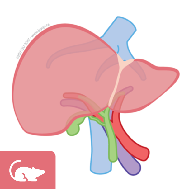
Midline incision: cut open the abdomen from the pubic symphysis to the xiphoid process. Flip over the intestines to locate the liver and its portal vein.
Please refer to Figure 14.4.1 of the following publication:
Citation
LINK
Cannulation
Cannulate the portal vein with the 22-gauge catheter. Secure it cannulation by suture or a micro serrefine.
Equipment
Insyte™ Autoguard™ BC
NAME
Intravenous Catheter
TYPE
BD
BRAND
382523
SKU
LINK
Non-winged, blue, 22 gauge, 1 inch, 0.9 × 25 mm
SPECIFICATIONS
Equipment
Schwartz Micro Serrefine
NAME
Micro Serrefine
TYPE
Fine Science Tools
BRAND
18052-03
SKU
LINK
Sharp Bend
SPECIFICATIONS
Collect blood: collect blood from the inferior vena cava (IVC) with a 20-gauge needle.
Cut the IVC and proceed to the next step immediately. Cutting the IVC allows blood and perfusate to drain.
Perfusion
Perfuse liver with calcium-free buffer without collagenase: 10 mL of Hanks' Balanced Salt Solution (HBSS) without calcium or magnesium + 0.5 millimolar (mM) EGTA at a flow rate of 2 mL/min until fluid drained from the IVC is no longer red (containing blood). This is done with a pump with no recirculation.
Note
This flow rate (2 mL/min) is slower than other liver perfusion protocols. We found that a slower perfusion rate results in hepatocytes with higher quality and quantity and therefore suitable for analyses such as single-cell RNA sequencing.
Equipment
Model EP-1 Econo Pump
NAME
Peristaltic Pump
TYPE
Bio-Rad
BRAND
7318140
SKU
LINK
5m
Perfuse liver with calcium-containing buffer with collagenase: 10 mL of Hanks' Balanced Salt Solution (HBSS) with calcium and magnesium + 0.1 mg/mL type IV collagenase at the same flow rate (2 mL/min). This is done with the same pump with no recirculation.
Collagenase from Clostridium histolyticumMerck MilliporeSigma (Sigma-Aldrich)Catalog #C5138
A well-perfused liver shows drastic discoloration due to the lack of blood. A well-dissociated liver shows white patches.
5m
Harvest
Excise the dissociated liver out of the animal and place it in DMEM containing 10% FBS, which inactivates the collagenase enzyme.
Dulbecco’s Modified Eagle’s Medium - high glucoseMerck MilliporeSigma (Sigma-Aldrich)Catalog #D5796
Cell Isolation
Gently cut the excised liver with a scalpel to release the dissociated cells.
Note
Good dissociation can be visualized by abundance of cells released (in pink) with little cutting. Shake the tissue gently to release cells. Avoid tearing the tissue with the scalpel (horizontal movement); only cut the tissue vertically.
Filter dissociated cells with a 70 µm filter:
Equipment
70 µm Cell Strainer
NAME
Cell Strainer
TYPE
Falcon
BRAND
352350
SKU
LINK
White, Sterile, Individually Packaged
SPECIFICATIONS
Cell Enrichment
Enrich dissociated cells for hepatocytes by centrifugation 50 x g Room temperature for 5 minutes. The supernatant can be further purified in the next step. Resuspend the pellet (hepatocyte-rich) with DMEM.
5m
Enrich supernatant for non-parenchymal cells (NPC) by centrifugation 400 x g Room temperature for 5 minutes. Discard the supernatant. Resuspend the pellet (NPC-rich) with DMEM.
5m
Citations
Step 5
Froh M, Konno A, Thurman RG. Isolation of liver Kupffer cells.
https://doi.org/10.1002/0471140856.tx1404s14