Oct 08, 2021
Microdialysis guide cannula implantation surgery
- Christiana.bjorkli 1
- 1The Norwegian University of Science and Technology
- Sandvig lab


Protocol Citation: Christiana.bjorkli 2021. Microdialysis guide cannula implantation surgery. protocols.io https://dx.doi.org/10.17504/protocols.io.bszxnf7n
License: This is an open access protocol distributed under the terms of the Creative Commons Attribution License, which permits unrestricted use, distribution, and reproduction in any medium, provided the original author and source are credited
Protocol status: Working
We use this protocol and it’s working
Created: March 04, 2021
Last Modified: October 08, 2021
Protocol Integer ID: 47895
Keywords: insert microdialysis guide cannula, cma microdialysis ab, microdialysis guide, microdialysis probe, insertion of the guide cannula, lateral ventricle of mice, lateral ventricle, implantation surgery, guide cannula, implantation surgery implantation surgery, csf from the animal, arteries in close proximity
Abstract
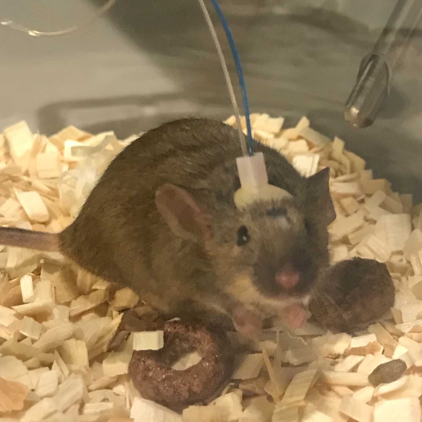
Implantation surgery to insert microdialysis guide cannulas (CMA 7; CMA Microdialysis AB, Kista, Sweden) into the lateral ventricle of mice. In animals that are implanted in the lateral ventricle, CSF surfaces shortly after the insertion of the guide cannula during implantation surgeries. There are arteries in close proximity of the lateral ventricle that can be inadvertently hit, and minor bleeding can therefore occur. This needs to be avoided, because the blood could clog the microdialysis probe and make it difficult to sample CSF from the animal.
Materials
Chemicals and solutions
- Isoflurane for anesthesia
- Metacam, temgesic, and marcain for analgesia and local anesthetics
- 70% Ethanol
- Klorhexidin and NaCl for disinfecting and cleaning
- Simplex eye salve
Equipment and accessories for stereotaxic surgery
- Stereotaxic frame
- Absorption triangles
- Dental cement
- Surgical instruments
- Dummy cannula for temporal occupation for the guide cannula
- Stereotaxic adaptor
- Dental drill
- Clipper
- Tape
- Tooth picks
- Heating pad
- Super glue
- Porridge/diet gel
- Q tips
Troubleshooting
Pre-surgery
Habituate animals to food and housing they will receive post-surgery. Check the health status of the animal and autoclave your instruments the day prior to surgery.
Preparation of stereotaxic surgery for microdialysis guide implantation
Tidy and clean the surgery table. Disinfect the surgery table and the surgery close to it with 70% ethanol. Prepare steel cups with 70% ethanol and one with sterile saline. Prepare a 5ml syringe with sterile saline to be used to keep tissue moist during the surgery.
Check the level of isoflurane in the vaporizer, and refill if necessary. Check that all tubes in the anesthesia setup are connected properly and that there is no leakage or compression of the tubes. Check the pressure of the medical air.
Turn on the heat pad for the animal.
Prepare analgesic drugs.
Stereotaxic surgery for microdialysis guide implantation
Weigh the animal.
Place animal in induction chamber pre-filled with isoflurane; start at 3% and slowly move down to 1.5%, and keep this concentration throughout the surgery. Set oxygen to 0.2-0.3%.
Move the animal to the small suction table mask; use tool to push the tongue of the animal down while attaching its teeth to the mask of the stereotaxic frame.
Fixate the skull in the stereotaxic frame using ear bars. A trick to be sure that the ear bars are well placed is to see if the ears are draped over the ear bars. The ear bars should be placed on the bone in front of the ears after going through the ear canal. Dial in coordinates to make sure that the animal is located correctly in the stereotaxic frame.
Shave head of the animal and use tape to remove the hair, start from in-between ears to in-between eyes. Put on eye salve to protect the cornea of the animal.
Give subcutaneous injections of analgesics, give temgesic and metacam. Give local anaesthetic marcain by subcutaneous injection in the area where the incision is to be made. Give a subcutaneous injection of sterile saline to keep the animal hydrated during surgery.
Wait a few minutes for the drugs to take effect. While waiting, set up an assembly for a guide implantation: the microdialysis guide cannula is attached to the stereotaxic frame using a guide clip and connection rod for the clip (CMA Microdialysis AB, Kista, Sweden).
Disinfect the surface of the head with 70% ethanol and klorhexidin. Use small scissors to cut a triangle of skin on top of the skull.
Remove periosteum with absorption triangles to reveal the skull. Identify lambda, position the guide cannula above bregma and lower it until the guide cannula gently touches the skull and record the ventral coordinate. Move the guide cannula posteriorly from bregma and position it above lambda. Lower the guide cannula until it gently touches the skull and record the ventral coordinate. Equal heights of bregma and lambda were measured to ensure that the skull was level for each animal (with ±0.1 mm tolerance), as well as 2 points equally distant from the midline.
After levelling the skull, the stereotaxic coordinates were derived to target the lateral ventricle (A/P -0.1 mm, M/L +1.2 mm, D/V -2.75 mm; Fig. 11). The skull was drilled through at these coordinates and the guide cannula was slowly lowered into the drilled hole.
The guide cannula was attached to the skull with super glue and dental cement (Dentalon Plus; Cliniclands AB, Trelleborg, Sweden).
Post-surgery
Following surgery, the animal was placed in a heated chamber until awake and moving normally. Post-surgery, Metacam and Temgesic were administered within 24 hours.