May 29, 2026
MetaSPR Assay Protocol for Binding Interaction Between Recombinant Human IL21 and IL21R Proteins (Ligand Capture Method)
- Rosie Liu1
- 1CUSABIO
- CUSABIO TECHNOLOGY LLC


Protocol Citation: Rosie Liu 2026. MetaSPR Assay Protocol for Binding Interaction Between Recombinant Human IL21 and IL21R Proteins (Ligand Capture Method). protocols.io https://dx.doi.org/10.17504/protocols.io.5jyl83redv2w/v1
License: This is an open access protocol distributed under the terms of the Creative Commons Attribution License, which permits unrestricted use, distribution, and reproduction in any medium, provided the original author and source are credited
Protocol status: Working
We use this protocol and it's working
Created: May 29, 2026
Last Modified: May 29, 2026
Protocol Integer ID: 318176
Keywords: Recombinant Human Interleukin-21 ; IL21; Recombinant Human Interleukin-21 receptor; IL21R; biologically active; binding interaction and affinity; Association rate constant (Kₐ); Dissociation rate constant (Kd); Equilibrium dissociation constant (Kᴰ), il21r protein, recombinant human il21, metaspr assay protocol for binding interaction, metaspr assay protocol, ligand capture method, ligand capture, metaspr technology on the wespr, based metaspr technology, affinity between cusabio
Abstract
This protocol is designed to measure the binding interaction and affinity between CUSABIO recombinant human Interleukin-21 (IL-21) (CSB-MP872537HU) and recombinant human IL-21R (CSB-MP862061HU) using the ligand capture-based MetaSPR technology on the WeSPR™ 200 system. In this protocol, hFc-tagged IL-21 is captured on a Protein A biosensor chip, and soluble His-tagged IL-21R is used as an analyte to determine kinetic parameters (Ka, Kd, and KD).
Guidelines
Principle:
MetaSPR (Metasurface Surface Plasmon Resonance) is a label-free, real-time biosensing technology that detects changes in the refractive index at the surface of a nanostructured metasurface chip when biomolecules bind to it. In this ligand capture assay:
- The Protein A-coated chip specifically and reversibly binds to the Fc region of the hFc-tagged Human IL21 Protein, immobilizing the ligand on the chip surface in a highly oriented manner. This orientation maximizes the accessibility of the IL21 receptor-binding domain, ensuring accurate measurement of binding kinetics.
- Different concentrations of the His-tagged Human IL21R Protein (analyte) are introduced over the immobilized IL21 ligand.
- The binding of IL21R to IL21 alters the local refractive index at the chip surface, which is converted into a proportional electrical signal (response unit, RU) by the WeSPR‱ 200 system's optical detector.
- The system continuously records the RU signal during both the association (binding) and dissociation phases. By fitting the resulting sensorgrams to a 1:1 Langmuir binding model, the kinetic parameters (Kₐ, Kd) and equilibrium dissociation constant (Kᴰ) are calculated. The Kᴰ value is a direct measure of the binding affinity between the two proteins, with lower values indicating stronger binding.
Precautions:
- Bubble Prevention: Avoid introducing air bubbles into the chip wells during reagent loading. Bubbles will disrupt the optical path and cause severe signal artifacts. If bubbles form, gently tap the side of the chip plate to dislodge them or reload the reagent.
- Chip Handling: Handle the chip plate only by the edges. Do not touch the active chip surface with fingers, pipette tips, or any other objects, as this will damage the Protein A coating and render the chip unusable.
- Protein Stability: Prepare all protein dilutions immediately before use. Diluted proteins are less stable and may lose activity if stored for extended periods at room temperature.
- Regeneration Efficiency: Ensure complete regeneration of the Protein A chip surface after each assay cycle. Incomplete regeneration will leave residual bound protein on the chip, leading to elevated baselines and inaccurate results in subsequent runs.
- Incubation Timing: Strictly adhere to the specified incubation times for all steps (ligand fixation, analyte binding, dissociation, and regeneration). Even small variations in incubation time can significantly affect the measured kinetic parameters.
- Replicate Consistency: Perform all experiments in at least duplicate to ensure the reliability and reproducibility of the results. All replicates should be run in the same experimental session to minimize variability.
- Protective Solution: Do not let the chip dry. After the assay, always add protective solution and seal to preserve the chip for reuse.
Materials
- Recombinant Human IL-21 (Active) – Fc-tagged (hFc), 3e90% purity (Cusabio, Cat. #CSB-MP872537HU). Lyophilized powder (contains PBS, 6% trehalose).
- Recombinant Human IL-21R (partial, Active) – His-tagged, 3e95% purity (Cusabio, Cat. #CSB-MP862061HU). Lyophilized powder (contains PBS, 6% trehalose).
- MetaSPR Instrument – WeSPR™ 200 (LifeDisc, China) with Protein A sensor chip (LifeDisc biosensor kit, Cat. #G21003). Kit includes 10X sample diluent, 5X elution/regeneration solution, and protective solution.
- Buffers/Reagents: Sterile deionized water, phosphate-buffered saline (PBS, pH 7.4), glycerol (molecular biology grade).
- Consumables: Low-binding pipette tips, 1.5 mL microcentrifuge tubes, new 96-well microplate (compatible with the SPR instrument). Parafilm or sealing film.
- Equipment: Analytical balance, centrifuge, vortex mixer, ice bucket, thermal incubator or water bath (set to 25°C), timer.
- Personal Protective Equipment: Gloves, lab coat, eye protection.
Troubleshooting
Problem
Low maximum response signal: Possible causes: 1. Incorrect ligand or analyte concentration 2. Protein denaturation or loss of activity 3. Insufficient ligand fixation time 4. Damaged Protein A chip surface.
Solution
Solutions: 1. Verify the protein stock concentration and prepare fresh dilutions 2. Thaw a new aliquot of reconstituted protein; test protein activity using an alternative assay if available 3. Increase the ligand fixation time to 15 minutes 4. Replace with a new Protein A biosensor chip.
Problem
High Background/Noise: Possible causes: 1. Air bubbles 2. dirty fluidics 3. nonspecific binding.
Solution
Solutions: 1. Degas buffers thoroughly; ensure no bubbles at pipetting. 2. Clean instrument optics if recommended 3. Add surfactant (e.g., 0.005% Tween-20) to buffer if nonspecific binding is suspected, or include a reference channel.
Problem
Regeneration Incomplete: Possible causes: Regenerant too weak or too short exposure, leaving bound analyte.
Solution
Solutions: Extend the regenerant incubation (e.g., 2–5 min) or increase its strength per the kit guidelines. However, avoid harsh conditions that strip off IL-21. Test regenerant on control runs.
Problem
Unstable baseline or significant baseline drift: Possible causes: 1. Reagents not equilibrated to room temperature 2. Chip plate not properly seated in the instrument 3. Incomplete chip regeneration 4. Air bubbles trapped in the chip wells.
Solution
Solutions: 1. Equilibrate all reagents to room temperature for an additional 15 minutes 2. Power off the instrument, correctly remove and reinsert the chip plate, then restart system 3. Repeat the regeneration step with fresh 1× Elution Regenerant, extending the incubation time by 30 seconds 4. Remove bubbles by gently tapping the chip plate or carefully aspirating and reloading the reagent.
Problem
Poor curve fitting or inconsistent kinetic parameters between replicates: Possible causes: 1. Inappropriate analyte concentration range 2. Insufficient data acquisition rate 3. Variations in incubation times or temperatures 4. Incorrect binding model used for analysis.
Solution
Solutions: 1. Adjust the analyte concentration range to ensure that the response signals cover 10–90% of the maximum binding capacity 2. Set the instrument to acquire data at a rate of at least 1 point per second 3. Use a timer for all incubation steps; perform all replicates in a single experimental run 4. Use the 1:1 Langmuir binding model, which is the correct model for this monovalent ligand-receptor interaction.
Safety warnings
- Biological Hazards: IL-21 and IL-21R are recombinant proteins but handle them with standard biohazard precautions. Wear gloves and lab coat. Dispose of used wells and pipette tips as biological waste.
- Chemical Hazards: The elution/regeneration solution may be acidic or contain detergents. Consult the kit MSDS; use gloves and eye protection when handling.
- Waste Disposal: Dispose of all biological waste (used pipette tips, protein solutions, and chip plates) in designated biohazard waste containers according to your institution's biosafety guidelines.
- Single-Use Chips: The Protein A biosensor chips are designed for single use only. Reusing chips will result in degraded performance, increased non-specific binding, and unreliable data. Dispose of used chips as biohazard waste.
- Instrument Safety: Follow the instrument manual for electrical safety and proper shutdown. Ensure liquids do not spill into the device.
- Data Integrity: Label all reagents clearly. Do not use expired or contaminated reagents. Maintain a clean workspace to avoid sample cross-contamination.
Before start
All reagents, samples, and the chip plate must be equilibrated to room temperature for 30 minutes before starting the experiment. Temperature fluctuations are the most common cause of baseline drift in SPR assays.
Pre-experiment Sample Processing
Bring samples to room temperature:
Remove IL-21 and IL-21R vials from –20°C/–80°C storage and allow to warm to room temperature. Briefly centrifuge the vials to collect any powder at the bottom.
Reconstitute proteins:
Reconstitute each lyophilized protein in sterile water to a high concentration (e.g., 0.5–1.0 mg/mL) as recommended. For IL-21, consider a final stock of 0.5 mg/mL; for IL-21R, 0.5 mg/mL. Add sterile water directly to the vial and gently swirl to dissolve.
Note: The datasheet suggests adding glycerol for long-term storage (up to 50%). For immediate use, glycerol is optional, but can be added to a final 20–50% (v/v) concentration if storing aliquots at –20°C.
Remove particulates:
After dissolution, centrifuge each protein solution at high speed (e.g., 10,000×g for 5 min) to pellet any insoluble material. Carefully transfer the supernatant to a new tube. If needed, filter the solution through a 0.22 µm filter.
Prepare working aliquots:
Dilute and aliquot stocks so that freeze-thaw cycles are minimized. Store aliquots at –20°C or –80°C until use. Thaw an aliquot on ice before the assay, and keep it on ice to prevent degradation.
Reagent Preparation
1X Sample Diluent:
Dilute the 10X sample diluent (from the LifeDisc kit) 1:10 with sterile water to prepare 1X working diluent. Mix well. Equilibrate to room temperature (25°C) before use.
1X Elution/Regeneration Solution:
Dilute the 5X regenerant concentrate 1:5 with sterile water to make 1X. Prepare fresh as needed or according to kit instructions.
Protective Solution:
Use as supplied by the kit; no dilution needed.
Ligand (IL-21) Solution:
Dilute IL-21 stock into 1X sample diluent to a final concentration of 10 µg/mL. For example, add 10 µL of 0.5 mg/mL IL-21 stock to 490 µL of 1X diluent. Prepare immediately before use. Mix gently by pipetting; keep on ice until use.
Analyte (IL-21R) Solutions:
Prepare seven concentrations by diluting IL-21R stock into 1X sample diluent: 0 (blank), 1.25, 2.5, 5, 10, 20, and 40 µg/mL. For instance, to make 40 µg/mL, dilute 20 µL of 1.0 mg/mL stock into 480 µL diluent. Similarly, prepare each dilution (use serial dilution for accuracy). Prepare at least 50 µL of each for filling wells. Keep all analyte solutions on ice briefly before dispensing.
Additional Buffers:
Have an extra 1X sample diluent and water available. Degas all solutions if your SPR instrument requires bubble-free fluids (e.g., by vacuum or sonication).
Assay Procedures
Instrument and Chip Setup:
Power on the WeSPR™ 200 instrument. Insert a clean Protein A biosensor chip into a new 96-well plate provided with the kit. Prime the instrument lines according to manufacturer instructions, ensuring all lines are filled with 1X sample diluent and that no air bubbles are trapped. Equilibrate the system temperature to 25°C (room temperature).
Baseline Stabilization:
If required, run a baseline by pipetting 100 µL of 1X sample diluent into a well and monitoring until the signal stabilizes. Wipe any spills and equilibrate the chip surface (with protective solution if recommended by the kit).
Ligand Capture (IL-21):
- Pipette 50 µL of the 10 µg/mL human IL-21 solution into each designated well on the Protein A chip plate using a multichannel pipette. Ensure each well over the chip surface receives IL-21.
- Incubate at 25°C for 10 minutes to allow the hFc-tagged IL21 to bind specifically to the Protein A-coated chip surface.
- After 10 minutes, gently pat the edge of the chip plate on absorbent paper to remove unbound ligand. Do not touch or scratch the active chip surface with the pipette tip or absorbent paper.
Analyte Binding (IL-21R):
- Pipette 50 µL of each diluted Human IL21R Protein solution (0, 1.25, 2.5, 5, 10, 20, and 40 µg/mL) into the corresponding chip wells above the captured IL-21 on the chip. The 0 µg/mL well will serve as a blank (diluent only).
- Incubate all wells at 25°C for 10 minutes to allow IL-21R to bind to the immobilized IL-21.
- Gently pat the chip plate dry with absorbent paper to remove unbound analyte.
Dissociation Phase:
- Load 150 µL of 1× Sample Diluent into each chip well.
- Incubate at room temperature for exactly 20 minutes. The WeSPR™ 200 system will continuously record the real-time response unit (RU) changes during the entire dissociation phase.
Chip Regeneration:
- Load 150 µL of 1× Elution Regenerant into each chip well to completely strip all bound proteins (both IL21 ligand and IL21R analyte) from the Protein A chip surface.
- Incubate for the time specified in the LifeDisc™ Protein A Biosensor Kit instructions (typically 30–60 seconds) to ensure complete regeneration.
- Gently pat the chip plate dry with absorbent paper.
Post-Regeneration Washing:
- Wash the chip wells twice with 150 µL of 1× Sample Diluent per well, patting the plate dry after each wash.
- Wash the chip wells twice with 150 µL of sterile deionized water per well, patting the plate dry after each wash.
Chip Storage (If Required):
- Load 150 µL of Protective Solution from the LifeDisc™ Protein A Biosensor Kit (G21003) into each chip well.
- Seal the chip plate tightly with a sealing film and store at 4°C for up to 1 week if not analyzed immediately.
Result Analysis
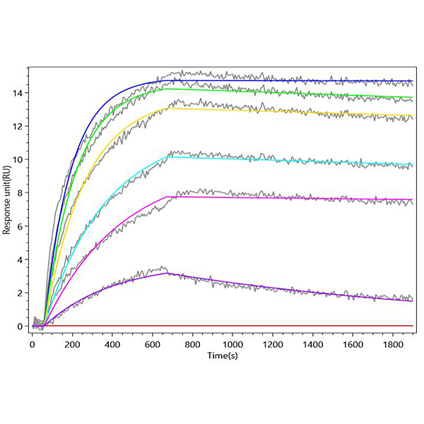
Binding Curves:
Use the MetaSPR software to plot sensorgram curves (Response Units vs time or concentration). For each IL-21R concentration, verify that the response increases with higher analyte concentration. Subtract the blank (0 µg/mL) curve to correct for nonspecific signal.
Kinetic Fitting:
- Fit the association and dissociation phases of the curves to a 1:1 binding model to extract kinetic parameters Ka (association rate) and Kd (dissociation rate). The equilibrium dissociation constant is KD = Kd/Ka.
- The following validated kinetic and affinity parameters are obtained: Association rate constant (Ka): 1.53 × 10^4 1/M·S, Dissociation rate constant (Kd): 2.86 × 10^−5 1/S, Equilibrium dissociation constant (KD): 1.87 × 10^−9 M (1.87 nM).
Sensorgram Interpretation:
Human IL21 captured on Protein A Chip can bind Recombinant Human IL21R (CSB-MP862061HU) with an affinity constant of 1.87 nM as detected by MetaSPR Assay (WeSPRTM 200).
- The sensorgrams exhibit a clear dose-dependent increase in maximum response units (RU) with increasing IL21R concentrations, confirming specific binding between immobilized IL21 and soluble IL21R.
- The association phase (0–600 seconds) shows a rapid rise in RU as IL21R binds to IL21, reaching a plateau at higher analyte concentrations, indicating saturation of available binding sites.
- The dissociation phase (600–1800 seconds) shows a slow, gradual decrease in RU, indicating the formation of a stable IL21-IL21R complex with a relatively long half-life.
- The blank control 0 µg/mL IL21R shows no significant RU change throughout the experiment, confirming the absence of non-specific binding to the Protein A chip surface.
Protocol references
Protocol references
○ Product Name: Recombinant Human Interleukin-21 (IL21) (Active) (CSB-MP872537HU, hFc1-tag)
Recombinant Human Interleukin-21 receptor (IL21R), partial (Active) (CSB-MP862061HU, 10xHis-tag)
○ Immunogen Species: Homo sapiens (Human)
○ Target Analyte: IL21RSample