Aug 05, 2025
Version 3
Marine mammal sex determination using epidermal tissue recovered from suction-cup tags V.3
- Sadie Novak1,
- Jacob Linsky2,
- Kaitlyn Knapp1,
- Dana Adcock1,
- Susan Parks1,
- David Wiley3,
- Dana Cusano1
- 1Syracuse University;
- 2The University of Queensland;
- 3Stellwagen Bank National Marine Sanctuary


Protocol Citation: Sadie Novak, Jacob Linsky, Kaitlyn Knapp, Dana Adcock, Susan Parks, David Wiley, Dana Cusano 2025. Marine mammal sex determination using epidermal tissue recovered from suction-cup tags. protocols.io https://dx.doi.org/10.17504/protocols.io.5qpvor42dv4o/v3Version created by Dana Cusano
License: This is an open access protocol distributed under the terms of the Creative Commons Attribution License, which permits unrestricted use, distribution, and reproduction in any medium, provided the original author and source are credited
Protocol status: Working
We use this protocol and it's working
Created: July 30, 2025
Last Modified: August 05, 2025
Protocol Integer ID: 223688
Keywords: Polymerase chain reaction, trace DNA, sex determination, marine mammal, marine mammal sex determination, cup tags for many marine mammal species, dna extraction from recovered tag tissue, based sex determination, many marine mammal species, dna extraction, recovered tag tissue, protocol for tissue collection, ranging whale, genetic information, epidermal tissue from the underside, using epidermal tissue, genetic analysis, epidermal tissue, dna, valuable source of genetic information, extraction, accuracy of pcr, using suction cup tag, sampled skin, tissue collection, suction cup tag, pcr, tissue, species, minimal sexual dimorphism
Funders Acknowledgements:
Volgenau Foundation
Grant ID: NA
International Fund for Animal Welfare
Grant ID: NA
Bureau of Ocean Energy Management
Grant ID: NA
Stellwagen Bank National Marine Sanctuary
Grant ID: NA
Abstract
For many marine mammal species, minimal sexual dimorphism means that sex cannot be reliably identified through observation. Instead, genetic analysis of sampled skin and blubber are required to determine an individual's sex. Studies using suction cup tags on free-ranging whales may retrieve exfoliated epidermal tissue from the underside of the tag’s suction cups, offering a valuable source of genetic information. This study provides a protocol for tissue collection and DNA extraction from recovered tag tissue and describe the accuracy of PCR-based sex determination using these methods.
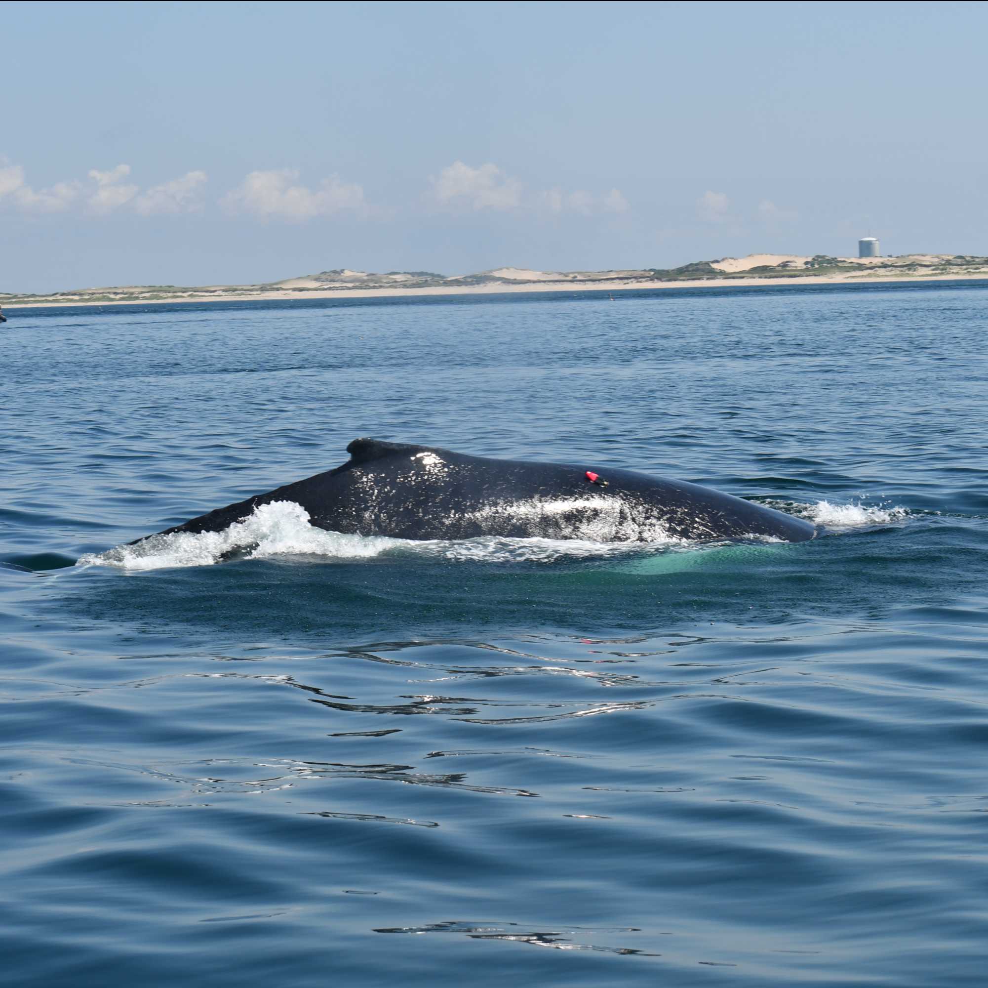
Image Attribution
Image taken under NMFS permit 18059. Image credit: Laura J. Howes.
Materials
MyTaq Extract-PCR Kit (Meridian Bioscience, cat. no. BIO-21126)
DNEasy Blood and Tissue Kit (Qiagen, cat. no. 69504)
Protocol-specific primers:
Forward SRY: 5’ TGT GAA CGG TGA GGA TTA 3’
Reverse SRY: 5’ GTG CAT GGC TCG TAG TCT 3’
Forward ZFX: 5’ ATA GGT CTG CAG ACT CTT CTA 3’
Reverse ZFX: 5’ CAC TTA TGG GGG TAG TCC TTT 3’
Field Sampling
Retrieve the tag from the water. While retrieving the tag, ensure that nothing touches the suction cups with the exception of the net used for retrieval and wear gloves to prevent contamination.
Using fresh nitrile gloves, wipe the inside of the suction cups with a sterile swab, avoiding the outside of the suction cup and any other part of the tag. This reduces the chances of obtaining a sample that is contaminated with the genetic material of another animal that may have come into contact with the tagged animal. Note that suctions cups may not have visible skin.
Swab two suction cups with one swab, then the other two suction cups with a new sterile swab. Retrieving two samples from the same tag allows for more testing as well as sample security in cases of contamination.
Place the swab tip first into a plastic vial, trimming the handle as needed in order to close or screw on the lid.
Clearly label the vial with the date and the identifying information that is associated with that specific deployment. Make sure that there is enough information on the label to ensure that all samples are easily and accurately associated with the respective deployment data. This includes, at a minimum, the ID associated with the tag deployment. For example 'bb22_115a', indicating the two letter species ID (bb), julian day (115), and the order of deployment (a, the first tag of the day).
It is also important to note this information on a datasheet designed to store this data along with other metadata that will be needed later. Of particular importance are:
- Date the sample was collected
- Time the sample was collected
- Location (latitude and longitude) the sample was collected
- Group size/proximity to other animals, as these factors may be a source of contamination
Move the vial immediately into a -80 °C freezer if possible. If no -80 °C is available on board the vessel, store on dry ice until the sample can be moved into an appropriate freezer.
Repeat steps 3 through 6 for the remaining pair of suction cups.
DNA Extraction
11h 25m 45s
Note
DNA extraction was performed with a commercially available kit (DNease Blood & Tissue Kit, Qiagen, cat. no. 69504).
To begin DNA extraction, remove swab tip from plastic stick. This can be done by tracing a clean razor blade along the connection point from the swab tip to the swab stick. Transfer the swab tip to
a clean 1.5-mL microcentrifuge tube for each sample.
Note
Be sure to use a clean razor blade for each sample to avoid cross-contamination of any genetic material.
Add 180 µL of Buffer ATL directly to each tube containing swab as directed by the manufacturer's protocol. Pipette solution up and down and over entirety of swab thoroughly, at least 20 times.
Add 20 µL of Proteinase K to each tube containing the swab and previous buffer solution as directed by the manufacturer's protocol. Pipette the solution up and down over the swab to mix. Vortex each sample for 00:00:15 on high to ensure thorough mixing.
15s
Incubate samples at 56 °C in a hot water bath for 03:00:00 .
3h
Add 200 µL of Buffer AL to each sample as directed by the manufacturer's protocol and mix thoroughly by quickly vortexing for 00:00:15 . Immediately add 200 µL of 96 % (v/v) EtOH and mix again by vortex for 00:00:15 .
30s
Pipette solution into the DNeasy Mini spin column and place column in a 2 mL collected tube (included in kit). Incubate samples at Room temperature for 00:05:00 before centrifuging samples at 6000 x g for 00:05:00 . Discard flowthrough and collection tube. Place spin column in a clean 2 mL collection tube (included in kit).
Note
Do not discard flowthrough in this step or any step in the downstream processing into bleach. Chloroform gas will be generated.
10m
Add 500 µL of Buffer AW1 to each sample as directed by the manufacturer's protocol and centrifuge samples at 6000 x g for 00:05:00 . Discard the resulting flowthrough and collection tube. Place the spin column in a clean 2 mL collection tube (included in kit).
5m
Add 500 µL of Buffer AW2 to each sample as directed by the manufacturer's protocol and centrifuge samples at 17000 x g for 00:03:00 to completely dry the column membrane. Discard the resulting flowthrough and collection tube. Place spin column in a clean 2 mL collection tube (included in kit).
Note
This step is important to ensure no residual ethanol is carried in downstream processing steps of the DNA extraction. If the bottom of the spin column comes in contact with any flowthrough solution, place spin column in a clean 2 mL collection tube and repeat the centrifugation step (17000 x g for 00:03:00 )
3m
Place the dried spin column into a clean 1.5 mL eppendorf tube and apply 100 µL Buffer AE onto the center of the column membrane. Be sure not to touch the membrane with the pipette tip. Allow the buffer to incubate on the membrane for 00:05:00 at room temperature. Elute DNA by centrifuging the column at 6000 x g for 00:01:00 . Elute DNA from the column again by applying 100 µL Buffer AE onto the center of the column membrane and centrifuging at 6000 x g for 00:01:00 .
Note
The elution step can be performed once with 200 µL of buffer. Eluting DNA step-wise will provide an increased DNA yield.
Do not exceed an elution volume of 200 µL to ensure no flowthrough is refluxed into the bottom of the spin column. This may yield samples with lower purity.
7m
Dry DNA samples in a speed vac overnight at room temperature. Re-solubilize DNA in 20 µL of ultrapure water. Measure sample concentration (ng/uL) and purity (260/280 value) using a Nanodrop.
8h
PCR Amplification
2h 15m
Note
PCR amplification was performed with OneTaq DNA polymerase (New England BioLabs, cat. no. M0480S) and the standard reaction buffer supplied with the enzyme.
PCR amplification was performed on extracted DNA samples using primers specific to the SRY (sex-determining region Y) and ZFX/ZFY (sex chromosomal zing-finger) genes. Oligonucleotide primers for both of these genes have been validated in other species (Bérubé et al., 1996; Suárez-Menéndez et al., 2024). Primers were ordered from IDT DNA.
Forward SRY: 5' TGT GAA CGG TGA GGA TTA 3'
Reverse SRY: 5' GTG CAT GGC TCG TAG TCT 3'
Forward ZFX: 5’ ATA GGT CTG CAG ACT CTT CTA 3’
Reverse ZFX: 5’ CAC TTA TGG GGG TAG TCC TTT 3’
Note
Primers updated 5/29/2025. These primers yield better results for baleen whales compared to Version 1. This also provides two confirmations of males (SRY and ZFY). PCR conditions and bpd are adjusted accordingly.
Citation
LINK
Citation
LINK
Prepare the following reaction mixture for each sample of interest in 0.2 mL PCR-grade tubes:
| Reagent | Volume/reaction (uL) | Final reaction concentration | |
| OneTaq 5X Standard Buffer | 5.0 uL | 1X | |
| 10 mM dNTPs | 0.5 µL | 200 µM | |
| 20 uM forward ZFX/ZFY primer | 0.5 µL | 0.4 µM | |
| 20 uM reverse ZFX/ZFY primer | 0.5 µL | 0.4 µM | |
| 20 uM forward SRY primer | 0.5 µL | 0.4 µM | |
| 20 uM reverse SRY primer | 0.5 µL | 0.4 µM | |
| deionized water | 15.0 µL | � | |
| Template DNA (extracted genomic DNA) | 2.0 µL | ||
| OneTaq DNA polymerase | 0.5 µL | 2.5 U |
Table 1: Reaction components for PCR amplification reaction.
Note
If necessary (to increase band intensity during analysis by gel electrophoresis), the volume of template DNA added to the reaction can be increased. To keep the final volume of the reaction at 25 µL , offset the added volume from the template DNA by subtracting the same volume of deionized water when calculating the reaction components before mixing.
Amplify DNA fragments of interest with 30 cycles of PCR at the following conditions:
| Step | Temperature (°C) | Time (seconds) | Number of cycles | |
| y | m | Cycles | ||
| Initial Denaturation | 94 °C | 120 | 1 | |
| Denaturation | 94 °C | 30 | 35 | |
| Annealing | 55 °C | 90 | ||
| Extension | 72 °C | 30 | ||
| Final extension | 68 °C | 600 | 1 | |
| Hold | 4 °C | Infinite (as needed) | 1 |
Table 2: PCR amplification cycles
2h 15m
Analysis of PCR product by gel electrophoresis
1h 7m
The PCR product will be assessed by gel electrophoresis using the known length of the SRY (332 bpd), ZFX (245), and ZFY (212). This protocol is written for use with an EasyCast B1 Mini Gel Electrophoresis system (ThermoFisher).
Cast a 2 % volume agarose gel dyed with SYBR Safe DNA gel stain.
Prepare mold by placing gel tray in the buffer chamber of the electrophoresis apparatus so that the gaskets of the tray form a seal with the walls of the chamber. Use a level to adjust the tray position so that the gel is cast evenly.
Prepare the 2 % volume agarose solution by combining the appropriate mass of agarose with the appropriate volume of 1X tris-acetate EDTA (TAE) buffer (i.e. for a 75 mL agarose gel, combine 1.875 g agarose in 75 mL 1X TAE buffer).
Melt agarose by microwaving solution in 00:00:15 - 00:00:30 time increments, swirling flask between heatings.
2m
Allow solution to cool at room temperature for 00:05:00 before adding appropriate volume of SYBR Safe dye to the solution to dilute to 1X SYBR Safe DNA gel stain (i.e. for a 75 mL agarose gel, add 7.5 µL 10,000X SYBR Safe DNA gel stain to solution).
Note
If solution polymerizes in its melting vessel before it is stained and cast, the solid agarose solution can be re-melted as described in step 21.3. If the solid agar was stained and polymerized before casting, the gel can be melted, but should be re-stained to a final concentration of 1X stain.
Note
Other DNA stains such as GelRed or Ethidium Bromide (EtBr) may be used for this step, at appropriate concentrations. SYBRSafe is recommending for its biosafety properties and resulting ease of use.
5m
Once agarose solution is stained, cast immediately in appropriate gel mold. Place a comb in the mold to create a row of wells. Allow gel to polymerize at Room temperature for 00:20:00 under dark conditions to protect the DNA stain.
20m
Re-position gel tray with gel in the buffer chamber of the gel electrophoresis apparatus to the running position (wells should be placed in the chamber so that they are positioned at the negative electrode). Fill the buffer chamber with 1X TAE buffer so that the gel is completely submerged. Remove comb.
Dilute running dye in PCR product mixture to a final concentration of 1X xylene cyanol (runs at ~4000 kbp) by adding 4 µL of a 6X xylene cyanol solution to each sample product.
Load 2.5 µL of 100 bp DNA molecular weight marker (New England Biolabs, cat. no. NEB#B7025) prepared to contain 1X bromophenol blue running dye into the first well (bromophenol blue runs at ~500 bp). Load 10 µL of the sample(s) in the following wells.
Place the lid on the electrophoresis apparatus and ensure that cables are connected to the power supply. Run the gel at 140 V under dark conditions. Monitor gel run until the bromophenol blue running dye line has migrated about 1 cm from the midpoint of the gel (~00:35:00 -00:45:00 ).
40m
Once the gel run is complete, resolved DNA fragments may be visualized with a UV transilluminator.
Note
In this protocol, a ChemiDoc imaging system (Bio-Rad) was used to image the resolved PCR products following separation by gel electrophoresis. The blue tray and the protocol pre-programmed in the instrument for use with SYBR Safe DNA stain was used.
Product analysis
The expected molecular weight of the PCR product associated with the SRY gene is 332 bp and the expected molecular weight of the PCR product associated with the ZFX and ZFY genes are 245 and 212 bp, respectively.
The presence of the SRY gene suggests that the DNA sample came from a male individual. Females will not possess this gene, and thus this DNA fragment will not be amplified in DNA extracted from a female. However, absence of this gene may be caused by issues in the experimental workflow, such as an insufficient amount of template DNA added, which would result in a false result. To validate the PCR reaction and minimize the chance a false sexing, DNA is also screened simultaneously for a gene that is present in both male and female individuals. The ZFX gene serves as a positive control in this protocol, and the ZFY gene serves as an additional indicator for males.
An expected profile for DNA extracted from a male individual would show the presence of DNA fragments from both the SRY gene (332 bp) and the ZFX and ZFY genes (245 and 212 bp). An expected profile for DNA extracted from a female individual would show presence of only the DNA fragement from the ZFX gene (245 bp). A sample that showed presence of only the DNA fragment from the SRY gene would suggest non-specific priming and would be considered an inconclusive result.
Expected result
Figure 1: The results of three samples of extracted DNA, amplified following the protocol described above, and resolved via gel electrophoresis. Samples run in lanes 1 and 2 show an amplicon profile indicative of a male individual, with three bands at 332 bp (solid purple arrow), 245 bp (dashed blue arrow), and 212 bp (dotted green arrow). The sample run in lane 3 also displayed the amplicon profile of a male but only displayed a band at 332 bp (solid purple arrow). Samples with an amplicon profile indicative of a female individual showed one band resolved at 245 bp (dashed blue arrow), only.
Acknowledgements
Data were collected under NMFS permits #27272 and #18059 (issued to Stellwagen Bank National
Marine Sanctuary) and #21371 (issued to Northeast Fisheries Science Center). The authors thank
the crew of the R/V Auk, the Miss Emily, and the Warren Jr.; the Whale Ecology Branch at Northeast
Fisheries Science Center; the North Atlantic Right Whale Consortium for providing the sex data on
North Atlantic right whales; the Center for Coastal Studies for providing the sex data on the
humpback whales; and all contributors to the tagging efforts in Stellwagen Bank National Marine
Sanctuary, Southern New England, and Cape Cod Bay. The authors thank Heather Coleman and
James Hougland for the use of their equipment; Sara Tennant, and Julia Zeh for
contributing to the analyses; and Jooke Robbins and Vania Rivera Leon for reviewing the draft and
providing comments on the methodology.