Sep 29, 2023
Manual extraction of High Molecular Weight DNA from single mosquitoes using the Qiagen MagAttract HMW DNA kit
- 1Wellcome Sanger Institute
- High molecular weight DNA extraction from all kingdoms
- Darwin Tree of Life


Protocol Citation: Fiona Teltscher, Harriet Johnson, Mara Lawniczak 2023. Manual extraction of High Molecular Weight DNA from single mosquitoes using the Qiagen MagAttract HMW DNA kit. protocols.io https://dx.doi.org/10.17504/protocols.io.n92ldp6ool5b/v1
License: This is an open access protocol distributed under the terms of the Creative Commons Attribution License, which permits unrestricted use, distribution, and reproduction in any medium, provided the original author and source are credited
Protocol status: Working
We use this protocol and it's working
Created: March 16, 2023
Last Modified: September 29, 2023
Protocol Integer ID: 78911
Keywords: magattract, HMW DNA, arthropods, insects, high molecular weight, reference genome, long read, qiagen magattract hmw dna kit, dna from insect, extracted hmw dna, dna extraction, chromiumtmgenome reagent kits user guide, dna extractions in parallel, high molecular weight dna, using qubit hs dna kit, single mosquito, qubit hs dna kit, dna yield, typical weight of an anopheles mosquito, hmw dna in the field, hmw dna, anopheles mosquito, mosquito, ng of dna, quantity of dna, single fresh anophele, mb genome, more dna, most reagent, qiagen magattract kit, dna, sheared dna, high molecular weight, genome, insects for preservation, manual extraction, extraction, reagent
Funders Acknowledgements:
Bill and Melinda Gates Foundation
Grant ID: INV-009760
Wellcome
Grant ID: 220540/Z/20/A
Abstract
This is a protocol for the manual extraction of high molecular weight (HMW) DNA from insects. It uses the Qiagen MagAttract kit and factors in modifications described in the ChromiumTMGenome Reagent Kits User Guide pages 6-8 (https://support.10xgenomics.com/genome-exome/library-prep/doc/user-guide-chromium-genome-reagent-kit-v2-chemistry). It includes further modifications that benefited two particular needs. First, most reagents are halved per extraction (apart from beads) due to small specimen size (2 mg is a typical weight of an Anopheles mosquito, which is far less than the 25 mg of tissue the MagAttract kit can support). Second, due to this small specimen size, we need to maximize DNA yield, so we also perform a second elution to release more DNA.
DNA resulting from this protocol can be further sheared and cleaned for successful PacBio HiFi sequencing. In our experience, a single fresh Anopheles gambiae mosquito weighing 2-3 mg yields about 600-800 ng of DNA (quantified using qubit HS DNA kit) using this protocol. Following shearing using G-tubes or the MegaRuptor and SPRI based clean up, we typically retain about 200 ng of sheared DNA, which is just about sufficient to reach 25x coverage PacBio HiFi of a 250 Mb genome. The quality and quantity of DNA is best when starting with a snap frozen from living specimen. However, we have also successfully extracted HMW DNA using this protocol from ethanol and DESS preserved specimens held at room temperature for as long as a week, as long as the specimen was punctured or gently squished to ensure rapid penetration of the preservative. See: Squishing insects for preservation of HMW DNA in the field https://www.protocols.io/view/squishing-insects-for-preservation-of-hmw-dna-in-t-cyp3xvqn
We normally perform up to 8 DNA extractions in parallel (this will also depend on which magnetic rack is being used). Please add a comment to let the wider community know if it has worked or not on your species.
NB -- an automated version of this protocol that works on the Kingfisher Apex is also available here on protocols.io
Guidelines
This protocol is an adaptation of Chromium™Genome Reagent Kits User guide and Qiagen's MagAttract® HMW DNA Handbook. The Chromium protocol suggests the use of PBS at the beginning. Also, both SOPs recommend using a thermomixer for wash steps, but we find doing this by hand works fine. Finally, we use 1.5 mL microcentrifuge tubes because the plastic pestles that we use to grind insect tissue do not support effective grinding in the 2 mL microcentrifuge tubes recommended by both original protocols. There are, however, pestles available to use in 2 mL microcentrifuge tubes.
Materials
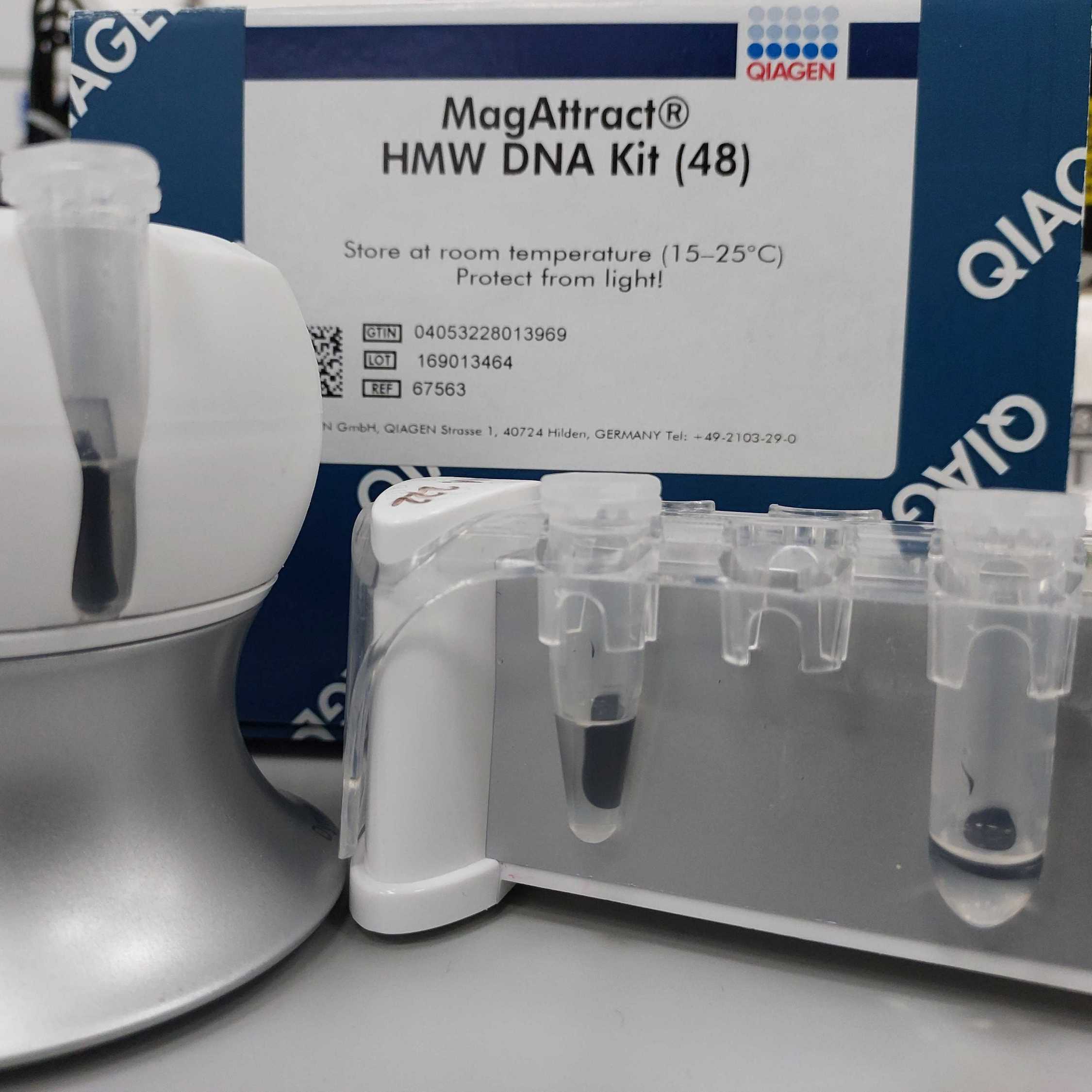
MagAttract HMW DNA kitQiagenCatalog #67563
Ethanol absoluteMerck Millipore (EMD Millipore)Catalog #107017 1X PBS (Phosphate-buffered saline ) Dry Ice
Equipment
Mini-Centrifuge 100-240V, 50/60Hz Universal Plug, Grey
NAME
minicentrifuge
TYPE
Fisherbrand™
BRAND
16617645
SKU
LINK
Equipment
DNA LoBind® Tubes
NAME
microcentrifuge tubes
TYPE
Eppendorf
BRAND
0030108051
SKU
LINK
Equipment
Pestle for 1.5 mL Microtube, 100/pk
NAME
pellet pestle
TYPE
Cole-Parmer Essentials
BRAND
WZ-44468-19
SKU
LINK
Equipment
1,000 µl graduated TipOne® Filter Tip, Natural, Racks (sterile), Case
NAME
Pipette tips
TYPE
Starlab
BRAND
S1126-7810
SKU
LINK
Equipment
200 µl Filter Tip / Wide Orifice
NAME
Pipette tips
TYPE
Starlab
BRAND
E1011-8618
SKU
LINK
Equipment
DynaMag™-2 Magnet
NAME
Magnetic tube rack
TYPE
Invitrogen™
BRAND
12321D
SKU
LINK
Protocol materials
Ethanol absoluteMerck Millipore (EMD Millipore)Catalog #107017
Dry Ice
MagAttract HMW DNA kitQiagenCatalog #67563
1X PBS (Phosphate-buffered saline )
MagAttract Suspension GQiagenCatalog #67563
Buffer MW1Catalog #67563
Buffer PEQiagenCatalog #67563
Nuclease-Free WaterQiagenCatalog #67563
Buffer AEQiagenCatalog #67563
RNase AQiagenCatalog #67563
Buffer ALQiagenCatalog #67563
Proteinase KCatalog #67563
Buffer MBCatalog #67563
Buffer AEQiagenCatalog #19077
Qubit® dsDNA HS Assay KitThermo Fisher ScientificCatalog #Q32854
Quant-iT™ PicoGreen™ dsDNA Assay KitInvitrogen - Thermo FisherCatalog #P11496
gDNA 165kb Analysis Kit 275 SamplesAgilent TechnologiesCatalog #FP-1002-0275
Safety warnings
Buffers AL, MB, and MW 1 contain guanidine hydrochloride/guanidine thiocyanate, which can form highly reactive compounds when combined with bleach. DO NOT add bleach or acidic solutions directly to the sample preparation waste. Waste needs to be collected in a suitable vessel and disposed of in accordance with local regulations.
Before start
Ensure all surfaces have been cleaned with 70-80% Ethanol (and ideally bleach before that). Have cleaning wipes for forceps. All kit components, buffers, and RNase A stock solution can be stored at room temperature (15–25°C) for up to 1 year. The box should be labelled with received date. Mix Buffer AL thoroughly by shaking before use. Buffers MW1 and PE are supplied as a concentrate. Before using for the first time, be sure to add the appropriate amount of ethanol (96–100%) as indicated on the bottle. Many components of the kit are also available from Qiagen separately.
Procedure
2h 24m
Prepare an open insulated box of dry ice to store sample tubes on whilst working through steps 2-4.
Make mastermix of reagents for lysis.
Note
Make sure to choose the right size of tube for preparing the mastermix.
Calculate the mastermix volumes for the number of samples plus 1 for spare pipetting volume. Volumes per sample are: 100 µL 1X PBS (Phosphate-buffered saline ) ; 10 µL Proteinase KCatalog #67563 (Mix by inverting the tube 5 times); 2 µL RNase AQiagenCatalog #67563 ; 75 µL Buffer ALQiagenCatalog #67563 (mix by inversion)
For each sample, add 187 µL of the mastermix from step 2 into a new 1.5 mL DNA LoBind tube.
Note
A 2 mL DNA LoBind tube can also be used but an appropriate size pestle will have to be used.
Carefully remove the Sample from the sample tube using clean forceps. If the Sample has been stored in a preservation liquid, lightly make contact on a clean piece of tissue to remove surface liquid from the sample. Submerge the Sample into the mastermix in a tube (see step 3) with clean forceps. Insert a sterile pestle in the tube and smash, smear, squash, twist, grind the tissue against the wall of the tube for 00:01:00 . There should be no recognisable body parts visible following pestle smashing, only flakes. Place the sample in a tube rack on the bench. Clean forceps with 100% ethanol.
Mosquito in lysis buffer.
Tissue disruption with an autoclavable pestle.
Mosquito debris after tissue disruption.
1m
Repeat step 4 for the remaining samples.
Briefly spin all samples in a minicentrifuge or similar to collect solution at bottom before next step.
Incubate the sample at 25 °C for 02:00:00 .
2h
Briefly spin samples to collect solution at the bottom of the tube.
Vortex the MagAttract Suspension GQiagenCatalog #67563 for 00:01:00 and add 15 µL to each sample.
Note
If this is the first time usingMagAttract Suspension GQiagenCatalog #67563 , increase the vortexing time to
00:03:00 . Briefly vortexMagAttract Suspension GQiagenCatalog #67563 before adding to each subsequent sample.
1m
Add 140 µL Buffer MBCatalog #67563 to each sample. Mix by gentle inversion, fully invert but don't shake. If you see the beads making flakey clumps, a little like gold leaf, this is a good sign. This is difficult to do simultaneously as you want to see the mixing. If you are doing 8 samples, give all samples another gentle inversion after the last one. Leave at least 00:01:00 for the beads to bind. Doing multiple samples will often take more time than this.
Flakes of beads building in the lysis buffer after addition of buffer MB and careful inverting of tube.
1m
Centrifuge the tube briefly and place on a DynaMagTM-2 Magnetic Rack for 00:01:00 to allow bead capture.
Beads are collecting on the magnetic side.
Note
We use a DynaMagTM-2 Magnetic Rack but other magnetic racks suitable for 1.5 or 2 mL microcentrifuge tubes will work as well.
1m
Remove and discard the supernatant. Take care not to disturb the bead pellet.
Supernatant being removed without disturbing the pellet.
Remove the Sample from the magnetic rack. Add 350 µL Buffer MW1Catalog #67563 directly to the bead pellet. Mix by inversion, ensuring that the beads have come away from the side of the tube. This often requires tapping the tube or swilling the contents. Try to be as gentle as possible. Again, this is difficult to do simultaneously as you need to check each sample.
Centrifuge the tube briefly and place on a DynaMagTM-2 Magnetic Rack for 00:01:00 to allow bead capture.
1m
Remove and discard the supernatant. Take care not to disturb the bead pellet.
Repeat steps 13-15 for a total of 2 washes.
Remove the sample from the magnetic rack. Add 350 µL Buffer PEQiagenCatalog #67563 directly to the bead pellet. Mix by inversion, ensuring that the beads have come away from the side of the tube. This often requires tapping the tube or swilling the contents. Try to be as gentle as possible. Again, this is difficult to do simultaneously as you need to check each sample.
Centrifuge the tube briefly and place on a DynaMagTM-2 Magnetic Rack for 00:01:00 to allow bead capture.
1m
Remove and discard the supernatant. Take care not to disturb the bead pellet.
Repeat steps 17 and 18 for a total of 2 washes but do not remove supernatant immediately, proceed to step 21.
If you have more than four tubes, split them into groups of four or fewer. Perform steps 22-24 (water wash) on the first group of samples, whilst the remaining samples wait in Buffer PEQiagenCatalog #67563 on the magnet. [While the first group of samples are eluting in step 25-26 below, it is possible to perform the water wash on the second group of samples.]
Remove and discard the supernatant. Take care not to disturb the bead pellet. With a P20 pipette remove any remaining supernatant. Leave the sample on the magnetic rack for the next step. Do not pipette water directly onto the beads.
Note
The timing of the next step is extremely important. If a multichannel pipette is not available, ensure that each tube has the exact same incubation time. Do not exceed 00:01:00 .
Water being pipetted against the side opposite of the magnetic beads to avoid disturbing beads.
Carefully add 350 µL Nuclease-Free WaterQiagenCatalog #67563 down the side of the tube opposite the magnetic pellet. Start a timer counting up from zero. After 00:00:15 add water to the second sample, after 00:00:30 add to the third sample, after 00:00:45 add to the fourth sample. At 00:01:00 remove and discard the water from the first sample, at 00:01:15 remove and discard water from the second sample, at 00:01:30 the third, and 00:01:45 the fourth. This will enable multiple samples to be incubated for exactly 00:01:00 .
8m
Repeat step 23 for a total of 2 washes.
Remove the samples from the magnetic rack. Add 100 µL Buffer AEQiagenCatalog #67563 directly to the bead pellet of each sample. Ensure that the pellet is submerged and has come away from the side of the tube. Incubate at 25 °C and 1400 rpm shaking for 00:03:00 .
3m
During this 00:03:00 incubation perform steps 22-25 on the second group of samples if present.
3m
Centrifuge each tube briefly and place them on a magnetic rack for 00:01:00 to allow bead capture.
1m
Using a wide-orifice pipette tip, carefully transfer the supernatant containing purified gDNA to a new labelled 1.5 mL LoBind microcentrifuge tube or barcoded (e.g. FluidX) tube.
Second elution: remove the samples from the magnetic rack. Add 100 µL Buffer AEQiagenCatalog #67563 directly to the bead pellet of each sample. Ensure that the pellet is submerged and has come away from the side of the tube. Incubate at 25 °C and 1400 rpm shaking for 00:03:00 .
Note
Due to the second elution step, an additional bottle of Buffer AEQiagenCatalog #19077 will be necessary if you buy the kit.
3m
Using a wide-orifice pipette tip, carefully transfer the supernatant containing purified gDNA to the same 1.5 ml LoBind microcentrifuge tube or Fluidx tube with a final volume of 200 µL .
Store the extracted gDNA sample at 4 °C . Assess the quantity of DNA extracted using the Qubit® dsDNA HS Assay KitThermo Fisher ScientificCatalog #Q32854 or Quant-iT™ PicoGreen™ dsDNA Assay KitInvitrogen - Thermo FisherCatalog #P11496 and assess the quality of the DNA using the Femto PulsegDNA 165kb Analysis Kit 275 SamplesAgilent TechnologiesCatalog #FP-1002-0275 .
Example of a Femtopulse profile of DNA extracted from a single snap frozen Anopheles mosquito with this protocol.
Protocol references
ChromiumTMGenome Reagent Kits User Guide pages 6-8 (https://support.10xgenomics.com/genome-exome/library-prep/doc/user-guide-chromium-genome-reagent-kit-v2-chemistry).
The Qiagen MagAttract SOP can be found here