Feb 13, 2026
Macrophage-Based Assay for Evaluating Compound Activity Against Leishmania Amastigotes
- 1Microbiology and Parasitology Department, University of Navarra
- Parasitolab


Protocol Citation: Paolo Edoardo Ginatta Arosemena 2026. Macrophage-Based Assay for Evaluating Compound Activity Against Leishmania Amastigotes. protocols.io https://dx.doi.org/10.17504/protocols.io.dm6gp1545gzp/v1
License: This is an open access protocol distributed under the terms of the Creative Commons Attribution License, which permits unrestricted use, distribution, and reproduction in any medium, provided the original author and source are credited
Protocol status: Working
We use this protocol and it's working
Created: February 11, 2026
Last Modified: February 13, 2026
Protocol Integer ID: 243061
Keywords: Leishmania, amastigote, macrophage infection assay, drug screening, metacyclic promastigotes, Giemsa staining, antiparasitic compounds, in vitro assay, infection, macrophage, compound activity against leishmania amastigote, leishmania amastigote, leishmaniaparasite, intracellular amastigote, derived macrophage, macrophage, relevant amastigote stage within the host cell environment, macrophage differentiation, metacyclic promastigotes from laboratory culture, based assay, assay, infected mice, compound efficacy, evaluating compound activity, parasite, selected metacyclic promastigote
Funders Acknowledgements:
Spanish Ministry of Science, Innovation and Universities
Grant ID: FPU23/01618
Disclaimer
This protocol is commonly used in Leishmania screening. I have added two references to the protocol: the first one is on of the first articles that describes in-vitro macrophage infection. The second one is the first article describing PNA selection of infective parasites.
Abstract
This protocol describes a macrophage-based assay for evaluating anti-leishmanial compound activity against intracellular amastigotes. BALB/c mouse-derived macrophages are seeded in 8-well chamber slides and infected with Leishmaniaparasites using either parasites isolated from infected mice or PNA-selected metacyclic promastigotes from laboratory cultures (for L. major only). Following infection, macrophages are treated with test compounds, then fixed and stained with Giemsa for microscopic evaluation. This assay assesses compound efficacy against the clinically relevant amastigote stage within the host cell environment. The complete process takes 4-5 days, with an additional 8 days required for macrophage differentiation.
Materials
Materials
Cells and Parasites
- BALB/c mouse-derived macrophages
- Infective Leishmania parasites (isolated from infected mice or laboratory culture)
Culture Media and Reagents
- DMEM (Dulbecco's Modified Eagle Medium) + 10% Fetal Bovine Serum (FBS) + 1% Streptomycin + Penicillin
- Schneider's medium
- Phosphate Buffered Saline (PBS), pre-warmed to 37°C
- Peanut agglutinin (PNA), 5000 µg/mL stock (for L. major selection)
Staining Reagents
- Methanol (MetOH)
- Giemsa stain
- Distilled water
- Mounting glue
- Cover slides
Consumables
- 8-well chamber slides (Lab-Tek)
- 50 mL centrifugation tubes
- Pasteur pipettes
- Aluminum foil (for light-sensitive steps)
Equipment
- Centrifuge
- Incubator (37°C, 5% CO₂)
- Neubauer counting chamber
- Microscope
- p1000 micropipette
- Ice bath
- Three beakers (for washing steps)
Test Compounds
- Compound solutions at desired concentrations
- Positive and negative controls
Safety warnings
DO NOT WASH MACROPHAGES WITH COLD PBS. IT SHOULD ALWAYS BE WARM (37 C). If not, they can detach. Also, make sure it does not have
Follow closely the timing, since more incubation time with methanol or Giemsa could spoil the staining.
Before start
Before starting, make sure macrophages were obtained correctly from Balb/C mice for optimal infection. Keep in mind the whole process of monocyte isolation and differentiation into macrophages takes approximately 8 days.
Also, you need to have infective parasites already in culture. If they are isolated directly from infective mice then the infection will be optimal. You can also opt to select infective L. major parasites from a lab culture wt strain if you add Peanut agglutinin (PNA) to the culture (see other protocols for this). Normally, 10% of a lab culture wt strain will be infective after 2 days of culture without renovation of medium (to let them enter the metacyclic phase).
You will need a considerable amount of PBS so check you have it available. Also, remember to pre-warm it before the start of the experiment (so that the macrophages do not detach from the labtecks).
Macrophage seeding in 8 well slides (Labtecks)
1d 0h 45m
Place the Balb/C derived Macrophages in ice for 6 minutes
6m
Scrape them gently and pass the contents to a 50 mL centrifugation tube
Centrifuge the cells at500 x g, 4°C, 00:07:00
7m
Discard the supernatant and resuspend the pellet with 1 mL of DMEM + 10% FBS + 1 % Streptomycin + Penicillin (from now on, whnever DMEM appears, it refers to DMEM + FBS + Streptomycin and penicillin)
Count the cells in a Neubauer chamber
7m
Calculate how much DMEM you should add to the pellet to obtain a 5 x 10
5m
Seed 100 µL of the 5 x10⁵ cells/mL solution into each well.
20m
Incubate for24:00:00 at 37 °C
1d
The following steps depend on the Leishmania species you are working with and if they are lab strains or extracted from infected mice
Leishmania obtained from infected mice (infective)18 steps
Follow this path if you are using Leishmania parasites isolated from infective mice. The species does not matter. It could be L. major, L. infantum, L. amazonensis... Since they have infected mice before, they will be able to infect your cultured macrophages.
If you are working with L. major you can go further and select them with PNA. Go to the other path if you choose to do so, but is not necessary.
If you are working with any Leishmania species isolated from infected mice then wait until the parasites acquire an elongated shape. This means they are ready for infection. Normally, it takes around 10 days of culturing in 10 mL of Schneider (add medium progressively, every day, until you reach 10 mL) since isolation to acquire this infective form.
The day of Labteck infection centrifuge the cells at 3000 rpm, 4°C, 00:10:00 . Stay with the pellet and discard the supernatant.
10m
Resuspend the pellet in 1 mL of DMEM and count in a Neubauer.
10m
Calculate how much DMEM you need in total (100 µL per well). Then how many µL you should take from the pellet to obtain a concentration of 1 x10⁷ parasites/mL. This means we are infecting in a 1:20 ratio (20 parasites per macrophage)
Add 100 µL of the 1 x10⁷ parasites/mL solution to each well (this means we are adding 1 x 10⁶ parasites per well).
20m
Incubate for at 37 °C for 24:00:00
1d
Treatment with compounds
2d 1h
Before treatment, pre warm PBS and DMEM at 37 °C
Also before proceeding, calculate how much compound you should dilute in warm DMEM to obtain the desired concentration at a final volume of 200 µL per well. Take in mind you need negative control wells (with no treatment), positive controls and replicates.
Proceed to remove the 200 µL of DMEM from each well with a pasteur pipette. Do it gently and from one of the corners of each well. Be consistent: if you chose the lower left corner for the first well, do the same for the rest.
10m
Now add warm PBS to each well for washing. Remove it and repeat the process 3 times (a total of 3 PBS washes). The last wash you should do with a p1000 micropipette to remove all the content and leave no trace.
Before proceeding, go to the microscope and check that there are no parasites left in one of the wells (the rest should be simmilar). If there is a considerable amount, then repeat the washing until there are almost no parasites left floating.
30m
Add 200 µL of compound dilution to each well. Don't forget negative controls (200 µL of DMEM per well) and positive controls.
20m
Incubate for 48:00:00 at 37 °C (or whatever treatment regime you choose, 12h, 24h...).
2d
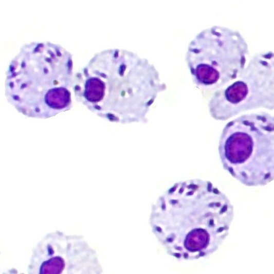
Staining
1h 5m
When the treatment incubation time ends, wash the wells with warm PBS (see steps 22 and 23)
30m
Add 300 µL MetOH to each well and leave it 5 minutes
5m
Dilute Giemsa in water 1:20 (20 mL of water per mL of Giemsa).
Add 200 µL of the 1:20 Giemsa solution to each well and incubate for 00:20:00
20m
Wash with distilled water. You can use three beakers full with water and simply submerge each Lab-tek in a stepwise manner. Submerge each lab-tek 5 in the first beaker, then pass to the next beaker (5 times) and then to the third beaker (5 more times).
10m
Wait until the slides are dry. Follow the instruction from Lab-tek to remove the chamber walls. Then, mount the cover slides with mounting glue.
Protocol references
Lab-teck-cultured macrophage infection has long been used in Liehsmania experiments:
Zilberstein, Dan, and Dennis M. Dwyer. “Antidepressants Cause Lethal Disruption of Membrane Function in the Human Protozoan Parasite Leishmania.” Science, vol. 226, no. 4677, 1984, pp. 977–79. JSTOR, http://www.jstor.org/stable/1693371. Accessed 13 Feb. 2026.
PNA selection was first described in:
Sacks, D. L., et al. "Identification of Cell Surface Carbohydrate and Antigenic Changes between Noninfective and Infective Developmental Stages of Leishmania Major Promastigotes." The Journal of Immunology, vol. 135, no. 1, July 1985, pp. 564-69, https://doi.org/10.4049/jimmunol.135.1.564.