Aug 04, 2023
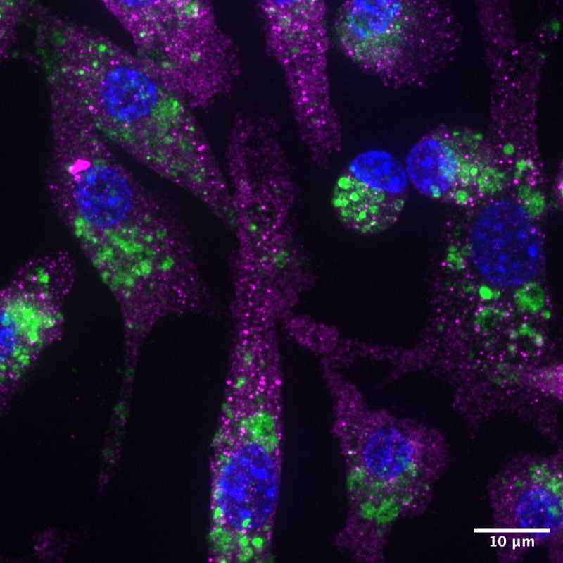
LRRK2 and LAMP1 immunofluorescence staining in various cell lines
- Siyu Chen1,
- eva karasmanis1
- 1UC San Diego, HHMI


Protocol Citation: Siyu Chen, eva karasmanis 2023. LRRK2 and LAMP1 immunofluorescence staining in various cell lines. protocols.io https://dx.doi.org/10.17504/protocols.io.bp2l6x991lqe/v1
License: This is an open access protocol distributed under the terms of the Creative Commons Attribution License, which permits unrestricted use, distribution, and reproduction in any medium, provided the original author and source are credited
Protocol status: Other
After multiple rounds of optimization, anti-LRRK2 antibody generates significant non-specific signals in the LRRK2 KO cell lines so this antibody may not be reliable for Immunofluorescence staining experiments. The protocol can be used for general IF experiments.
Created: August 03, 2023
Last Modified: May 31, 2024
Protocol Integer ID: 85948
Keywords: IF, LRRK2, RAW264.7, LAMP1, ASAPCRN, lrrk2 ko cell line, lamp1 immunofluorescence, reliable for immunofluorescence, immunofluorescence, various cell lines this protocol, antibody, staining experiment, lrrk2, various cell line, cell line
Disclaimer
DISCLAIMER – FOR INFORMATIONAL PURPOSES ONLY; USE AT YOUR OWN RISK
The protocol content here is for informational purposes only and does not constitute legal, medical, clinical, or safety advice, or otherwise; content added to protocols.io is not peer reviewed and may not have undergone a formal approval of any kind. Information presented in this protocol should not substitute for independent professional judgment, advice, diagnosis, or treatment. Any action you take or refrain from taking using or relying upon the information presented here is strictly at your own risk. You agree that neither the Company nor any of the authors, contributors, administrators, or anyone else associated with protocols.io, can be held responsible for your use of the information contained in or linked to this protocol or any of our Sites/Apps and Services.
Abstract
This protocol is being used to test the antibody Recombinant Anti-LRRK2 antibody [MJFF2 (c41-2)] (ab133474)Abcam , as well as Anti-LAMP1 antibody [1D4B] (ab25245)Abcam . Please note that after multiple rounds of optimization, anti-LRRK2 antibody generates significant non-specific signals in the LRRK2 KO cell lines so this antibody may not be reliable for Immunofluorescence staining experiments. The protocol can be used for general IF experiments.
Guidelines
GDB buffer was used as blocking buffer in the optimized protocol.
This protocol uses ibidi 8 Well Chamber µ-Slides and will coat them with Fibronectin. Standard volume would be 300 uL for each well. Volumes will need altering for wells, other plates and slides.
All steps are performed at room temperature (RT) on the lab bench, except for methanol permeabilization.
Materials
- (0.1M) NaPi pH 7.4 3.1 g of NaH2PO4•H2O 0.9 g of Na2HPO4 (anhydrous) distilled H2O to make a volume of 1 L The pH of the final solution will be 7.4. This buffer can be stored for up to 1 mo at 4°C
- GDB buffer 30mM NaPi (sodium phosphate) pH 7.4 0.45mM NaCl 0.2% porcine (or fish) gelatin In ddH2O
- Kim wipes
- Ethanol (100%, stored in dark chemicals cupboard)
- Water (double-deionised H2O from Milli-Q, “MQ-H2O”)
Protocol materials
Recombinant Anti-LRRK2 antibody [MJFF2 (c41-2)] (ab133474)Abcam
Anti-LAMP1 antibody [1D4B] (ab25245)Abcam
Chloroquine diphosphate saltMerck MilliporeSigma (Sigma-Aldrich)Catalog #C6628-25G
Leu-Leu methyl ester hydrobromideMerck MilliporeSigma (Sigma-Aldrich)Catalog #L7393-500MG
Ammonium chlorideMerck MilliporeSigma (Sigma-Aldrich)Catalog #254134
Triton X-100 Merck MilliporeSigma (Sigma-Aldrich)Catalog #X100
GelatinMerck MilliporeSigma (Sigma-Aldrich)Catalog #G2500
Goat Anti-Rabbit IgG H&L (Alexa Fluor® 647) (ab150079)Abcam
Goat Anti-Rat IgG H&L (Alexa Fluor® 488) (ab150157)Abcam
DAPI Thermo Fisher ScientificCatalog #D1306
FluorSave™ ReagentMerck MilliporeSigma (Sigma-Aldrich)Catalog #345789
Safety warnings
N/A
Before start
Important note:
While LAMP1 immunofluorescence staining robustly gives expected results, after multiple rounds of optimization, anti-LRRK2 antibody generates significant non-specific signals in the LRRK2 KO cell lines in our hand. This is a warning that this antibody may not be reliable for Immunofluorescence staining experiments. However, this protocol can be used widely for IF experiments.
Please check This google sheet to learn more about the cell lines and conditions tested for the antibody
Day 0: : Seed cells
1h
Add 300 µL of 11 ug/mL fibronectin into each ibidi well. Incubate at RT for 01:00:00 .
1h
Rinse the wells with PBS for 3 times
Make GDB buffer if necessary
| Stock | Amount needed | C | Final conc. | E | |
| (0.1M) NaPi pH 7.4 | 15 | mL | 30 | mM | |
| (5M) NaCl | 0.0045 | mL | 0.45 | mM | |
| Gelatin | 0.1 | g | 0.2 | % | |
| H2O | 34.9955 | mL | |||
| Total | 50 | mL | � |
Recipe to make GDB buffer
Seed adherent cells to 40-80% confluency in each well. Incubate at 37 °C Overnight to get optimal seeding.
Note
For RAW264.7 cells, 6x10^4 in 300uL is a good starting point. Less would be needed for other typical cell lines since macrophage cells are smaller.
It is suggested to start with two different cell concentrations for the first time. Please refer to this page for more information
Day 1: Drug treatment
3h
Apply any drug treatments or controls and note time of additions before proceeding with fixing. As an example, Chloroquine diphosphate saltMerck MilliporeSigma (Sigma-Aldrich)Catalog #C6628-25G and Leu-Leu methyl ester hydrobromideMerck MilliporeSigma (Sigma-Aldrich)Catalog #L7393-500MG can be added at desired concentrations for 03:00:00
| Drugs | MW - g/mol | mM | weight in 1 mL | E | |
| LLOME | 339.27 | 1000 | 339.27 | mg/1mL | |
| CQ | 515.86 | 100 | 51.586 | mg/1mL |
Drug stock recipe and concentrations
In each well, add 1 in 1000 ( 1 millimolar (mM) for LLOME and 0.1 millimolar (mM) for CQ)
3h
Staining
25m
Put 100% ethanol on ice before proceeding with next steps.
Bring GDB buffer to Room temperature . Prepare and prewarm fixation buffer (4% sucrose, 3% PFA in 1xPBS) at 37 °C
Note
3% PFA is preferred from 4% as it reduces autofluorescence
Need 375 µL Pierce™ 16% Formaldehyde (w/v) Methanol-freeThermo Fisher ScientificCatalog #28906 and 0.08 g SucroseMerck MilliporeSigma (Sigma-Aldrich)Catalog #S0389 , dissove with 2 mL PBS.
Get cells, aspirate media and immediately add prewarmed fixation buffer. Incubate for 00:10:00 at 37 °C
10m
Aspirate PFA, rinse 2x with PBS and wash two times with PBS for 5 minutes each, 00:10:00 in total
10m
Quenching: Only necessary when staining the day of fixation. Incubate 3 times of 10 minutes (00:30:00 in total) using 0.4% Ammonium chlorideMerck MilliporeSigma (Sigma-Aldrich)Catalog #254134 (75 millimolar (mM) )
30m
Optional step: For half of the samples, choose to add another permeablisation step with 300 µL 100% ethanol at -20°C for 00:20:00 . Leave the rest at Room temperature in GDB buffer.
Note
This step helps to increase contrast when imaging Recombinant Anti-LRRK2 antibody [MJFF2 (c41-2)] (ab133474)Abcam , but doesn't help with the issue of non-specific signals when LRRK2 KO cell lines are used.
Note
ethanol/methanol permeablisation at -20 °C will disrupt the microtubule staining. Apply this step with caution when other antibodies are used.
20m
Apsirate ethanol, wash 2x with PBS.
Prepare GDB + 0.05% Triton X-100 Merck MilliporeSigma (Sigma-Aldrich)Catalog #X100 freshly. Add 300 µL for 00:10:00 .
5 µL in 10 mL GDB buffer. Can be stored at 4 °C for a couple of days.
10m
Aspirate Triton X-100, wash 2x with GDB.
Block cells with 300 µL GDB for 00:30:00 at Room temperature
30m
During blocking, prepare final concentration of 1 ug/mL for bothRecombinant Anti-LRRK2 antibody [MJFF2 (c41-2)] (ab133474)Abcam and Anti-LAMP1 antibody [1D4B] (ab25245)Abcam solution with GDB and 50000 rpm, 4°C, 00:10:00 - each ibidi dish well requires 150 µL
Note
150 µL is the minimum required amount. 200 µL would be sufficient and optimal to cover the entire well
10m
Take most of the supernatant and place in new tube. Mix to get even concentration (due to GelatinMerck MilliporeSigma (Sigma-Aldrich)Catalog #G2500 in there, there will be a small clear precipitate)
Aspirate blocking solution and incubate cells with 150 µL of primary antibody solution Overnight at 4 °C on a table-top shaker
Note
Primary antibody incubation can be as short as 2 hours without affecting the final outcome
Note
When incubating overnight, consider wrapping up the dish with parafilm and/or put the dish in a humidity chamber to prevent the well from drying up
Day 2
15m
Bring GDB to Room temperature . Aspirate antibody solution and rinse 2x with GDB.
Wash 3x 5min Room temperature RT with GDB, 00:15:00 in total
15m
Prepare 1:500 Alexa-flour conjugated secondary antibody solution with GDB - each ibidi well requires 200 µL .
When LRRK2 and LAMP1 are co-stained, Goat Anti-Rabbit IgG H&L (Alexa Fluor® 647) (ab150079)Abcam and Goat Anti-Rat IgG H&L (Alexa Fluor® 488) (ab150157)Abcam are used at final concentration of 4 ug/mL . Lower concentration did not help with the issue of non-specific LRRK2 signal.
Incubate cells with 200 µL of secondary antibody solution for 01:30:00 at Room temperature and protect from light with an ice box.
1h 30m
15 minutes before the incubation is finished, add DAPI Thermo Fisher ScientificCatalog #D1306 to a final concentration of 1 ug/mL and incubate until the last step finishes.
Rinse cells with 1xPBS for 5 times
Apply 2-4 drops of FluorSave™ ReagentMerck MilliporeSigma (Sigma-Aldrich)Catalog #345789 hard mounting media in each well and swirl to make sure the bottom is fully covered.
Allow to air-dry for 00:10:00 at Room temperature .
10m
Image within 48 h of mounting or the sample will begin to deteriorate (bright debris impeding imaging) and visibly autofluoresce in red.