Sep 26, 2025
Low-Cost Sucrose Gradient Protocol for GFP Purification from Recombinant E. coli: A Visual Fluorescence-Based Method for Resource-Limited Laboratories
- Joseph shenekji1,2,
- Kamar Shayah1,
- Suad Ali Hassna1,
- Wjoud Kahel1,
- Hind Alnaem1,
- Najwa Hasn Bek1
- 1Department of biotechnology engineering, faculty of technical engineering, university of Aleppo;
- 2Coordinator of Biotechnologysy.org
- Joseph shenekji: PhD in Biotechnology Engineering - [email protected];
- Kamar Shayah: PhD in Biotechnology Engineering
- Suad Ali Hassna: MSc in Biotechnology Engineering
- Wjoud Kahel: Graduate students in Biotechnology Engineering
- Hind Alnaem: Graduate students in Biotechnology Engineering
- Najwa Hasn Bek: Graduate students in Biotechnology Engineering
- Reclone.org (The Reagent Collaboration Network)Tech. support email: [email protected]


Protocol Citation: Joseph shenekji, Kamar Shayah, Suad Ali Hassna, Wjoud Kahel, Hind Alnaem, Najwa Hasn Bek 2025. Low-Cost Sucrose Gradient Protocol for GFP Purification from Recombinant E. coli: A Visual Fluorescence-Based Method for Resource-Limited Laboratories. protocols.io https://dx.doi.org/10.17504/protocols.io.n92ld62pxg5b/v1
License: This is an open access protocol distributed under the terms of the Creative Commons Attribution License, which permits unrestricted use, distribution, and reproduction in any medium, provided the original author and source are credited
Protocol status: Working
We use this protocol and it's working
Created: September 26, 2025
Last Modified: September 26, 2025
Protocol Integer ID: 228235
Keywords: purifying green fluorescent protein, cost sucrose gradient protocol for gfp purification, green fluorescent protein, gfp purification, visual fluorescence, fluorescence, cost sucrose gradient protocol, sucrose gradient, gfp, gene expression education laboratory, molecular tagging, expensive chromatography system, biomedical imaging
Disclaimer
This protocol was developed and executed in a laboratory with limited infrastructure. The confirmation of GFP purification was based solely on visual fluorescence under UV light. No spectrophotometric, electrophoretic, or biochemical validation was performed to assess concentration and contaminants. As such, the results should be considered preliminary. Further studies are necessary to quantitatively assess purity, yield, and functional integrity of the extracted GFP.
Abstract
This protocol describes a simple, cost-effective method for purifying Green Fluorescent Protein (GFP) from E. coli using a sucrose gradient. It avoids toxic chemicals and expensive chromatography systems, making it accessible and environmentally friendly. The method preserves GFP's fluorescence and is suitable for applications in biomedical imaging, molecular tagging, and gene expression education laboratories.
with further improvements, it could be used in research studies.
Image Attribution
Biotechnology Engineering Department - University of Aleppo
Materials
Genetically modified E. coli BL21 (DE3) pLysS with pRSET plasmid
Sucrose (analytical grade)
Sterile distilled water
Falcon tubes (15 ml)
Microcentrifuge tubes (1.5 ml)
Ethanol (70%)
Micropipettes and tips
Centrifuge (R5810 and CF-10 models or equivalents)
UV light source
Preparation of Sucrose Gradient
7m
Prepare four sucrose solutions:
weigh 1, 2, 3, 4 g of sucrose
Figure (1): Prepared sucrose weights for the experiment
to be out in 10 mL sterile water in order to make
10% (Tube 1) 20% (Tube 2) 30% (Tube 3) 40% (Tube 4)
Figure (2): Sucrose powder in tubes before adding distilled water
Dissolve the appropriate sucrose amounts in sterile distilled water and mix until homogeneous.
5m
Layer 2 ml of each sucrose solution sequentially into a Falcon tube using a micropipette. Begin with 40% at the bottom, followed by 30%, 20%, and finally 10% at the top. Layer gently to avoid mixing between layers.
Figure (3): The gradient of sucrose layers after preparation- the layers could be visible if you spotlight a smartphone flash and move it, this can help you know if the layers interfaced. in case layers interfaces you need to redo the process.
2m
Sample Loading and Initial Centrifugation
5m 30s
Centrifuge 5 ml of E. coli culture at 7000 rpm for 5 minutes in a falcon tube to pellet the bacteria.
Figure (4): The bacterial culture after centrifugation. the GFP is secreted to the media.
5m
Carefully collect 1 mL of the supernatant, which contains secreted proteins, and gently layer it on top of the prepared sucrose gradient.
Figure (5): The process of loading the supernatant onto the sucrose
gradient
Figure (6): The sucrose gradient tube after bacterial supernatant
addition
30s
GFP Purification
32m
Centrifuge the gradient tube at 10000 rpm for 00:30:00 using an R5810 centrifuge. After centrifugation, observe the tube under UV light. GFP will localize between the 10- 20% sucrose layer, visible as a fluorescent band.
Figure (7): Protein localization and fluorescence between 10-20% sucrose
gradient concentration
30m
Carefully aspirate the fluorescent layer using a micropipette and transfer it to a 1.5 ml microcentrifuge tube. Centrifuge this tube at 12000 rpm for 2 minutes using a CF-10 centrifuge.
2m
Final Purification and Concentration
4m
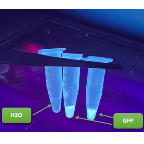
Examine the pellet under UV light to confirm GFP fluorescence.
Figure (8): The purified GFP exhibited higher fluorescence intensity
compared to the control water sample
Add 20 µL of 70% ethanol and 20 µL of distilled water to the GFP sample. Mix gently and centrifuge again at 12000 rpm for 4 minutes.
4m
After centrifugation, visualize the sample under UV light. The ethanol precipitation enhances fluorescence intensity, confirming successful concentration of GFP.
Figure (9): The ethanol-precipitated GFP sample demonstrated significantly higher
fluorescence compared to the untreated control when examined under identical UV
exposure conditions
Results
GFP is localized between 10-20% sucrose layers.
Ethanol precipitation significantly enhances fluorescence.
The final product is visibly fluorescent and suitable for downstream applications.
Protocol references
Fernández-Martínez, J., & Rout, M. P. (2016). Density gradient ultracentrifugation for the isolation of protein complexes. Cold Spring Harbor Protocols. Retrieved from https://www.ncdir.org/wp-content/uploads/2018/03/Cold-Spring-Harb-Protoc-2016-Fernandez-Martinez.pdf
Joseph shenekji, Dr. Kamar Shayah, Sidra Latfo, Roukaia Hariri, Walaa Alothman, Dr. Zaher Samman Tahhan, Mai Al-Ansary 2025. manufacturing dried cell reagents using the genetically modified bacterial strain E.coli pLysS expressing GFP protein. protocols.io https://dx.doi.org/10.17504/protocols.io.rm7vzk9j4vx1/v1
Joseph shenekji 2024. Using Skimmed milk as a low-cost alternative to IPTG in plasmid expression induction (LacZ Promoter) . protocols.io https://dx.doi.org/10.17504/protocols.io.j8nlk81z6l5r/v1