May 29, 2023
Version 2
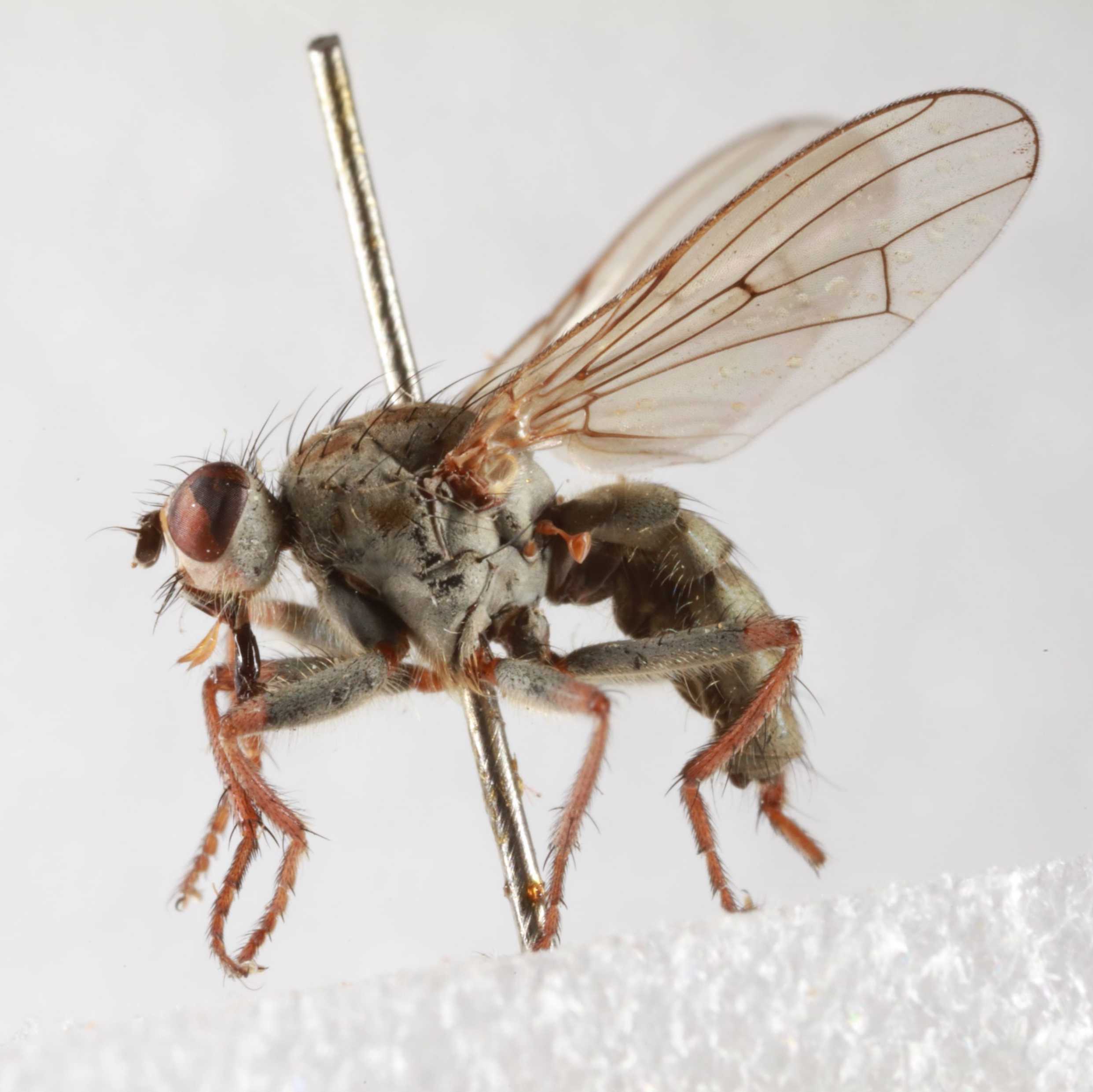
Low-cost museum DNA extraction using magnetic beads V.2
- Andie Hall1,
- owain.powell 1,
- Piotr Cuber1,
- Ben Price1
- 1Natural History Museum


Protocol Citation: Andie Hall, owain.powell , Piotr Cuber, Ben Price 2023. Low-cost museum DNA extraction using magnetic beads. protocols.io https://dx.doi.org/10.17504/protocols.io.4r3l27ebxg1y/v2Version created by Ben Price
License: This is an open access protocol distributed under the terms of the Creative Commons Attribution License, which permits unrestricted use, distribution, and reproduction in any medium, provided the original author and source are credited
Protocol status: Working
We use this protocol and it's working
Created: May 29, 2023
Last Modified: October 27, 2023
Protocol Integer ID: 82583
Keywords: DNA extraction, historical, aDNA, museum specimen, silica, magnetic bead, dna extraction, high throughput dna extraction, cost museum dna extraction, biosciences bead, using magnetic bead, v2 replaces serasil beads with qiagen magattract bead, qiagen magattract bead, magnetic bead, extraction, historical specimens in natural history collection, v2 replaces serasil bead, historical specimen, dna, specimen
Abstract
Modified from Korlevic et al 2021& Rohland et al 2018 for high throughput DNA extraction from historical specimens in natural history collections. Please cite these papers when using this method.
An in depth SOP by Korlevic can be found here: https://dx.doi.org/10.17504/protocols.io.ewov1o3molr2/v1
Post lysis the protocol can be run by hand or on a magnetic bead based robot (e.g. Kingfisher Flex / Apex).
v2 uses 5x lysate volume of binding buffer as tests have shown improved recovery over 10x volume.
v2 replaces SeraSil beads with Qiagen MagAttract beads which provide a much cheaper option to G-Biosciences beads.
Materials
Lysis buffer C:
200mM Tris pH8, 25mM EDTA pH8, 0.05% (vol/vol) Tween 20, 0.4mg/ml PK
Binding buffer D:
5M guanidine hydrochloride, 40% (vol/vol) 2-propanol, 0.12M sodium acetate, 0.05% (vol/vol) Tween 20.
Wash buffer:
Option 1: Qiagen PE wash buffer
Option 2: DIY wash buffer:
10mM Tris pH8, 80% (vol/vol) ethanol
Elution buffer:
10mM Tris pH8, 1mM EDTA pH8, 0.05% (vol/vol) Tween 20
Silica magnetic beads:
Option 1: G-Biosciences Silica magnetic bead suspension (GENO786-915)
Option 2: MagAttract Suspension G magnetic beads (Qiagen: 1026901)
buffer preparation
Lysis buffer C
200mM Tris pH8, 25mM EDTA pH8, 0.05% Tween 20, 0.4mg/ml PK
To make 10 mL mix together: 7295 µL molecular grade water, 2000 µL 1M Tris pH8,
500 µL 0.5M EDTA (pH 8), 200 µL Proteinase K (20mg/ml), and 5 µL Tween 20.
This is sufficient for 1 extraction plate of 95 samples and 1 negative control (90 µL each).
Binding buffer D
5 M guanidine hydrochloride, 40% (vol/vol) 2-propanol, 0.12 M sodium acetate and 0.05% (vol/vol) Tween 20.
In a glass or plastic bottle, weigh 124.2 g of guanidine hydrochloride and fill up with molecular water to 150 mL . Heat briefly in a microwave until the buffer is warm to the touch and shake until the salt is fully dissolved. Add 104 mL of 2-propanol, 10.4 mL of 3M sodium acetate buffer solution (pH 5.2) and 130 µL of Tween 20.
This is sufficient for 6 extraction plates of 95 samples and 1 negative control (450 µL each). Store in a fridge for up to 4 weeks. Seal the bottle with Parafilm to avoid evaporation.
Wash buffer
Option 1: To 100 mL of Qiagen buffer PE concentrate, add 400 mL of 96-100% Ethanol.
Option 2: To 5 mL 1M Tris-HCl, add 412 mL 97% Ethanol and 83 mL molecular grade water.
Note: Kevin Beentjes (Naturalis) has compared the Qiagen PE and "DIY" wash buffers with no apparent difference in fragment recovery.
This makes 500 mL of wash buffer, sufficient for 6 extraction plates of 96 wells (750 µL each). This buffer can be stored at room temperature for at least 1 year.
Elution buffer
To make 50 mL of elution buffer, combine 49.4 mL of molecular water, 500 µL of 1 M Tris-HCl (pH 8.0), 100 µL of 0.5 M EDTA (pH 8.0) and 25 µL of Tween 20.
This is sufficient for 10 96-well extraction plates 50 µL each). This buffer can be stored at room temperature for at least 1 year.
silica bead preparation
8s
Option 1: G-Biosciences Silica magnetic beads (GENO78 6-915)
Option 2: MagAttract Suspension G magnetic beads (Qiagen: 1026901)
Note: MagAttract beads are much cheaper and have very similar recovery for fragments 50bp and above, with minor loss of 35bp fragments. SeraSil beads only recover from 75bp (SeraSil 700) or 100bp (SeraSil 400).
The original Rohland protocol washes the beads before use, but we have found unwashed beads work just as effectively. If washing follow these steps:
- Fully resuspend the stock suspension of silica beads by vortexing.
- Transfer 10 µL of silica bead suspension per reaction to a 2.0-ml lobind tube, including an excess of 5% (e.g., for a plate take 1 mL ).
- Place the tube on a magnet to collect the beads.
- Pipette off and discard the supernatant.
- Remove the tube from the magnet, add 500 µL of elution buffer and resuspend the beads by vortexing for 8 seconds.
- Spin the tubes briefly in a microcentrifuge to collect the suspension at the bottom.
- Place the tube back on the magnet to collect the beads.
- Pipette off and discard the supernatant.
- Repeat steps 5 to 8 for a total of two washes.
- Resuspend the beads in a volume of elution buffer equivalent to the initial volume used (e.g., for a plate add 1 mL ).
Manual protocol
Tissue can be added to the plate/tubes either before or after the addition of lysis buffer, however tissue from dried specimens (e.g. pinned insects) should be added to wells/tubes with lysis buffer to prevent static displacement. If tissue was previously stored in ethanol this should be dried off before lysis.
- Add 90 µL of lysis buffer C to each sample tube / well. Volume can be adjusted to ensure coverage of tissue, however be mindful that 5x volume of binding buffer is required further down the protocol.
- Incubate overnight at 56 °C on a shaking incubator. The longer the incubation the more the tissue / specimen is digested. Shorter incubations are recommended for better voucher recovery with whole body lysis.
- Pipette lysate into a new tube / deep-well plate. If tissue / voucher is to be recovered then add appropriate volume of 80% ethanol to cover sample in original tube / well.
- Add 450 µL of binding buffer D (= 5x volume of lysate) to lysate in tube / deep-well plate.
- Add 10 µL of silica magnetic bead suspension to lysate and binding buffer.
- Vortex for 5sec.
- Rotate (mix) the tubes / plates for 15min at room temperature.
- Spin briefly in a microcentrifuge to collect the suspension at the bottom and place them on an appropriate magnet.
- Once solution clears pipette off the supernatant and discard.
- Remove from the magnet, add 250 µL of wash buffer and vortex for 8sec.
- Spin briefly to collect the suspension at the bottom and place back on the magnet.
- Repeat wash (steps 9 - 11) twice for a total of three washes.
- Aspirate any remaining drops of liquid and dry the beads at room temperature by leaving them on the magnet with open lids. Usually a few minutes is sufficient.
- Remove from the magnet, add 50 µL - 100 µL of elution buffer depending on original sample size and anticipated DNA recovery.
- Vortex until all beads have been resuspended and then briefly spin down.
- Incubate for 2min at room temperature.
- Place back on the magnet, wait until the solution clears and transfer the supernatant to a fresh tube / plate.
Proceed to library preparation or freeze extracted DNA.
Automated protocol
The manual protocol has been tested on the Kingfisher Flex and Apex platforms. The protocol starts after step 5 above, i.e. prepare the deep-well plate with 90 µL lysate + 450 µL binding buffer D and 10 µL silica beads.
The kingfisher protocol uses onboard "tip mixing" rather than vortexing / mixing to resuspend beads and skips all centrifugation steps.
Protocol references
Petra Korlević, Erica McAlister, Matthew Mayho, Alex Makunin, Paul Flicek, Mara K N Lawniczak, A Minimally Morphologically Destructive Approach for DNA Retrieval and Whole-Genome Shotgun Sequencing of Pinned Historic Dipteran Vector Species,Genome Biology and Evolution, Volume 13, Issue 10, October 2021, evab226, https://doi.org/10.1093/gbe/evab226
Rohland, N., Glocke, I., Aximu-Petri, A.et al.Extraction of highly degraded DNA from ancient bones, teeth and sediments for high-throughput sequencing.Nat Protoc13, 2447–2461 (2018). https://doi.org/10.1038/s41596-018-0050-5