Feb 28, 2022
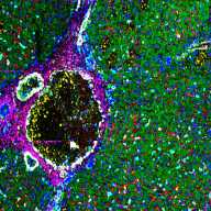
Liver tissue staining with multiple lanthanides-tagged antibodies
- Hua Tian1
- 1Department of Chemistry, Pennsylvania State University
- Liver Ln-Abs staining protocol


Protocol Citation: Hua Tian 2022. Liver tissue staining with multiple lanthanides-tagged antibodies . protocols.io https://dx.doi.org/10.17504/protocols.io.b5qmq5u6
License: This is an open access protocol distributed under the terms of the Creative Commons Attribution License, which permits unrestricted use, distribution, and reproduction in any medium, provided the original author and source are credited
Protocol status: Working
We use this protocol and it’s working
Created: February 28, 2022
Last Modified: February 28, 2022
Protocol Integer ID: 58861
Keywords: lanthanides-tagged antibody, immunostaining, imaging mass spectrometry, secondary ion mass spectrometry, C60-SIMS, hydrated tissue with antibodies cocktail, consistent antibody affinity, optimized immunostaining, tagged antibody, multiple lanthanide, antibodies cocktail, subcellular resolution, ims image quality, imaging
Funders Acknowledgements:
NIH
Grant ID: UG3CA256962
Abstract
Imaging mass spectrometry (IMS) is able to image the multiple lanthanides-tagged antibodies (up to 40) simultaneously at a subcellular resolution on a single tissue section. This protocol is developed for optimized immunostaining of frozen-hydrated tissue with antibodies cocktail (10-25 Abs), which produces consistent antibody affinity and improved IMS image quality using C60-secondary ion mass spectrometry (C60-SIMS).
Materials
• Five micrometer fresh snap-frozen human liver sections on Au-coated Silicon wafer (2.2cm*2.2cm)
• 1% Paraformaldehyde (diluted from 16% solution from Thermo Fisher Scientific)
• Methanol (Sigma)
• 3% Goat serum (diluted from 10% stock from Thermo Fisher Scientific)
• DPBS (Corning)
• Wash buffer [DPBS supplemented with 0.05% Tween(Thermo Fisher Scientific) and 1% BSA(Sigma)]
• Metal-conjugated antibodies as in Table 1 below. Please refer to Fluidigm Maxpar Antibody Labeling User Guide Chapter 3 for detailed conjugation steps. Maxpar® X8 Antibody Labeling Kit with all the metal tags in Table 1 are purchased from Fluidigm
Table 1 Antibody panel for human liver tissue
| A | B | C | D | E | |
| Metal | Target | Clone/Host | Cell/Pathway | Manufacturer/Catalog No. | |
| 89Y | CD45 | D9M8I, Rabbit IgG | Pan leukocyte | Cell Signaling/13917S | |
| 113 In | CD4 | RPA-T4, mouse monoclonal | T cell | Novus/NBP2-25199 | |
| 141Pr | SMA | 1A4, Mouse IgG2a | Vascular walls/Hepatic Stellate cells/fibroblasts | Fluidigm/3141017D | |
| 143Nd | GFAP | EPR1034Y, Rabbit monoclonal | Ito Stellate Cells | Abcam/ab218309 | |
| 145Nd | Heppar-1 | HepPar1, mouse monoclonal | hepatocytes | Novus/NBP3-08970 | |
| 147Sm | Glul | ab240193,Rabbit monoclonal | Pericentral hepatocytes (Zone 3) | Abcam/ab240193 | |
| 148Nd | CD31 | JC/70A, Mouse monoclonal | Endothelial cells | Abcam/ab264090 | |
| 151Eu | CD68 | D4B9C,Rabbit IgG | Macrophages | Cell Signaling/76437S | |
| 153Eu | CD32 | FUN-2, Mouse IgG2b | Macrophages | Fluidigm/3153018B | |
| 158Gd | Arginase1 | D4E3M™,Rabbit IgG | Zone 1-2 hepatocytes | Cell Signaling/93668S | |
| 161Dy | Albumin | EPR20195, Rabbit monoclonal | Periportal (Zone 1) | Abcam/Ab271979 | |
| 166Er | CK19 | SPM561, Mouse monoclonal | Cholangiocytes (Portal triad) | Abcam/ab212569 | |
| 168Er | Ki-67 | Ki-67, Mouse IgG1 | Proliferation (Midlobular) | Cell Signaling/9449S | |
| 169Tm | CD34 | QBEND/10, Mouse / IgG1 | Endothelial cells | Abcam/ ab198395 | |
| 170 Er | EpCAM | E6V8Y, Rabbit IgG | Hepatic stem/progenitor cells | Cell Signaling /93790S | |
| 171Yb | LYVE1 | EPR21857, Rabbit monoclonal | Sinusoidal endothelial cells | Abcam/ab232935 | |
| 176Yb | EGFR | EP38Y, Rabbit monoclonal | cell membrane | Abcam/ ab272293 | |
| 176Yb | Na/K ATPase | D4Y7E, Rabbit IgG | cell membrane | Cell Signaling/ 23565S | |
| 191Ir | Nuclear DNA | Nuclei | Fluidigm/ 201192B | ||
| 196Pt | Human collagen I | EPR7785, Rabbit monoclonal | Collagen | Abcam/ab215969 |
• Intercalator-Ir (500µM, Fluidigm)
• DI water (Sigma)
• Water-Repellent Slides Marker Pen (Dando Sangyo Co Ltd. Japan)
• glass petri dish (diam 40mm, Sigma)
Safety warnings
Wear proper PPE when handling the agents. Discard the waste in the biohazard waste container
Cut the fresh frozen sections at 5µm using a cyromicrotome and mount them on Au-coated silicon slides
Place the slides at -20 ◦C for 1h and then at 4 ◦C for 30 min for temperature equilibrium
Fix the tissue slides in 1% PFA for 5 min at 4 ◦C, then in prechilled methanol for 5 min at -20 ◦C
Wash the slides twice for 5 min in a glass petri dish by submerging the slides in 25 ml wash buffer, following by rehydrating the slides for 5 min in a glass container with 25ml DPBS and rinsing the slides for 5 min in a glass container with 25ml wash buffer
Use the Liquid-Repellent Slide Marker Pen to draw a circle around the tissue section to create a barrier to contain the solutions on the tissue sections
Apply 100 µl 3% goat serum solution to each slide for 1 h at RT and then remove excess block solution by tapping on a tissue
Prepare the antibody cocktail. Vortex all the antibodies for 15 s and then centrifuge at 12,000G for 2 min, followed by diluting the upper liquid of each antibody in 0.5% PBS as desired concentration
Add 100 µl of the antibody cocktail to each section and incubate overnight at 4◦C in a sealed glass petri dish in a fridge
After the incubation, wash the sections two times for 5min in a glass petri dish with 5 ml wash buffer with slow agitation
Incubate the slides with 100 µl 1:100 dilution of Intercalator-Ir in DPBS for 30 min at RT
After nuclei staining, wash the slides for 5 min in a glass container with 5 ml DPBS
Quickly dip the slides in a container with 5 ml DI water for 15 s, repeat three times
Dry the slides in a fume hood at RT for 20 min
Store the slides at 4◦C until C60-SIMS imaging.