Mar 17, 2026
Leafy Green Phenolic / Polyphenolic Extraction
- Alexander Hernandez1,
- Elaine Le1,
- Alvin Tak1,
- Michael Dzakovich1
- 1USDA-ARS Children's Nutrition Research Center


Protocol Citation: Alexander Hernandez, Elaine Le, Alvin Tak, Michael Dzakovich 2026. Leafy Green Phenolic / Polyphenolic Extraction. protocols.io https://dx.doi.org/10.17504/protocols.io.36wgqpprkvk5/v1
Manuscript citation:
Dzakovich, M.P.; Le, E.A.; Tak, A.L.; Chacko, S.K. A Comprehensive UHPLC-MS/MS and Extraction Method Reveals Flavonoid Profile and Concentration Are Diverse in Spinach (Spinacia oleracea L.). Front. Plant Sci. 2025, 16. doi:10.3389/fpls.2025.1496200.
License: This is an open access protocol distributed under the terms of the Creative Commons Attribution License, which permits unrestricted use, distribution, and reproduction in any medium, provided the original author and source are credited
Protocol status: Working
We use this protocol and it's working
Created: October 21, 2025
Last Modified: March 17, 2026
Protocol Integer ID: 230418
Keywords: Spinach, Flavonoids, Polyphenolics, Phenolics, Water-soluble, Polar metabolites, Phytochemicals, Extraction, Leafy greens, polyphenolic extractions from plant material, polyphenolic compounds across plant matrice, quantification of diverse phenolic, leafy green phenolic, polyphenolic extraction, polyphenolic extraction this protocol, polyphenolic compound, flavonoid, diverse phenolic, optimized extraction solvent, liquid chromatography, extraction solvent, tandem mass spectrometry, plant matrice, plant material
Funders Acknowledgements:
USDA-ARS CRIS Funds
Grant ID: 3092-10700-066-001S
Abstract
This protocol describes a streamlined method for phenolic and polyphenolic extractions from plant material and subsequent preparation for analysis by liquid chromatography–tandem mass spectrometry (LC-MS/MS). Previously optimized extraction solvents, sample disruption techniques (e.g. sonication), and the use of internal standards primarily targeted at flavonoids are covered in detail. A workflow for diluting, filtering, and otherwise preparing samples for analysis is also included. This approach enables sensitive, reproducible profiling and quantification of diverse phenolic and polyphenolic compounds across plant matrices.
Materials
3D printed lab items:
Sample organizer: https://3d.nih.gov/entries/3DPX-020366
Universal vial holder:https://3d.nih.gov/entries/3DPX-020367
15 mL tube rack: https://3d.nih.gov/entries/3DPX-021167
Solutions:
98:2 MeOH:formic acid
1:1 naringin:naringenin (each starting 20 µM - final conc. 10 µM each) mixed stock solution
1:1 MeOH:H2O +0.1% formic acid (LC-MS grade)
Equipment:
Sonicator
Centrifuge (swinging bucket)
Repeating pipette
Seripettor
5 mL pipette
1 mL pipette
200 µL pipette
Titer plate shaker
Nitrogen evaporator
RapidVap (sample concentrator)
Consumables:
15 mL flip-cap tubes (one per sample)
16 x 125 mm glass borosilicate culture tube (one per sample)
15 mL tube rack
8 mL glass vials (one per sample)
1.5 mL HPLC vial and cap (one per sample; or other volume as necessary)
3 mL syringe
0.2 μm PTFE or regenerated cellulose syringe filter
Tubs of ice
Safety warnings
Please ensure you are wearing proper PPE while performing these extractions (gloves, goggles, lab coat). Methanol and formic acid, in particular, are harmful to human health.
Preparation
Pre-weigh 20 mg of freeze dried plant powder (± 1 mg) into a 15 mL snap-cap tube.
Pre-cool centrifuge to 4 ℃. Also pre-heat both the nitrogen sample concentrator water bath and the RapidVap to 35 ℃. Ensure there is sufficient nitrogen gas available for sample drying.
Label 1 glass borosilicate culture tube (16x125 mm) for each sample tube you have. In addition, label 3 extra tubes that will only contain your internal standards.
Extraction
Embed all sample tubes and all glass collection tubes in a tub of ice to keep the samples cool.
Typical setup for keeping samples on ice.
The sample organizer seen above can be download and 3D printed from the following link:
Add 200 μL of 1:1 20 µM naringin:20 µM naringenin to each sample as your internal standard.
In addition, add 200 μL of 1:1 naringin:naringenin to your 3 extra internal standard-only glass collection tubes. Dry these tubes immediately and store at -80 until analysis.
Add 4 mL of 98:2 methanol:formic Acid to each sample tube. You can save time by using a Seripettor for this step.
Image showing solvent being added to sample tubes.
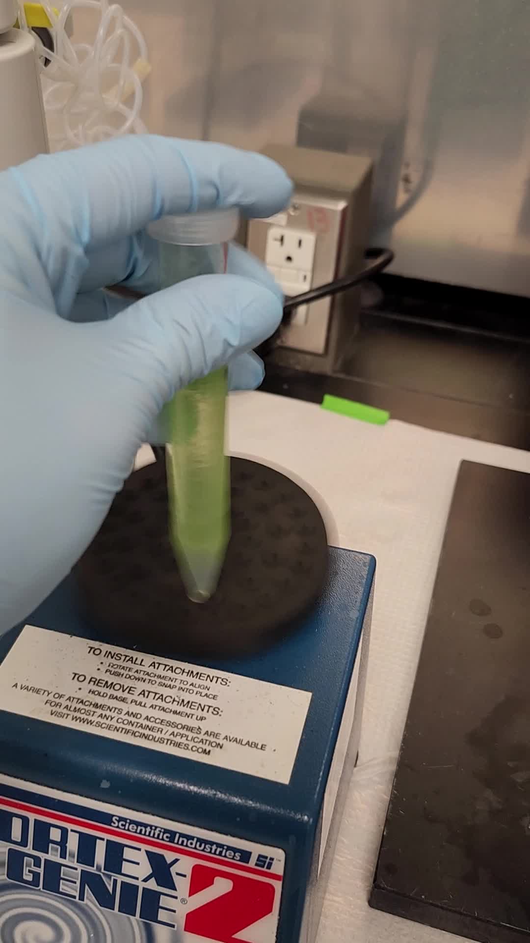
Cap each sample, then vortex for ~30 seconds until samples are mixed
If the sample pellet remains stuck at the bottom of the tube, alternate between vortexing and vigorous handshaking until no pellet remains stuck at the bottom of the tub. Keep your index finger on the tube cap to minimize the chance of dropping a tube or liquid leaking out of the cap.
Sample pellet stuck in the bottom of the tube will not be properly extracted as solvents cannot efficiently penetrate this tissue.
Place samples in sonicator water bath. Sonicate the samples for 20 minutes for the first extraction round. For subsequent extraction rounds, sonicate for 10 minutes.
Image showing a typical setup used to sonicate samples
The modified rack seen above can be downloaded and 3D printed from the following link:
Add ice to the water bath to keep the samples cool
Move the location of samples around each extraction to minimize effects of "hot spots" on extraction efficiency.
Centrifuge at 3700 rpm (~3200 x g) for 5 minutes at 4 ℃
Decant supernatant of each sample into their respective glass collection tube.
Repeat Steps 6 through 10 two more times for a total of 3 extractions.
By the end of the third round, each glass collection tube should have ~12 mL of sample supernatant.
Sample Drying
Open the nitrogen evaporator luer valves. Make sure to close luer valves that will not be used.
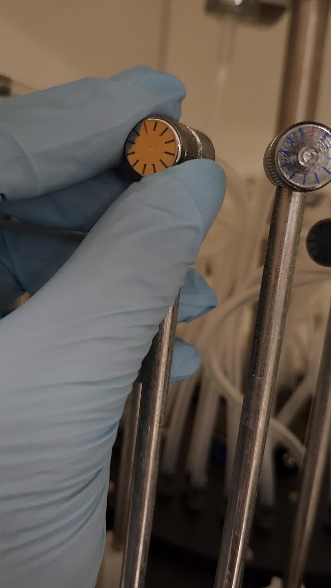
Note: this video shows the luer being moved from the closed position to the ideal open position (1.5 turns counterclockwise)
Ensure valves for each sample are turned 1.5 times counterclockwise from the closed (12 o'clock) position. The red dot will now be at the 6 o'clock position. This practice ensures even nitrogen flow from each luer.
Wipe each needle with a KimWipe soaked in IPA to clean any potential residue from a previous user.
Load glass collection sample tubes (including the 3 internal standard only tubes) into nitrogen evaporator. Lower each luer to be within the glass sample tube but still above the sample supernatant.
The luer does not need to be particularly close to the surface of the liquid for effective evaporation. Often, the end of the luer will be 1 - 2" away from the liquid surface.
Lower nitrogen evaporator sample rack into the warm bath water (pre-heated to 35 ℃) if faster evaporation is desired. Ensure that locking knob is fully tightened once the sample rack is lowered.
Slowly open nitrogen gas regulator to provide flow of nitrogen gas to each sample. Increase the flow until it is high enough to form dimples on the supernatant surface, but not high enough to cause splashing.
Ensure that the nitrogen regulator is in the lowest flow position before opening the tank to prevent sudden influx of gas that can cause samples to splash out of tubes/cross-contaminate. Should this happen, the extracts are no longer quantitative and cannot be used for analysis.
We highly recommend using a two-stage regulator to ensure consistent gas delivery pressure as the internal pressure of the tank decreases.
Wait for samples to dry down, but check on the samples' progress once every ~30 min. The samples should take about 3 hours to dry in the evaporator.
As liquid level goes down, you can increase the flow rate to speed up the drying process, but make sure not to cause splashing/sputtering.
Once the liquid level of the sample collection tubes is very low (a few millimeters; mostly residual water and formic acid), turn off the nitrogen flow and unload the samples and internal standards.
Wipe the luers again with a KimWipe soaked in IPA to clean off any residue that may have been left behind by previous samples.
Load samples and internal standards into RapidVap. Dry samples with a speed setting of 35 and a starting pressure of 200 mbar.
Stair-step the RapidVap pressure down in 20 mbar steps every ~10 minutes.
If the RapidVap is very easily going below the set target pressure, you can go ahead and proceed to a lower pressure sooner than 10 minutes.
Alternatively, if the RapidVap continues to face resistance to reaching the target pressure, leave it set to that pressure until it no longer faces significant resistance.
After stair-stepping down, remove the remaining moisture by setting the pressure to 10 mbar for 15 minutes.
Unload and then cap samples and internal standard tubes. Store dried-down samples and internal standards at -80 ℃ until ready for analysis.
Dried and closed flavonoid samples that are ready to be placed in the -80.
Ensure that each sample is dry by manually inspecting the bottom of each vial. Residual liquid can throw-off final volume estimates leading to quantification errors. If any moisture remains, place it back into the RapidVap for an additional 10 minutes at 10 mbar.
Resolubilize and Run on HPLC
Allow samples to warm slightly (Temperature should be around ~4 ℃). Attempting to open tubes while they are at -80 ℃ can result in cracking or shattering of tubes and causes condensation to rapidly form inside.
To each sample tube, add 5 mL of LC-MS grade 1:1 methanol:water + 0.1% formic acid
Sonicate each sample for ~10 seconds. Rapidly swirl each tube by hand while sonicating to help get dried sample off walls of the tube.
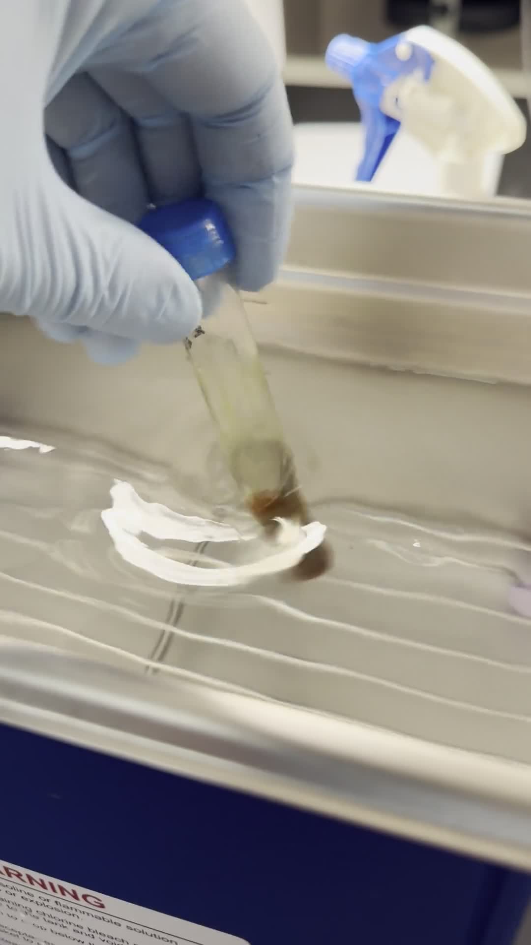
Vortex each sample for ~5 seconds ensuring liquid reaches the upper parts of the tube without getting to the top.
For each sample, label an 8 mL glass vial and add 200 μL of 20 μM taxifolin to each 8 mL vial
Image of the sample tube, 8 mL dilution tube, LC vial, syringe, and filter needed for the following steps.
The universal sample holder seen above can be downloaded and 3D printed from the following link:
Add an additional 3800 μL of LC-MS grade 1:1 methanol:water + 0.1% formic acid
Briefly mix original redissolved sample and pipette 1 ml of redissolved sample into the mixture described above in the 8 mL vial.
Total volume in the 8 mL vial is now 5 mL with a final composition of 4% taxifolin and the concentration of plant flavonoids diluted 5-fold. This dilution brings the concentration of plant flavonoids into the linear range of the LC-MS/MS platform, dilutes potential matrix compounds that interfere with analysis, and allows the user to work with a reasonable amount of starting sample to lower the contribution of weighing errors. For the purposes of quantification, the true redissolve volume is equivalent to 25 mL given the starting volume of 5 mL and 5-fold dilution.
Graphical depiction of 5x dilution strategy described above.
With a 3 ml syringe, aspirate ~2.5 ml of the diluted sample in addition to a little bit of air.
Image of a 3 mL syringe with aspirated sample and additional ~0.5 mL of air.
Safely recap the needle, unscrew it from the syringe, and attach a 13 mm 0.20 μm PTFE filter
Filter sample into a labeled 2 ml glass LC vial. Cap each vial, then run samples on LC-MS/MS
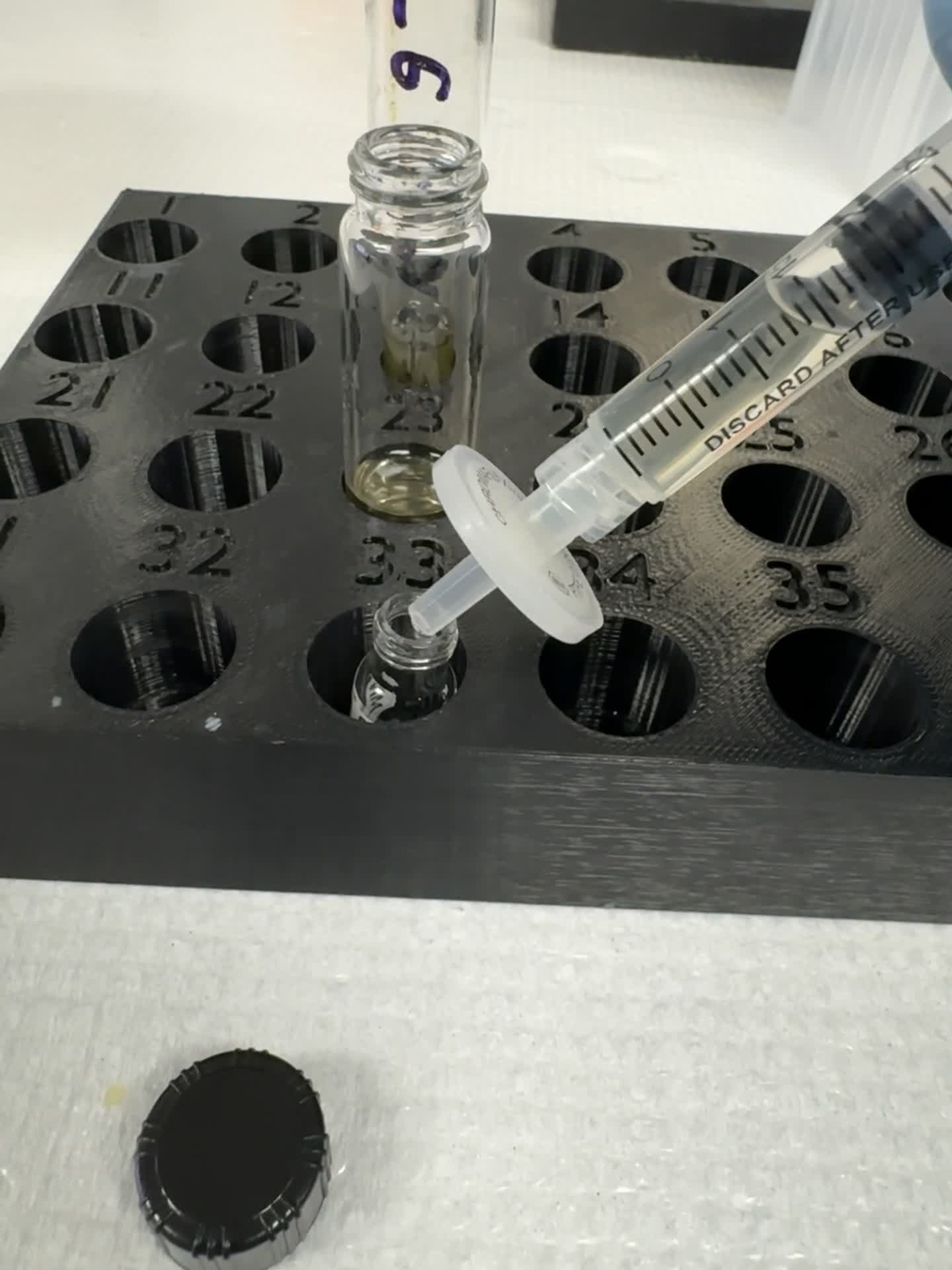
Use the air bubble inside the syringe to push out any liquid trapped in the filter, if needed.
If samples are not run immediately, store in a -20 ℃ freezer to keep samples in a cold, dark place. Samples will not freeze at this temperature due to methanol, but the cold and darkness will minimize oxidation. Long term storage in a -80 ℃ freezer is possible as samples will freeze at this temperature.
Protocol references
Mohamedshah, Z., Hayes, M., Chadwick-Corbin, S., Neilson, A. P., and Ferruzzi, M. G. (2022). Bioaccessibility, gut microbial metabolism and intestinal transport of phenolics from 100% Concord grape juice and whole grapes are similar in a simulated digestion and fecal fermentation model. Food Funct. 13, 4315–4330. doi: 10.1039/D1FO04226B
Dzakovich, M.P.; Le, E.A.; Tak, A.L.; Chacko, S.K. A Comprehensive UHPLC-MS/MS and Extraction Method Reveals Flavonoid Profile and Concentration Are Diverse in Spinach (Spinacia oleracea L.). Front. Plant Sci. 2025, 16. doi:10.3389/fpls.2025.1496200.