May 20, 2025
Labeling of Zebrafish Organs and Substructures in Light Data in Amira 3D Classic Segmentation Workroom
- Alyson Petruncio1,
- Aparna Dev1,
- Virginia Ruetten1,
- Gudrun Ihrke1,
- Aubrey Weigel1,
- Project Technical Resources1,
- CellMap Project Team1
- 1HHMI Janelia Research Campus
- PTR collaborations


Protocol Citation: Alyson Petruncio, Aparna Dev, Virginia Ruetten, Gudrun Ihrke, Aubrey Weigel, Project Technical Resources, CellMap Project Team 2025. Labeling of Zebrafish Organs and Substructures in Light Data in Amira 3D Classic Segmentation Workroom. protocols.io https://dx.doi.org/10.17504/protocols.io.yxmvm3xobl3p/v1
License: This is an open access protocol distributed under the terms of the Creative Commons Attribution License, which permits unrestricted use, distribution, and reproduction in any medium, provided the original author and source are credited
Protocol status: Working
We use this protocol and it's working
Created: June 27, 2023
Last Modified: May 20, 2025
Protocol Integer ID: 84112
Keywords: Zebrafish, Expansion Microscopy, Anatomy, labeling of zebrafish organ, light microscopy dataset, variable light microscopy dataset, use on light microscopy dataset, expansion into variable light microscopy dataset, general amira segmentation tip, zebrafish organ, amira 3d classic segmentation workroom, segmentation summary amira project, segmentation setup mask segmentation overview, zebrafish, ultimate segmentation goal, fluorescent protein, segmentation, based fluorescent protein, segmentation technique, substructures in light data, light data, fluorescent, labeling, membrane
Abstract
Intended for use on light microscopy datasets of larval zebrafish ubiquitously expressing soma- and membrane-based fluorescent proteins. Methods are subject to change based on ultimate segmentation goals and expansion into variable light microscopy datasets.
Segmentation Summary
- Amira Project and Segmentation Setup
- Mask Segmentation
- Overview of Class Types
- Glossary and Segmentation Techniques
See protocol in References for general Amira segmentation tips.
Materials
Amira 3D 2022.2
Amira Project and Segmentation Setup
After opening a new Amira project, select File > Open Data, or click+drag the raw data tiff to the empty space under the Project tab. Confirm coordinates and voxel size in the Image Read Parameters window.
For higher label resolution: Select the arrow of the raw data object in the Project tab. In the window search bar, type ‘resample’. Select the Resample object. With the Resample object selected, adjust Voxel Size: under Properties to half of the original resolution value. Select Apply, and select the newly generated raw data object as the new working raw data object.
Select the Segmentation tab to automatically generate a label field. Confirm Image: dropdown under Segmentation Editor is the raw data. Under Materials, ensure the Exterior (Not Assigned) material remains unlocked. The existing Inside material may be renamed and used as the first cell segmentation material.
Mask Segmentation
Before segmenting organ classes, creating a mask of the zebrafish sample will aid in determining organ segmentation boundaries and later mesh visualization.
Under the Segmentation tab, adjust the 2D data contrast range to the data histogram by selecting the 'Data Histogram' option in the Display Control window (Figure 1). This will present the general shape of the sample, which can be roughly segmented in XY, interpolated through the Z plane, and globally smoothed (Figure 2). The final fish mask should include all signal in the data (Figure 3).
Figure 1.
Figure 2.
Figure 3.
Create a new label field for each class type (see Overview of Organ Class Types) to organize each class and avoid overwriting segmentations.
Note: Since class types have a range of signal across multiple light channels, multiple channel datasets were used across class types. The best channel dataset was chosen for each class type segmentation, and segmentations in different channels were overlayed after Amira export.
Overview of Class Types
All substructure and organ classes fall under a class type. Amira label fields may be organized by class type to track classes with similar patterns in the data.
Muscle
Trunk Muscles
The fish contains two main truck muscles, the epaxial and hypaxial muscles separated by the transvese septum.
Pectoral Girdle Muscles
Three muscles take their origin at the ventral tip of the posterior cleithrum, the first bony structure to emerge. This is just lateral to the caudal edge of the heart.
Abdominal muscle
The abdominal muscle (in the literature called posterior hypaxial muscle) sends long muscle fibers caudo-dorsally to join the ventral boundary of the hypaxial muscle where there is a fascial thickening. It attaches to the ventral side of the epaxial myotomes at the level of myotomes 4, 5, 6.
Sternohyoid muscle
The sternohyoid muscle courses anteriorly and is formed of two consecutive myotomes reaching the urohyal cartilage.
Sternobranchial muscle
Sternobranchial muscle (my name). The third muscle of the group courses dorsally and attaches to the base of the skull at the level of the base of 5th ceratobranchial arch.
Branchial Muscles
Branchial Levator 5.
This muscle attaches at the caudal rim of the ear bone and project ventro-medially to attach to the fifth branchial arch. This is one of the muscles which keep together the cranium and the jaw.
Figure 4. All muscle subtypes in a single XY slice.
Cartilage
Neurocranium
The cranium is formed of different cartilaginous structures. The base of the brain can be divided into the prechordal elements and the para and postchordal elements. The ethmoid plate connects to the basal plate (also parachordal cartilage) via trabeculae. This basal plate abuts the notochord. The basicapsule emanates from the basal plate encircling the base of the developing ear. It comprises the anterior, lateral and posterior basicapsular commissure. This is also sometimes referred to as the auditory capsule. At the anterior portion of the basal plate a perforated fossa is present formed by lateral commissure through which vessels run. The basal plate extends caudal to merge with the occipital plate which extends two dorsal elements, the occipital arches.
The cleithrum, a prominent vertical structure, acts as a central element of the zebrafish skeletal system.
Figure 5. All cartilage fragments in a single XY slice.
Notochord.
A structural and signaling element located directly under the spinal cord containing large vacuolated cells surrounded by notochord sheath cells.
The pharyngeal teeth pad is situated at the upper region of the pharynx.
Gills are protected by the operculum, a covering that shields the delicate structures underneath. The gills themselves are individually labeled.
Figure 6. All four gills in a single XY slice.
The pectoral fins are anterior limbs situated on each side of the fish.
Figure 7. One pectoral fin in a single XY slice.
Sensory organs include the eyes, with distinct labels for structures such as the iris and exterior, and the ears, which encompass the entire organ along with its internal compartments. Hair cells line the base of the ear in uniform lines.
Figure 8. Ear (left) and eye (right) in a single XY slice.
Organs include:
- Gastrointestinal tract: starts at the mouth, extending along the body cavity.
- Heart: includes the ventricle, a large, heavily lined cavity, the atrium, the more dorsal/caudal, thin cavity, and ductus arteriosus, the muscular outlet.
- Kidney: looping structure located in the midline and extending along the body cavity.
- Swim Bladder: characterized by its large size and dark interior pixels.
- Gallbladder: notable for its contrast between the muted interior and bright surrounding membrane.
- Pancreas: divided into exocrine regions, featuring wiry tubules near the swim bladder, and endocrine clusters of cells adjacent to the exocrine area.
- Liver: identified by its dark border and unique pattern, distinct from the surrounding muscle.
Figure 9. All organs in single XY slice.
Nervous system includes:
- Spinal Cord: Extends out from the brain, surrounded by epaxial muscle and dorsal to the notochord.
- Pineal gland: Located dorsally in the midline of the brain.
- Habenulae: A paired structure situated lateral to the pineal gland.
- Brain: Includes internal features while excluding surrounding muscle and nearby ears.
- Posterior lateral line ganglion: A dorsal ganglion directly behind the ear.
- Sympathetic Nervous System/Adrenals: Bright clusters of cells located near blood vessels.
Glossary and Segmentation Techniques
For all classes, carefully annotate intermittent slices of objects down the XY plane, then interpolate (ctrl+i) and add selection to the appropriate class material. Check the object segmentation in XZ and YZ planes to ensure segmentation does not overlap with any other data patterns relating to a different class.
Note: Smoothing may cause excessive material shrinking in datasets where the Z dimension is relatively short.
In many instances, multiple class patterns will have signal overlap in the same channel (Figure 12). Visible muscle fibers and cartilage should always be segmented in instances of overlap with other classes.
Figure 12. Gill class (green) contains some muscle fibers (blue) and cartilage (red)
in an XY slice.
Muscle
Epaxial and hypaxial muscles
Clearly defined dorsal and ventral bundles (top and bottom) are segmented separately and avoid the spinal cord and notochord, respectively.
Figure 13.
Abdominal muscle
More dispersed, streaking pattern extends across the ventral surface up towards the hypaxial muscles. Small, bright nerve signals can be included in the more sparse muscle patterns.
Figure 14.
Pectoral girdle muscles and cranial muscles
Short, small muscle bundles that wrap around organs like the gut and surround the gills, heart, and mouth.
Figure 15.
Cartilage
Fragments run throughout the body, primarly surrounding the ears, brain, and gills. Texture pattern is hollow, porous, and bright.
Figure 16.
Cleithrum
Small, bright bars pass behind the ears and gills, extending towards pectoral fins. The cleithrum segments do not have a distinct cartilage signal pattern.
Figure 17.
Pharyngeal teeth pad
Packed, bright, and webbed signal pattern towards the back of the pharynx, distinct from surrounding gut folds.
Figure 18.
Gills
Two sets of four gills have a soft, gray dotted signal pattern, with gill 4 (yellow) being closest to the cleithrum and gill 1 (green) being closest to the eye.
Figure 19.
Pectoral Fins
The pectoral fins have small muscle fiber and cartilage signal pattern. Muscle and cartilage may be segmented separately and combined to create the total pectoral fin segmentation.
Figure 20.
Sensory Organs
Eye
The most anterior organ, the eye has a soft, radial signal pattern with a thin, bright membrane.
Figure 21.
Ear and auditory hair cells
Ear signal pattern is primarily empty compartments with hair cells (blue) lining the base of the ear.
Figure 22.
Organs
Gastrointestinal tract
Extending from mouth to anus, the gut texture is hollow with irregular folds of soft, gray dots.
Figure 23.
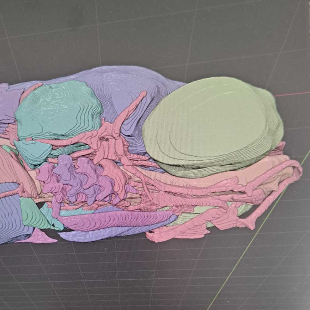
Heart
The ductus arteriosus (red) is the smallest compartment and closest to the eye with a signal pattern of soft gray dots throughout, the ventricle (blue) is most heavily lined with a visible valve separating the ventricle from the atrium (green), which is thinly lined with visible blood cells dispersing out of the compartment.
Figure 24.
Kidney nephron
The nephron and glomerulus with a thick, soft gray signal pattern that bundle dorsal to the liver, caudal to the ears, and dorsal to the gallbladder. Ducts are segmented separately to generally track nephric structure.
Figure 25.
Swim bladder
A mainly empty compartment with minimal signal, the swim bladder takes up a large portion of the body cavity and is directly above the gut.
Figure 26.
Gallbladder
Surrounded by the liver, the gallbladder often has a dark, soft interior signal pattern, lined with brighter, sparse dotted signal. While generally spherical, some invagination may be present.
Figure 27.
Swim-bladder duct and gall bladder duct
Figure 28.
Liver
Wedged between the gut and heart, the unique liver signal texture appears as a gradient of intensity and surrounds the gallbladder. Adjust data contrast as needed in order to include all liver signal.
Figure 29.
Nervous System and Notochord
Notochord
Surrounded by bright nerve signal, the notochord passes between epaxial and hypaxial muscles and into the main body cavity. Signal pattern is porous with thin bright strands passing vertically through a cross-section.
Figure 30.
Spinal cord
Directly dorsal and parallel to the notochord, the spinal cord has a varied soft gray dotted signal pattern with some bright nerve signal dispersed throughout.
Figure 31.
Pineal gland
Smaller, granular signal pattern is surrounded by slightly large gray cells located dorsally, between the eyes. The gland is physically separate from other brain regions.
Figure 32.
Habenula
Both lobes flank the pineal gland, with identical signal patterns to one another; dark soft interior is lined with slightly lighter gray cells.
Figure 33.
Brain
Composed of multiple signal patterns, the brain includes most gray signal in the dorsal cephalic region of the dataset. The brain connects directly to the spinal cord, which shares a similar signal pattern, and includes the pineal gland and habenula.
Figure 34.
Nodose Ganglion
Bundle of soft, gray dots clustered behind the ear, forming an upside down triangle shape.
Figure 35.
Autonomic Nervous System
Saturated signal is thresholded out using the Magic Wand tool (3) under the Segmentation tab. Once selection is added to a material, noise and excess signal is removed from the segmentation and significant nerve signals are refined.
Figure 36.
Figure 31.