Jun 20, 2023
Kidney Functional Tissue Unit (FTU) Segmentation
- Nathan Heath Patterson1,
- ellie.l.pingry 1,
- Katerina Djambazova1,
- Melissa Farrow1,
- Raf Van De Plas2,
- Jeff Spraggins1
- 1Vanderbilt University;
- 2Delft University of Technology
- VU Biomolecular Multimodal Imaging Center / Spraggins Research Group
- KPMP


Protocol Citation: Nathan Heath Patterson, ellie.l.pingry , Katerina Djambazova, Melissa Farrow, Raf Van De Plas, Jeff Spraggins 2023. Kidney Functional Tissue Unit (FTU) Segmentation. protocols.io https://dx.doi.org/10.17504/protocols.io.kxygx3mkog8j/v1
Manuscript citation:
Patterson, N. H.; Neumann, E. K.; Sharman, K.; Allen, J.; Harris, R.; Fogo, A. B.; De Caestecker, M.; Caprioli, R. M.; Van De Plas, R.; Spraggins, J. M. Autofluorescence Microscopy as a Label-Free Tool for Renal Histology and Glomerular Segmentation. https://doi.org/10.1101/2021.07.16.452703.
License: This is an open access protocol distributed under the terms of the Creative Commons Attribution License, which permits unrestricted use, distribution, and reproduction in any medium, provided the original author and source are credited
Protocol status: Working
We use this protocol and it's working
Created: June 16, 2023
Last Modified: October 18, 2023
Protocol Integer ID: 83532
Keywords: kidney functional tissue unit, functional tissue unit, segmentation model to autofluorescence, segmentation of glomeruli, microscopy image, distal tubule, ftus, kidney, ftu, autofluorescence, proximal tubule, glomeruli, tissue, segmentation, segmentation model
Abstract
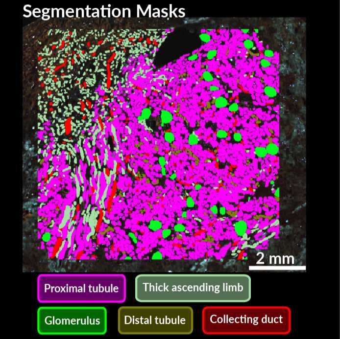
This protocol explains how to apply a segmentation model to autofluorescence microscopy images to find kidney functional tissue units (FTUs). Currently the model allows for segmentation of glomeruli, proximal tubules, thick ascending limb, distal tubules, and collecting ducts.
Materials
For performance, a workstation with at least 32 GB of memory is advised and a GPU with 12 GB memory is required for any GPU-based prediction.
Apply kidney FTU segmentation to autofluorescence images
Configure the segmentation prediction by creating the dataset .yaml file. Add the path to the saved model and images in the configuration file.
Run the model using the command line from the wsimap directory like so:
python scripts/config/instance-predict-from-config-newmodels-multi.py "/path/to/configuration-file-from-step2.yaml
Collect .geojson segmentations in output folder specified in .yaml file from Step 2. These can be visualized in QuPath.
Protocol references
Patterson, N. H.; Neumann, E. K.; Sharman, K.; Allen, J.; Harris, R.; Fogo, A. B.; De Caestecker, M.; Caprioli, R. M.; Van De Plas, R.; Spraggins, J. M. Autofluorescence Microscopy as a Label-Free Tool for Renal Histology and Glomerular Segmentation. https://doi.org/10.1101/2021.07.16.452703.