May 20, 2024
Version 3
KAT8 compound inhibition inhibits the initial steps of PINK1-dependant mitophagy V.3
- Capucine de Talhouet1,2,
- Benjamin O'Callaghan1,2,
- Helene Plun-Favreau1,2,
- Noemi Esteras3,
- Marc P. M. Soutar1
- 1UCL Queen Square Institute of Neurology, Department of Neurodegenerative Disease;
- 2Aligning Science Across Parkinson’s (ASAP) Collaborative Research Network;
- 3Department of Biochemistry and Molecular Biology, School of Medicine, Complutense University of Madrid


Protocol Citation: Capucine de Talhouet, Benjamin O'Callaghan, Helene Plun-Favreau, Noemi Esteras, Marc P. M. Soutar 2024. KAT8 compound inhibition inhibits the initial steps of PINK1-dependant mitophagy. protocols.io https://dx.doi.org/10.17504/protocols.io.eq2ly7m8qlx9/v3Version created by Capucine de Talhouet
License: This is an open access protocol distributed under the terms of the Creative Commons Attribution License, which permits unrestricted use, distribution, and reproduction in any medium, provided the original author and source are credited
Protocol status: Working
We use this protocol and it's working
Created: May 20, 2024
Last Modified: May 20, 2024
Protocol Integer ID: 100108
Keywords: KAT8, MG149, Parkinson's disease, mitophagy, autophagy, TMRM, mitoKeima, regulator of mitophagy, dependant mitophagy initiation, dual inhibition of kat5, kat8 compound inhibition, mitochondrial toxin, kat5 inhibition, partial downstream recruitment of the autophagy receptor p62, contrary to kat5 inhibition, dependant mitophagy process, autophagy receptor p62, kat8 via the mg149 compound, inhibitory effect of mg149, wide association study candidate risk gene for parkinson, dependant mitophagy, kat8 gene, autophagy process, mitochondrial membrane, following mitochondrial depolarisation, subsequent phosphorylation of parkin, mitophagy, mitochondrial delivery, impairing pink1 activation, mitochondrial depolarisation, additional support for kat8, increase in mitochondrial delivery, parkinson, mg149 treatment, mg149 treatment in the absence, lysine acetyltransferase, pink1 activation, subsequent phosphorylation, lysosome, kat8, mg149 compound, kat5
Funders Acknowledgements:
The Michael J. Fox Foundation for Parkinson’s Research (MJFF) and the Aligning Science Across Parkinson’s (ASAP) initiative.
Grant ID: ASAP-000478
Abstract
It has recently been shown that KAT8, a genome-wide association study candidate risk gene for Parkinson’s Disease, is involved in PINK1/Parkin-dependant mitophagy. The KAT8 gene encodes a lysine acetyltransferase and represents the catalytically active subunit of the non-specific lethal epigenetic remodelling complex. In the current study, we show that contrary to KAT5 inhibition, dual inhibition of KAT5 and KAT8 via the MG149 compound inhibits the initial steps of the PINK1-dependant mitophagy process. More specifically, our study shows that following mitochondrial depolarisation induced by mitochondrial toxins, MG149 treatment inhibits PINK1-dependant mitophagy initiation by impairing PINK1 activation, and subsequent phosphorylation of Parkin and ubiquitin. While this inhibitory effect of MG149 on PINK1-activation is potent, MG149 treatment in the absence of mitochondrial toxins is sufficient to depolarise the mitochondrial membrane, recruit PINK1 and promote partial downstream recruitment of the autophagy receptor p62, leading to an increase in mitochondrial delivery to the lysosomes. Altogether, our study provides additional support for KAT8 as a regulator of mitophagy and autophagy processes.
Materials
Mitochondrial toxins
Oligomycin (inhibitor of mitochondrial complex V) was purchased from Cayman Chemicals (11341) and from Sigma-Aldrich (O4876)
Antimycin A (inhibitor of mitochondrial complex III) was purchased from Sigma-Aldrich (A8674)
Antibodies used for immunoblotting
rabbit anti-phospho-ubiquitin (Ser65) (Cell Signaling, 37642, 1:1000)
mouse anti-TIM23 (BD Biosciences, 611223, RRID:AB_398755, 1:1000)
mouse anti-TOM20 (Santa Cruz, sc-11415, RRID:AB_2207533,1:1000)
mouse anti-GAPDH (Abcam, ab110305, RRID:AB_10861081, 1:1000)
rabbit anti-pParkin(Ser65) (Abcam/Michael J.Fox Foundation, MJF17, 1:1000)
rabbit anti-MFN2 (Cell Signaling, 9482, 1:1000)
mouse anti-OPA1 (BD Biosciences, 612607, 1:1000)
rabbit anti-LC3B (Novus Biologicals, NB100-2220, 1:1000)
IRDye 680LT donkey anti mouse (LI-COR Biosciences, 925-68022, RRID:AB_2814906, 1:20000)
IRDye 800CW donkey anti rabbit (LI-COR Biosciences, 925-32213, RRID:AB_2715510, 1:20000)
rabbit monoclonal anti-PINK1 antibody generated as described in the following manuscript: Sci Rep. 2015. DOI: https://doi.org/10.1038/s41598-018-26949-6
Antibodies used for immunofluorescence
mouse anti TOM20 (Santa Cruz, sc-17764, RRID:AB_628381 1:1000)
rabbit anti phospho-ubiquitin (Ser65) (Cell Signaling, 37642, 1:1000)
mouse anti-p62 (BD Biosciences, 610832, 1:1000)
AlexaFluor 488 goat anti rabbit (Invitrogen, A11008, 6 RRID:AB_143165,1:2000)
AlexaFluor 568 goat anti mouse (Invitrogen, A11004, 7 RRID:AB_2534072, 1:2000)
Cell Culture
Maintain cells in culture in a humified 5% CO2 incubator at 37 °C in Dulbecco's modified Eagle's medium (DMEM, Gibco, 11995-065) supplemented with 10% heat-inactivated foetal bovine serum (FBS, Gibco). Change cell medium every 3 days.
Note
Parkin over-expressing (POE) SHSY5Y cells are a gift from H. Ardley (Ardley, H., et al., 2003).
Mt-Keima POE SHSY5Y cells were a gift of C. Luft (Soutar, M., et al., 2019).
PINK1-HA overexpressing SHSY5Y cells were a gift from E. Deas (Deas, E.., et al., 2011).
Split cells at 75% to 90% confluency using 2 mL of Trypsin.
Cell Treatment (Small Molecule Modulator and Mitophagy Induction)
Dilute each compound in DMSO to reach concentrations determined according to their speculated toxicity and IC50, 10 micromolar (µM) for NU9056 and 100 micromolar (µM) for MG149.
Note
MG149 targets KAT5 and KAT8 with IC50 values of 74 micromolar (µM) and47 micromolar (µM) respectively; NU9056 targets KAT5 with an IC50 value of 2 micromolar (µM) (Li, L., et al., 2020).
Treat cells with DMSO, 10 micromolar (µM) NU9056, or 100 micromolar (µM) MG149 for 06:00:00 before collecting / fixing for downstream experiments.
6h
Treat cells with either DMSO or 10 micromolar (µM) oligomycin/ 10 micromolar (µM) antimycin for 03:00:00 to induce mitophagy before collecting / fixing for downstream experiments.
Note
Note that both small molecule modulator and O/A are often added to the same wells / cells to look at the impact of MG149 on mitophagy initiation.
3h
Mitochondrial Enrichment
2h 10m
Incubate cells Overnight at -80 °C in mitochondrial homogenization buffer (250mM sucrose, 1mM EDTA, 10mM Tris (pH 7.4) supplemented with protease and phosphatase inhibitors).
Thaw, scrape into 1.5mL centrifuged tubes, and triturate 20x, keeping samples On ice .
Freeze tubes at -80 °C for 01:00:00 , then thaw On ice .
1h
Remove cell debris / non-lysed cells by centrifugation 1500 rcf, 4°C, 00:20:00 .
20m
Centrifuge the supernatant 12500 x g, 4°C, 00:20:00 to collect the mitochondrial enriched pellet (aka the mitochondrial fraction) and save the supernatant (aka the cytoplasmic fraction).
20m
Centrifuge the cytoplasmic fraction 12500 x g, 4°C, 00:20:00 to remove membrane contamination and wash the mitochondrial fraction twice in 500 µL of fractionation buffer (12500 x g, 4°C, 00:10:00 x 2) to remove cytoplasmic contaminants.
30m
Determine the relative ratios of sample differences for the mitochondrial enriched fraction using the protein concentration of the cytoplasmic fraction (BSA assay). Re-suspend the mitochondrial enriched pellet accordingly using 1x NuPAGE® LDS sample buffer supplemented with 10 mM dithiothreitol (DTT).
Sonicate the mitochondrial fraction (10 microns power amplitude for 2 x 10 seconds, "flick" in
between) and heat to 70 °C for 00:10:00 .
10m
Cell Lysis
1h 30m
Lyse cells in whole-cell lysis buffer (50mM Tris (pH 7.4), 0.1mM EGTA, 1mM EDTA, 0.27M Sucrose, 1% triton-x-100, supplemented with protease and phosphatase inhibitors)On ice and incubated Overnight at -80 °C .
Cell Lysis
1h 30m
Centrifuge the lysates 1600 x g, 4°C, 00:20:00 and protein quantify the supernatant by BSA assay.
20m
Make LDS preps with 10 mM dithiothreitol (DTT), and heat to 70 °C for 00:10:00 .
10m
Western Blotting
1h 30m
Separate the LDS preps (either after cell lysis or mitochondrial enrichment) by SDS-PAGE and transfer them to nitrocellulose membrane.
Incubate the membrane Overnight at 4 °C with primary antibodies diluted in 3% milk in PBS-Tween.
Wash and incubate the membrane for 01:00:00 at Room temperature in secondary antibodies diluted in PBS-Tween.
1h
Detect the protein bands using Odyssey CLx LI-COR.
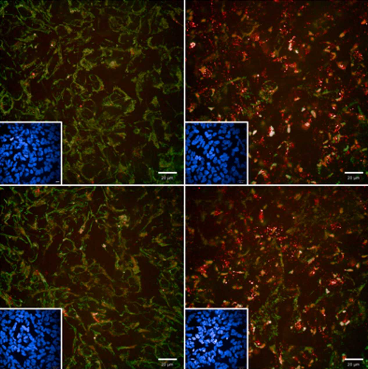
Immunofluorescence
4h 20m
Fix cells with 4% Paraformaldehyde (PFA, Sigma-Aldrich) for 00:20:00 atRoom temperature .
20m
Remove PFA solution, block and permeabilize the cells with a mix of 10% FBS, 0.25% Triton X-100 in PBS for 01:00:00 at Room temperature .
1h
Immunostain the cells with pUb(Ser65) and TOM20 primary antibodies diluted in 10% FBS/PBS for 02:00:00 at Room temperature .
Note
rabbit anti-pUb(Ser65) (Cell Signaling, 37642) and mouse anti-TOM20 (Santa Cruz, sc-17764) primary antibodies are both added at a 1:1000 dilution in 10% FBS/PBS.
2h
Wash wells three times using PBS 1X and incubate with secondary antibodies for 01:00:00 at Room temperature , before washing again three times using PBS 1X.
Note
AlexaFluor 568 anti-mouse, 488 anti-rabbit secondary antibodies, and Hoechst 33342 (Thermo Scientific) are all added at a 1:2000 dilution in 10% FBS/PBS.
1h
Image cells on the Opera Phenix (Perkin Elmer), acquiring confocal z-stacks for multiple fields of view across 3 individual wekks per experimental condition, using the 40X water objective.
Note
Analyse images using Columbus 2.8 analysis system (Perkin Elmer) to measure the integrated intensity of pUb(Ser65) within the whole cell.
For visualisation purposes, select brightness and contrast settings on control and apply the same settings to all other conditions.
Measuring Mitochondrial Membrane Potential using TMRM
40m
Seed POE-SHSY4Y cells in a 96-well CellCarrier Ultra plate.
Incubate live cells in25 nanomolar (nM) Tetramethylrhodamine, Methyl Ester, Perchlorate (TMRM) (Sigma Aldrich) diluted in cell media for 00:40:00 at 37 °C and 4% CO2.
40m
Image cells on the Opera Phenix (PerkinElmer) at 37 °C and 4% CO2, acquiring confocal z-stacks for multiple fields of view across 3 individual wells per experimental condition using the 40X water objective.
Note
Analyse images using the Columbus 2.8 analysis system (Perkin Elmer) to measure the TMRM signal intensity of maximum intensity projections.
Measuring Mitochondrial Delivery to Lysosomes using mitoKeima Reporter
40m
Seed mt-Keima expressing POE SHSY5Y cells in a 96-well CellCarrier Ultra plate and treat them according to desired conditions (MG149, NU9056, O/A as described in ).
Before imaging, replace cell media with phenol-free DMEM + 10% FBS containing the treatments previously administered , and adding Hoechst 33342 (1:10000) to stain the nuclei.
Image cells immediately on the Opera Phenix (PerkinElmer) at 37 °C with 5% CO2, acquiring multiple single-plane fields of view, using the 63X water objective.
Note
Analyse images in using the Columbus 2.8 analysis system (Perkin Elmer) to measure the mitophagy index.
To analyse:
- Identify cells using the Hoechst 33342 channel, before measuring the area of the cytoplasmic and lysosomal mitoKeima
- Calculate the mitophagy index = the ratio between the total area of lysosomal mitochondria and the total area of mitoKeima (cytoplasmic + lysosomal areas) per well.
RT-qPCR
Extract total RNA from cells using the Monarch Total RNA Miniprep Kit (New England Bioscience) with inclusion of the optional on-column DNAse treatment.
Reverse transcribe RNA with SuperScript™ IV reverse transcriptase and random hexamers (Invitrogen).
Dilute the cDNA product then subject to quantitative real-time PCR (qPCR) using Power SYBR™ Green Master Mix (Applied Biosystems) and gene specific primer pairs on a QuantStudio™ 7 Flex Real-Time PCR System (Applied Biosystems).
| A | B | C | D | |
| Sequence 5'-3' | ||||
| Gene Target | Forward | Reverse | Product Size / bp | |
| RPL18A | CCCACAACATGTACCGGGAA | TCTTGGAGTCGTGGAACTGC | 180 | |
| PINK1 | GTGGAACATCTCGGCAGGTT | CCTCTCTTGGATTTTCTGTAAGTGAC | 129 |
List of primer pairs used for RT-qPCR of target genes
Calculate relative mRNA expression levels using the 2−ΔΔCt method and RPL18A as the housekeeping gene.
Statistical Analysis
Use a minimum of 3 biological replicates for each experiments (N numbers shown in legends refer to the number of replicate experiments).
- For each imaging experiments, calculate the mean values of every condition from a minimum of 3 technical replicates.
- Normalise intensity and integrated density measurements to control conditions.
Subject data to a two-way ANOVA with Dunnett's post-hoc analysis for multiple comparisons unless stated otherwise.
- Express data as the mean ± standard deviation (SD) from replicate experiments.
- Set statistical significance at a p-value of <0.05.
Note
Use GraphPad Prism 6 (La Jolla, California, USA) for statistical analyses and graph production.
(Present data with error bars showing mean ± standard deviation (SD) from replicate experiments).
Measuring p62 recruitment to the OMM and mitochondrial delivery to lysosomes using mitoSRAI
3h
Seed mitoSRAI POE SHSY5Y cells in a 96-well CellCarrier Ultra plate and treat them according to desired conditions (MG149, NU9056, O/A as described in ).
Fix cells as described in to 22
Immunostain the cells with p62 primary antibodies diluted in 10% FBS/PBS for 02:00:00 at Room temperature .
Note
mouse anti-SQSTM1/p62 (Abcam, ab56416) primary antibody added at a 1:1000 dilution in 10% FBS/PBS.
2h
Wash wells three times using PBS 1X and incubate with secondary antibodies for 01:00:00 at Room temperature , before washing again three times using PBS 1X.
Note
AlexaFluor 568 anti-mouse secondary antibodies, and draq5 (Thermo Scientific) are all added at a 1:2000 dilution in 10% FBS/PBS.
1h
Image cells on the Opera Phenix (Perkin Elmer), acquiring confocal z-stacks for multiple fields of view across 3 individual wells per experimental condition, using the 63X water objective.
Note
Analyse images using Columbus 2.8 analysis system (Perkin Elmer) to measure the SRAI mitophagy index
- corresponds to the ratio between the area of lysosomal mitochondria (corresponding to the total YPet negative TOLLES area) and the total mitochondrial area (corresponding to the total TOLLES area) per well.
Analyse images using Columbus 2.8 analysis system (Perkin Elmer) to measure p62 recruitment to teh mitochondria using TOLLEs as a mitochondrial marker
- calculated as the ratio of p62 signal intensity inside vs. outside TOLLES
For visualisation purposes, select brightness and contrast settings on control and apply the same settings to all other conditions.
Generating PINK1 KO POE SH-SY5Y cell line
PINK1 was functionally knocked out from the POE SH-SY5Y cell line through CRISPR-Cas9 editing by adapting previously published protocols (DOI: 10.15252/emmm.202013579)
Note
Single guide RNA (sgRNA) sequences targeting Exon 1 of PINK1 (NM_032409) were designed using the Horizon Discovery CRISPR Design Tool (https://horizondiscovery.com/en/ordering-and-calculation-tools/crispr-design-tool )
| A | B | C | D | |
| SgRNA ID | 5’-3’ sgRNA Sequence (PAM) | 5’-3’ Forward ssDNA Oligo with BbSI Overhang | 5’-3’ Reverse ssDNA Oligo with BbSI Overhang | |
| PINK1_Seq1 | CGCCACCATGGCGGTGCGAC(AGG) | caccgCGCCACCATGGCGGTGCGAC | aaacGTCGCACCGCCATGGTGGCGc | |
| PINK1_Seq2 | ACCGGGCGCGGAGCCTCGCA(GGG) | caccgACCGGGCGCGGAGCCTCGCA | aaacTGCGAGGCTCCGCGCCCGGTc |
5’-3’ Forward and Reverse ssDNA sequences generated (Sigma Aldrich) inclusive of 5' overhangs suitable for cloning into BbSI digested pSpCas9(BB)-2A-GFP plasmid
Note
Materials:
pSpCas9(BB)-2A-GFP Addgene plasmid no. 48138 was a gift from Feng Zhang;(RRID:Addgene_48138)
IRDye 800CW donkey anti-rabbit secondary (LI-COR Biosciences, 925-32213, RRID:AB_2715510, 1:2000)
pUb(Ser65) (CST, 62802, RRID:AB_2799632, 1:1000)
A subconfluent 6-well dish of POE SH-SY5Ys were transfected with 1ug of each PINK1_Seq1 and PINK1_Seq2 sgRNA encoding pSpCas9(BB)-2A-GFP plasmid using lipofectamine 3000 (Invitrogen)
24 h post-transfection cells were trypsinised and subjected to fluorescence-activated cell sorting and clonal selection of GFP-positive cells: isolated clones were expanded and phenotypically screened for clones which did not show pUb(Ser65) signal after 1.5 h O/A treatment
Confluent wells of a 96-well were prepared for immunofluorescence staining of pUb(Ser65) with IRDye 800CW donkey anti-rabbit secondary
gDNA was extracted from a positive clone using the Wizard Genomic DNA Purification Kit (Promega) and subjected to whole genome sequencing (Novogene) which revealed homozygous 22 bp deletion and frameshift in exon 1 of PINK1 (c.156_177delGGGCGCGGAGCCTCGCAGGGTC, p.A54Sfs*46).
Generating mitoSRAI POE SH-SY5Y cell line
MitoSRAI cDNA was first PCR amplified from mitoSRAI_pcDNA3 with inclusion of 5′ EcoRI and 3′ NotI restriction sites
The PCR product was cloned into pLVX-EF1α-IRES-Puro using EcoRI and NotI restriction sites to generate pLVX-EF1α-mitoSRAI-IRES-Puro.
Note
Material:
mitoSRAI_pcDNA3: provided by the RIKEN BRC through the National BioResource Project of the MEXT, Japan; cat. RDB18223
pLVX-EF1α-IRES-Puro: Clontech, Takara Bio, 631988
pMD2.G (Addgene plasmid no. 12259, RRID:Addgene_12259) and pCMVR8.74 (Addgene plasmid #22036, RRID:Addgene_22036) were gifts from Didier Trono
Lenti-X 293 T HEK cells cultured in DMEM 10% FBS media were transfected with pMD2.G, pCMVR8.74 and pLVX-EF1α-mitoSRAI-IRES-Puro at a 1:1:2 molar mass ratio using Lipofectamine 3000 (Invitrogen)
The next day, a full media change was performed using DMEM 10% FBS and cells cultured for further 24
The lentivirus containing media was collected and diluted 1:2 with DMEM 10% FBS before filtering through 0.44 µm PES filters
1 × 10⁶ POE SHSY5Ys were reverse transduced with the 1:2 lentivirus supernatant dilution in the presence of 10 µg/mL polybrene (Sigma Aldrich)
Bulk populations of cells stably expressing mitoSRAI were established through selection with 1 ug/mL puromycin (M P BIOMEDICALS UK)
Puromycin was maintained during routine culture but withdrawn when seeding for experimental assays.
Protocol references
Ardley HC, Scott GB, Rose SA, Tan NG, Markham AF, Robinson
PA. Inhibition of proteasomal activity causes inclusion formation in neuronal
and non-neuronal cells overexpressing Parkin. Mol Biol Cell. 2003
Nov;14(11):4541-56. doi: https://doi.org/10.1091/mbc.e03-02-0078.
Epub 2003 Aug 22. PMID: 12937272; PMCID: PMC266771.
Soutar MPM, Kempthorne L, Annuario E, Luft C, Wray S,
Ketteler R, Ludtmann MHR, Plun-Favreau H. FBS/BSA media concentration
determines CCCP's ability to depolarize mitochondria and activate PINK1-PRKN
mitophagy. Autophagy. 2019 Nov;15(11):2002-2011. doi: https://doi.org/10.1080/15548627.2019.1603549.
Epub 2019 May 7. PMID: 31060423; PMCID: PMC6844515.
Deas E, Plun-Favreau H, Gandhi S, Desmond H, Kjaer S, Loh SH,
Renton AE, Harvey RJ, Whitworth AJ, Martins LM, Abramov AY, Wood NW. PINK1
cleavage at position A103 by the mitochondrial protease PARL. Hum Mol Genet.
2011 Mar 1;20(5):867-79. doi: https://doi.org/10.1093/hmg/ddq526.
Epub 2010 Dec 6. PMID: 21138942; PMCID: PMC3033179.