Nov 04, 2021
Intracellular Accumulation of HiLyte-488-Aβ42 in DU145 and DU145-TXR Cells
- Lauren Ammerman1
- 1University of Texas Southwestern Medical Center at Dallas
- Lauren Ammerman: PhD Candidate, Molecular and Cellular Biology
- McCormick Lab


Protocol Citation: Lauren Ammerman 2021. Intracellular Accumulation of HiLyte-488-Aβ42 in DU145 and DU145-TXR Cells . protocols.io https://dx.doi.org/10.17504/protocols.io.brvem63e
License: This is an open access protocol distributed under the terms of the Creative Commons Attribution License, which permits unrestricted use, distribution, and reproduction in any medium, provided the original author and source are credited
Protocol status: Working
LA designed and used this protocol for the 2021 publication in PLOS One. https://dx.doi.org/10.1371%2Fjournal.pone.0250371
Created: January 28, 2021
Last Modified: November 04, 2021
Protocol Integer ID: 46726
Keywords: HiLyte-488-Aβ42, DU145-TXR, DU145, Accumulation Assay, Amyloid Beta, amyloid, P-gp, P-glycoprotein, microscopy, ABC transporter, ABCB1, Intracellular accumulation, aβ42 in du145, aβ42, overexpressing human cancer, gp inhibitor, intracellular accumulation, txr cell, paclitaxel, human cancer, cell
Disclaimer
DISCLAIMER – FOR INFORMATIONAL PURPOSES ONLY; USE AT YOUR OWN RISK
The protocol content here is for informational purposes only and does not constitute legal, medical, clinical, or safety advice, or otherwise; content added to protocols.io is not peer reviewed and may not have undergone a formal approval of any kind. Information presented in this protocol should not substitute for independent professional judgment, advice, diagnosis, or treatment. Any action you take or refrain from taking using or relying upon the information presented here is strictly at your own risk. You agree that neither the Company nor any of the authors, contributors, administrators, or anyone else associated with protocols.io, can be held responsible for your use of the information contained in or linked to this protocol or any of our Sites/Apps and Services.
Abstract
Protocol to facilitate the imaging, and subsequent quantification, of the intracellular accumulation of HiLyte-488-Aβ42 in two cell lines - DU145 (paclitaxel sensitive, non-P-gp-overexpressing human cancer), and DU145-TXR (paclitaxel-resistant, P-gp-overexpressing human cancer) cells. Cells are grown on collagen-treated, sterile glass coverslips in complete medium for 24 hours at 37C in a humidified incubator. After the initial growth phase, complete medium is refreshed, and cells are treated with the following - 1) control solutions (DMSO and Ammonium Hydroxide vehicle controls), 2) 1μM HiLyte-488-Aβ42 and DMSO vehicle control, 3) 1μM Tariquidar (a potent P-gp inhibitor) and Ammonium Hydroxide vehicle control, or 4) 1μM HiLyte-488-Aβ42 and 1μM Tariquidar. Cells are incubated with treatments for 16 hours at 37C in a humidified incubator. After the 16 hour incubation, cells are washed, fixed with paraformaldehyde solution, stained with DAPI, washed twice more, and mounted on slides for imaging. Full details, including necessary calculations, reagent specifics, and notes are included in this protocol.
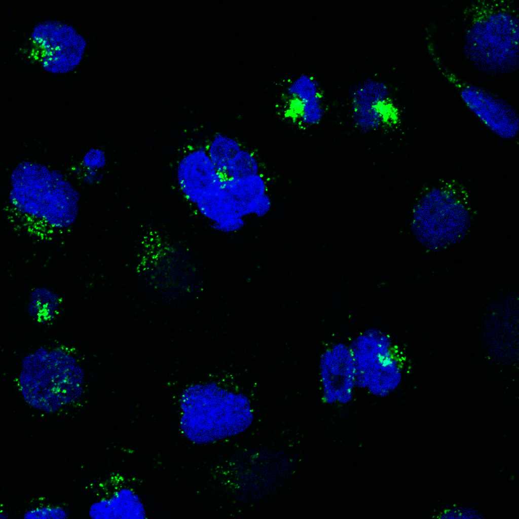
Image Attribution
Taken by L. Ammerman on a confocal microscope - DU145-TXR cells in the presence of both Tariquidar and HiLyte-488-Aβ42.
Materials with Info
| Reagent/Material | Manufacturer | Details | CAT# | |
| RPMI medium | HyClone | RPMI 1640 with L-glutamine and phenol red | SH3002701 | |
| Collagen I | Corning | Collagen I, High Concentration, Rat Tail | 354249 | |
| FBS (Fetal Bovine Serum) | Corning | Regular | 35-010-CV | |
| 24-well cell culture plate | FisherBrand | Sterile Surface Treated Tissue Cultur | FB012929 | |
| Trypan Blue Solution | Gibco | 0.4% Trypan Blue | 15250061 | |
| Pen-Strep | Gibco | Penicillin-Streptomycin | 15070063 | |
| Trypsin | Gibco | Trypsin-EDTA (0.05%), phenol red | 25300062 | |
| PBS (Phosphate Buffered Saline solution) | HyClone | 1X, 0.067M PO4 PBS | SH3025602 | |
| Coverslips | FisherBrand | 25 mm | 12-545-102P | |
| TC20 Automated Cell Counter | BioRad | Requires slides - Cat #1450011 | 1450102 | |
| HiLyte-488-Aβ42 | Anaspec | Beta - Amyloid (1 - 42), HiLyteᵀᴹ Fluor 488 - labeled, Human (LOT# 1958003) | AS-60479-01 | |
| Paraformaldehyde Solution | Alfa Aesar | 4% Paraformaldehyde in PBS | AAJ61899AP | |
| Fluoromount G | Southern Biotech | Water soluble, nonfluorescing mounting medium | OB100-01 |
Table of Reagents/Materials.
Preparation
1d
Prepare Coverslips and 24-well Plate.
notes - we chose 24-well plates because the wells were big enough to fit our circular coverglasses. This experiment could be done with larger plates; volumes/solutions should be scaled accordingly. Procedures should be performed in a Cell Culture Hood.
- Sterilize circular glass coverslips in UV light overnight, or using method of choice.
- Obtain a sterile 24 well cell culture plate.
- Using clean, sterilized tweezers, place one glass coverslip into each well of the 24 well plate.
| A | B | C | D | E | F | |
| coverslip | coverslip | |||||
| coverslip | coverslip | |||||
| coverslip | coverslip | |||||
| coverslip | coverslip |
Coverslip placement in a 24 well plate.
Collagen-Coat Plates.
notes - we utilized a light collagen treatment of our 24-well plates and slides to 'seal' the edges of the coverslip against the bottom of the flask, thereby preventing cells from growing on the wrong side of the coverslip; and to promote adhesion of cells to the coverslips, thereby preventing cells from washing off during step 7.
Follow the Sigma Aldrich protocol for coating culture ware with Collagen Type I with the following specifications:
- Add 500 μL of working Type I Collagen Solution to each well using a P1000.
- *Using a sterile pipette tip, gently press down on top of each coverslip to remove any bubbles, and ensure that the coverslip is flush with the bottom of the plate.
- Incubate at room temperature (~27C) for 10 minutes.
- Aspirate collagen solution with a P1000.
- Add 500 μL of PBS to each well
- if using immediately, replace the lid of the plate and set aside. Aspirate PBS prior to seeding.
- if using in 1-2 days, leave PBS in, replace the lid, seal the edges with parafilm, refrigerate.
Passage and Seed Cells.
1h
Passage Cells.
notes - our DU145 and DU145-TXR cell lines were a generous gift from Dr. Evan Keller, Univ. of Michigan. DU145 cancer cells are non-P-gp-overexpressing, and chemotherapy-sensitive (specifically Paclitaxel). DU145-TXR cells are P-gp overexpressing and paclitaxel-resistant, and are maintained under selection pressure by supplementing the growth medium with 10 nM Paclitaxel (final concentration in flask) during passaging. See PMID 17440963 for more information on these cell lines.
- Use 0.05% Trypsin-EDTA
- Incubate cells for 3 minutes with 0.05% Trypsin-EDTA at 37C.
- Add complete RPMI medium (RPMI 1640, 10% Corning FBS, 100 U/mL penicillin and 100 μg/mL streptomycin (Gibco)) to neutralize the trypsin.
- Spin cell suspension, and conditioned media, at 2000 rpm for 4 minutes.
- Aspirate media gently, to avoid disturbing the pellet.
- Re-suspend the pellet in 3 mL of new complete RPMI media.
- Combine a 15 μL sample with 15 μL of 0.4% Trypan Blue solution and count using the TC20 Automated Cell Counter from BioRad.
- Once 3 counts have been obtained, and an average live count and live % calculated, seed a flask in complete RPMI media to continue the culture, and move onto step 4.
Seed Plates
notes - this concentration gave us areas on the coverslip with ~80-95% confluency, after 24 hours growth + 16 hours incubation with treatments (40 hours total). This will need to be optimized/adjusted for your imaging preferences, and for different cell lines.
- Adjust the concentration of cell suspension to 60,000 cells / mL. 1 mL of cell suspension / well is required. Add a small amount ( ~1 or 0.5 mL) of conditioned media to reach the final appropriate volume. TC20 cell counter gives the count in cells/mL.
(Ci cells/mL)(Vi) = (60,000 cells / mL)((1 mL x # wells) + 0.5 mL extra)
Example: (5.3 x 10⁵ cells / mL)(Vi) = (60,000 cells / mL)(7.5 mL)
Vi = 0.85 mL cell suspension + 6.65 mL complete media
Final = 0.85 mL cell suspension + 1 mL conditioned media + 5.65 mL new complete media.
2. Add 1mL cell suspension to each well of the collagen treated plate using a P1000.
3. Incubate 24 hours at 37C in a humidified incubator with 5% CO2.
Prepare and Store ABeta Aliquots
30m
Preparation of HiLyte-488-Aβ42 for use in Cell Culture Experiments
notes
- Beta - Amyloid (1 - 42), HiLyteᵀᴹ Fluor 488 - labeled, Human. HiLyte™ Fluor 488 - DAEFRHDSGYEVHHQKLVFFAEDVGSNKGAIIGLMVGGVVIA; mw 4870.5 g/mol
- This is a fluorescent (HiLyte™ Fluor 488)-labeled ß-Amyloid peptide, Abs/Em=503/528 nm. Hilyte 488™ Fluor labeled Aß (1-42) has a brighter intensity than FAM-labeled Aß (1-42).
- Manufacturer recommends dissolving in a 1% NH4OH solution, but no longer includes it.
- Ryan et al. 2013 - "Ammonium hydroxide treatment of Abeta produces an aggregate free solution suitable for biophysical and cell culture characterization". PMID 23678397
- Dissolve 0.1 mg of the HiLyte-488-Aβ42 peptide in 30 μL of 1% (w/v) ammonium hydroxide in sterile water at room temperature (~27C). Vortex and mix thoroughly.
- Filter the HiLyte-488-Aβ42 solution using a sterile 0.45 μm pore filter and syringe.
- Once thoroughly dissolved and mixed, add 380 μL of PBS; final NH4OH concentration is 0.073%.
- (30 μL )(0.01 NH4OH) = (410 μL )(X NH4OH) ; = 0.00073 NH4OH, or 0.073% NH4OH
- After mixing thoroughly, the 50 μM peptide solution was aliquoted into 50 μL aliquots, and frozen until use.
- When using in cell culture experiments, the peptide aliquots were thawed and vortexed immediately before usage and dosing, and were not allowed to sit out for any length of time.
- Prepare a control solution for use in cell culture assays by adding 30 μL of 1% NH4OH in sterile water to 380 μL of PBS; use this to control for the presence of NH4OH in assays.
Preparing Dosing Solutions, Dose Cells.
17h
Make Compound Solutions (~23 hours post-seeding)
notes:
- working HiLyte-488-Aβ42 solution aliquots are thawed and used once; excess is discarded.
- never re-freeze/re-use an amyloid beta aliquot.
- Final volume of each well is 500 μL = 450 μL complete RPMI media + two 25 μL treatments.
- Tariquidar is dissolved in DMSO.
- Our protocol accounts for both the presence of DMSO and NH4OH in the dosage plan.
- final concentration of DMSO in each well is 0.1%; final concentration of NH4OH in each well is 0.0037%.
- We chose 1 μM Tariquidar (TQR) because this concentration is sufficient to inhibit most P-glycoprotein in the cells; while TQR has been shown to inhibit BCRP at concentrations near 1 μM,
1. Tariquidar Solution.
(1X10⁻³ M TQR)(Vi)=(1X10⁻⁶ M TQR)(20)(125 μL)
DMSO correction? = 125 μL (0.02) = 2.5 μL; we are adding 2.5 μL of 1mM TQR in DMSO, no extra DMSO needed.
( 2.5 μL 1mM TQR in DMSO) + (122.5 μL complete RPMI media)
Concentration factor = 500 μL total in well / 25 μL treatment = 20; working solution should be 20X final in well.
2. 2% DMSO Complete RPMI Media (controls for DMSO vehicle)
(980 μL complete RPMI media) + (20 μL DMSO).
3. HiLyte-488-Aβ42 Solution (make this last, as close to dosing time as possible)
(5X10⁻⁵ M HiLyte-488-Aβ42)(Vi)=(1X10⁻⁶ M HiLyte-488-Aβ42)(20)(125uL)
DMSO correction? = 125 μL (0.02) = 2.5 μL
(50 μL of 50 μM HiLyte-488-Aβ42 in 0.07% NH4OH in PBS) + (72.5 μL complete RPMI media) + (2.5 μL DMSO)
4. NH4OH complete RPMI Media (controls for NH4OH vehicle)
(50 μL of 0.073% NH4OH in PBS) + (72.5 μL complete RPMI media) + (2.5 μL DMSO).
(50 μL)(0.073% NH4OH) = (125 μL)(X) = 0.0292% NH4OH working solution.
(25 μL)(0.0292% NH4OH) = (500 μL)(X) = 0.0015% NH4OH final in well.
Dose 24 well plates (24 hours post-seeding).
notes - Keep the HiLyte-488-Aβ42 aliquot covered with foil and away from light.
- Gently aspirate the media from the plate.
- Add 450 μL of complete RPMI media to all wells – gently! - using a P1000. Pipet onto the side of the well.
- Add 25 μL of 2% DMSO media to “no treatment” and to “Aβ control”.– 4 wells total
- Add 25 μL of diluted ammonium hydroxide control solution to “no treatment” and “TQR only” wells.
- Add 25 μL of TQR solution to “TQR control” and “Aβ + TQR” wells.
- Add 25 μL of HiLyte-488-Aβ42 solution to the “Aβ control”, then “Aβ + TQR wells”.
- Add ~ 400 μL of PBS to wells surrounding the seeded wells, as an evaporation protection.
- Cover the plate with foil.
- Incubate for 16 hours at 37 C in a humidified incubator with 5% CO2.
| A | B | C | D | E | F | |
| no treatment | Aβ control | |||||
| no treatment | Aβ control | |||||
| TQR only | Aβ + TQR | |||||
| TQR only | Aβ + TQR |
Plate Plan for Aβ Accumulation Experiment
Processing Plates and Preparation for Imaging.
1h
Plate Processing (16 hrs post dosing).
Notes
- Keep plate protected from light if possible.
- Keep PBS and Paraformaldehyde solutions chilled.
- Aspirate/add solutions from the side of the well, not the top of the well (to avoid sucking up cells).
- Use a long glass aspirator connected to a gentle vacuum. Rinse tip in DI water when moving from fluorescent-containing wells to non-fluorescent, and vice versa.
- Start with wells that do not contain fluorescent compounds.
- Label your slides before starting this step.
- You can also use Molecular Probes™ SlowFade™ Diamond Antifade Mountant with DAPI (Molecular Probes™
S36964) and skip the DAPI stain.
Fixing/DAPI Staining
- Gently aspirate media from each well.
- Add 500 μL cold PBS to each well. Cover with foil. Wait 1 minute. Aspirate and discard.
- Add 4% Paraformaldehyde in PBS to each well. Cover with foil, wait for 20 minutes. Aspirate and discard.
- Add 500 μL cold PBS to each well, wait 1 minute. Cover. Aspirate and discard.
- Add 500 μL DAPI solution (1 to 10,000 DAPI stock in PBS) to each well. Cover. Wait 10 min. Aspirate and discard.
- Add 500 μL cold PBS to each well. Cover with foil. Wait 5 minutes. Aspirate and discard.
- Add 500 μL cold PBS to each well. Cover with foil. Wait 5 minutes. Aspirate and discard.
- Add ~300 μL PBS so that slides don’t dry out during transfer.
Transferring Coverslips to Microscope Slides
This is by far the trickiest step of the protocol.
- Sterilize long curved tweezers with ethanol.
- Sterilize the needle with a bent end using ethanol - we use a small syringe needle, the end (the very tip on the edge) has been flattened and bent to a 90 degree angle with tweezers or pliers.
- Set out microscope slides.
- Add ~10 μL of mounting fluid to each slide using a pipette tip – be careful not to make bubbles, and if you do make bubbles, use the side of the pipette tip to wipe them to one side.
- Using the tweezers and the bent syringe, lift the coverslip out of the plate.
- Flip it over, and place it on the mounting fluid.
- Give it a gentle tap with your tweezers to ensure it is flush with the slide (also expels any bubbles).
- Store slides, right-side-up, in a box protected from light.
- Parafilm the box to ensure that the slides do not dry out.
- Image the next day.
Imaging Notes
Notes
- The intracellular fluorescence of HiLyte-488-Aβ42 was not diffuse throughout the cytoplasm; see our manuscript for images.
- I developed a quantification method to measure the average brightness (in arbitrary units) of a green fluorescent pixel (HiLyte-488-Aβ42) in a cell of interest.
- As shown above, two slides (two samples) were performed per treatment; two trials were performed.
- 6 images were taken per slide, yielding 12 images per treatment in each individual trial.
- Quantification was performed on raw, unedited CZI files directly from the microscope.
- For more information, please see our manuscript McCormick & Ammerman et al. 2021 (in-review at PLOS One), "Transport of Alzheimer’s Associated Amyloid-β Catalyzed by P-glycoprotein"; BioRxiv https://doi.org/10.1101/2020.10.22.350777
Quantification with FIJI
If you use this code, please cite:
1. This protocol with the appropriate DOI
2. McCormick & Ammerman et al. 2021 (in-review at PLOS One), "Transport of Alzheimer’s Associated Amyloid-β Catalyzed by P-glycoprotein"; BioRxiv https://doi.org/10.1101/2020.10.22.350777
Relevant citations to include
1. FIJI - Schindelin J et al. "Fiji: an open-source platform for biological-image analysis", PMID 22743772
2. Bio-Formats - Linkert M et al. "Metadata matters: access to image data in the real world", PMID 20513764
// This code opens raw CZI files from the Microscope using the Bioformats plugin
// The Green Channel images are converted to a binary mask to facilitate quantification
// Then uses the 'Analyze Particles' function to measure the brightness, size, dimensions of fluorescent areas.
// Step 1. Open and load into FIJI
// uses a directory containing raw CZIs
dir1 = getDirectory("")
list = getFileList(dir1);
// takes the list of files and defines essential variables
for (i=0; i showProgress(i+1, list.length);
path = dir1 + list[i];
name = File.getName(path);
dir = File.getParent(path);
print(File.name);
print(name);
name2 = substring(name, 0, lastIndexOf(name, "."));
// on our files, channel 0 is the gfp channel.
name3 = name + " - C=0";
// import the file, we have variables defined, split into 3 channels. uses raw czi file.
// must have bio-formats importer plugin
run("Bio-Formats Importer", "open=[" + path + "] color_mode=Default split_channels view=Hyperstack");
selectWindow(name + " - C=1");
close();
selectWindow(name + " - C=2");
close();
// create a duplicate gfp channel image; use to create binary mask
run("Duplicate...","duplicate channels=0");
selectWindow(name + " - C=0-1");
// identify, threshold, and outline areas of green fluorescence
// subtract minor background
run("Subtract Background...", "rolling=10");
// Unsharp mask helps to define edges of fluorescent areas
run("Unsharp Mask...", "radius=1 mask=0.60");
// threshold the fluorescent areas
setAutoThreshold("Default dark");
run("Threshold...");
setThreshold(15, 255);
setOption("BlackBackground", false);
// save the binary mask as a TIF
// green fluorescent areas are black, all else is white.
run("Convert to Mask");
saveAs("Tiff", dir1+list[i]);
// now, load the original raw green channel.
selectWindow(name + " - C=0");
orig = getTitle();
quotedTitle = "'" + orig + "'";
print(orig);
print(quotedTitle);
selectWindow(name + " - C=0");
// use binary mask to define areas to measure on the raw channel
// redirect the measurements to the raw green channel
selectWindow(name2 + ".tif");
run("Set Measurements...", "area mean standard min integrated area_fraction limit display add decimal=3 redirect="+quotedTitle);
run("Analyze Particles...", "display exclude summarize in_situ");
close(name + " - C=0");
close(name2 + ".tif");
}
// Measures the average brightness of the background in the image so that it can be used to perform background subtraction. The overall change (in post-processing) was negligible, but important.
// Step2. Run after FIJI_step1_batch_auto_aBeta_analyzer.ijm
// uses a directory containing raw CZIs
dir1 = getDirectory("")
list = getFileList(dir1);
// takes the list of files and defines essential variables
for (i=0; i showProgress(i+1, list.length);
path = dir1 + list[i];
name = File.getName(path);
dir = File.getParent(path);
print(File.name);
print(name);
name2 = substring(name, 0, lastIndexOf(name, "."));
name3 = name + " - C=0";
// import the file, we have variables defined, split into 3 channels. uses raw czi file.
// close other channels, load and use the raw green channel.
run("Bio-Formats Importer", "open=[" + path + "] color_mode=Default split_channels view=Hyperstack");
selectWindow(name + " - C=1");
close();
selectWindow(name + " - C=2");
close();
selectWindow(name + " - C=0");
orig = getTitle();
quotedTitle = "'" + orig + "'";
print(orig);
print(quotedTitle);
selectWindow(name + " - C=0");
// uses the pre-made binary mask from step1
// creates an inverse selection of previously-identified aB regions
// Measures fluorescence of the "background"; convert to value / micron^2 to compare to particles.
open(dir1, name2 + ".tif");
selectWindow(name2 + ".tif");
run("Create Selection");
run("Make Inverse");
run("Set Measurements...", "area mean standard min integrated area_fraction limit display add decimal=3 redirect="+quotedTitle);
run("Measure");
close(name + " - C=0");
close(name2 + ".tif");
}