Feb 11, 2022
Inoculating mungbean with Curtobacterium flaccumfaciens pv. flaccumfaciens (Cff) for tan spot disease assessment
- Niloofar Vaghefi1
- 1University of Southern Queensland


Protocol Citation: Niloofar Vaghefi 2022. Inoculating mungbean with Curtobacterium flaccumfaciens pv. flaccumfaciens (Cff) for tan spot disease assessment. protocols.io https://dx.doi.org/10.17504/protocols.io.bym2pu8e
License: This is an open access protocol distributed under the terms of the Creative Commons Attribution License, which permits unrestricted use, distribution, and reproduction in any medium, provided the original author and source are credited
Protocol status: Working
We use this protocol and it’s working
Created: September 29, 2021
Last Modified: February 11, 2022
Protocol Integer ID: 53658
Keywords: Curtobacterium flaccumfaciens pv. flaccumfaciens, Tan spot of mungbean, mungbean phenotyping, disease assessment, xylem-inhabiting, Gram-positive, MSCFF medium, bacterial wilt of mungbean, mungbean with curtobacterium, tan spot disease assessment this protocol, inoculating mungbean, seedborne bacterial pathogen, tan spot disease assessment, curtobacterium, flaccumfacien, other fabaceous crop, cff, tan spot, vigna radiata
Funders Acknowledgements:
Department of Agriculture and Fisheries of the Queensland Government (DAF)
Grant ID: Broadacre Cropping Initiative (BACI)
Abstract
This protocol provides a method for inoculating mungbean (Vigna radiata) with Curtobacterium flaccumfaciens pv. flaccumfaciens (Cff); a seedborne bacterial pathogen that causes tan spot on mungbean and other fabaceous crops, worldwide.
Guidelines
The protocol and timeline described here is based on plant incubation in a Controlled Environment Room (CER) with 16h/8h photoperiod, 27°C/18°C day/night temperature, and 70% humidity.
Materials
Step 1: Mungbean seed sterilization
Equipment:
- Laminar flow cabinet
- A beaker, or preferably a container with a lid
- Forceps
Materials and regents:
- Autoclaved paper towels
- 70% ethanol
- 1% sodium hypochlorite (NaClO)
- Sterile water
- Nutrient Agar (Amyl Media)
- Petri plates
Step 2: Growing mungbeans
Equipment:
- Boiler for soil sterilization
Materials and regents:
- Potting mix (a 1:1 mixture of Searles® Premium Advanced Potting Mix and Hortico® Garden Soil was used)
- 14-cm-diamter or 8-cm-diameter pots
- A generic liquid fertilizer
Step 3: Inoculum preparation
Equipment:
- Temperature-controlled shaker-incubator
Materials and regents:
- Nutrient Broth (Amy Media) in McCartney bottles or Falcon tubes
- Sterile loop
Steps 4 and 5: Inoculation
Equipment:
- Centrifuge
- Spectrophotometer and cuvettes
- Micropipettes
Materials and regents:
- Sterile water
- 70% ethanol
- Acetone
Step 6: Disease assessment
- A camera or mobile phone
- Image analysis software (Leaf Doctor or Assess)
Step 7: Re-isolating Cff from symptomatic plants
Equipment:
- Laminar flow cabinet
- Compound microscope
Materials and regents:
- 70% ethanol
- A scalpel
- Microfuge tubes
- Sterile water
- Microscope slides and coverslips
- Sterile loops
- Nutrient Agar (Amyl Media) or Cff semi-selective medium (MSCFF: peptone – 5 g, meat extract – 3 g, sucrose – 5 g, agar 15 g, skim milk powder* - 5 g, Congo red* - 0.05 g-, chlorothalonil* – 0.01 g, thiophanate methyl* – 0.01 g, nalidixic acid* – 0.01 g, nitrofurantoin* – 0.01 g, oxacillin* 0.001 g, sodium azide* - 0.001 g and distilled water q.s. 1L; *added after autoclaving the basal medium, recipe from Maringoni et al. (2006b))
Troubleshooting
Mungbean seed sterilization
To ensure mungbean seeds are disease-free, germinate the seed on Nutrient Agar (NA) before planting, as described below.
Conduct this step 25 days before the planned inoculation date.
Note: The protocol and timeline described here is based on plant incubation in a Controlled Environment Room (CER) with 16h/8h photoperiod, 27°C/18°C day/night temperature, and 70% humidity.
Count the appropriate number of seeds for each mungbean genotype (2 to 3 times the number of the pots required) and transfer to a beaker or preferably a container with a lid.
In a laminar flow cabinet, add 70% ethanol to cover the seeds and wait for 30 s, shaking the container from time to time.
Empty the 70% ethanol. Add 1% sodium hypochlorite (NaClO) and wait for 3 min, shaking the container from time to time.
Note: Dilute commercial bleach in sterile water to obtain the required concentration of NaClO (Household bleach usually has 4.2% NaClO so 4× dilution should be ok).
Note: Shaking the container from time to time during ethanol and bleach sterilization will help with thorough sterilization of the seeds.
Rinse the seeds in sterile water three to five times by adding sterile water, shaking the container and emptying the water in a waste container in the laminar flow cabinet. Close the lid and shake the container vigorously for the last wash.
Transfer the seeds onto autoclaved paper towels using sterile forceps, and leave to air dry in the laminar flow cabinet.
Transfer the dry seeds onto labelled Nutrient Agar (NA) plates using sterile forceps (5 seeds per 90mm Petri plate).
Wrap the Petri plates in Glad wrap (wrap 10-12 plates together) and Aluminium foil and incubate in a plastic container on the bench at room temperature or in an incubator (22°C-27°C) for 3-4 days.
Growing mungbeans
After the seeds have germinated (approx. 3-4 days), sow two seeds per pot, as described below.
Conduct this step 21 days before the planned inoculation date.
Germinated mungbean seeds on Nutrient Agar (Amyl Media) after incubation for 3 days in the dark at 24°C.
Sterilize the potting mix at 75 °C for 00:45:00 one day before planting. The potting mix used in this protocol is a 1:1 mixture of Searles® Premium Advanced Potting Mix and Hortico® Garden Soil.
45m
On the day of planting, make sure the potting mix is moist. Use clean forceps to sow two germinated "clean" seeds (showing no bacterial or fungal growth) in each pot (~2cm deep) and water the pots.
Note: You can use 14-cm-diamter or 8-cm-diameter pots depending on the available space and the number of replicates. Try to have at least 5 replicates per treatment.
Check the pots daily and water as needed.
Note: For the first 7-10 days, make sure that the potting mix is moist at all times. After the plants have emerged, water every 2-3 days depending on the temperature and humidity of the growth chamber.
Note: Smaller pots dry up faster and may need watering more regularly.
When all plants have emerged (7-10 days after planting), thin all the pots to one plant per pot.
Mungbean plants 10 days after sowing, in a Controlled Environment Room (CER) with 16h/8h photoperiod, 27°C/18°C day/night temperature, and 70% humidity. Unifoliate leaves are visible and pots can be thinned to one plant per pot.
Fertilize the plants after emergence, and again after a week, using a generic liquid fertilizer for vegetables.
Seasol® seaweed concentrate or PowerFeed® liquid fertilizer can be used at the dosage recommended by the manufacturer.
Inoculum preparation
Prepare the inoculum, as described below.
Conduct this step 2 days before the planned inoculation date.
Transfer a one-week-old colony of Curtobacterium flaccumfaciens pv. flaccumfaciens (Cff) to autoclaved Nutrient Broth (NB).
Note: Since only small amounts of inoculum are needed, 10 mL Nutrient Broth (NB) for each bacterial isolate in a McCartney bottle or Falcon tube will be enough.
Incubate the broth cultures on a shaker-incubator at 180 rpm, 28°C, 48:00:00 .
Inoculation
On the inoculation date, prepare the bacterial suspension, as described below.
Centrifuge the broth culture at 5000 x g, 00:05:00 , discard the liquid broth, and resuspend the the bacterial pellet in sterile water.
Measure the Optical Density of the bacterial suspension at the wavelength of 600 nm (OD600) using a spectrophotometer, and adjust to 1.0 using sterile water if required.
Note: OD600 of 1.0 is approximately equivalent to 1 × 10⁸ CFU/ml for Cff.
Inoculate the mungbean plants at the first trifoliate stage, using the bacterial suspensions of OD600 = 1.0 prepared in the previous step, as described below.
Note: Plants will reach the first trifoliate stage between two to three weeks after sowing in a Controlled Environment Room (CER) with 16h/8h photoperiod, 27°C/18°C day/night temperature, and 70% humidity. This timeline may vary depending on the growth conditions of your mungbean plants.
Use a glass microsyringe (for example, Glenco Scientific Inc., Texas). Sterilize the microsyringe using 70% ethanol, and rinse with sterile water.
Inject 10 µL of the bacterial suspension into the stem on the first node (between the first two unifoliate leaves) using the microsyringe.
Inoculating mungbean with Curtobacterium flaccumfaciens pv. flaccumfaciens using the stem injection method.
Note: Inject the inoculum very slowly into the stem, taking care that the tip of the needle does not protrude from the other side of the stem. You may see a droplet starting to form at the point of inoculation, which is a mixture of plant sap and the inoculum. Take extra care not to dislodge the droplet so that it stays on the stem.
After inoculation, clean the microsyringe by drawing in and ejecting 70% ethanol. Give a final rinse with acetone and leave the syringe to dry.
Disease assessment
Disease assessment can be done at 25-26 days after inoculation.
Assess the plants daily for symptom development. On susceptible genotypes like Berken or Opal-AU, first symptoms may be visible within 5 to 7 days as small chlorotic patches or partial wilting of leaves.
Record the number of days to visible symptoms for each pot.
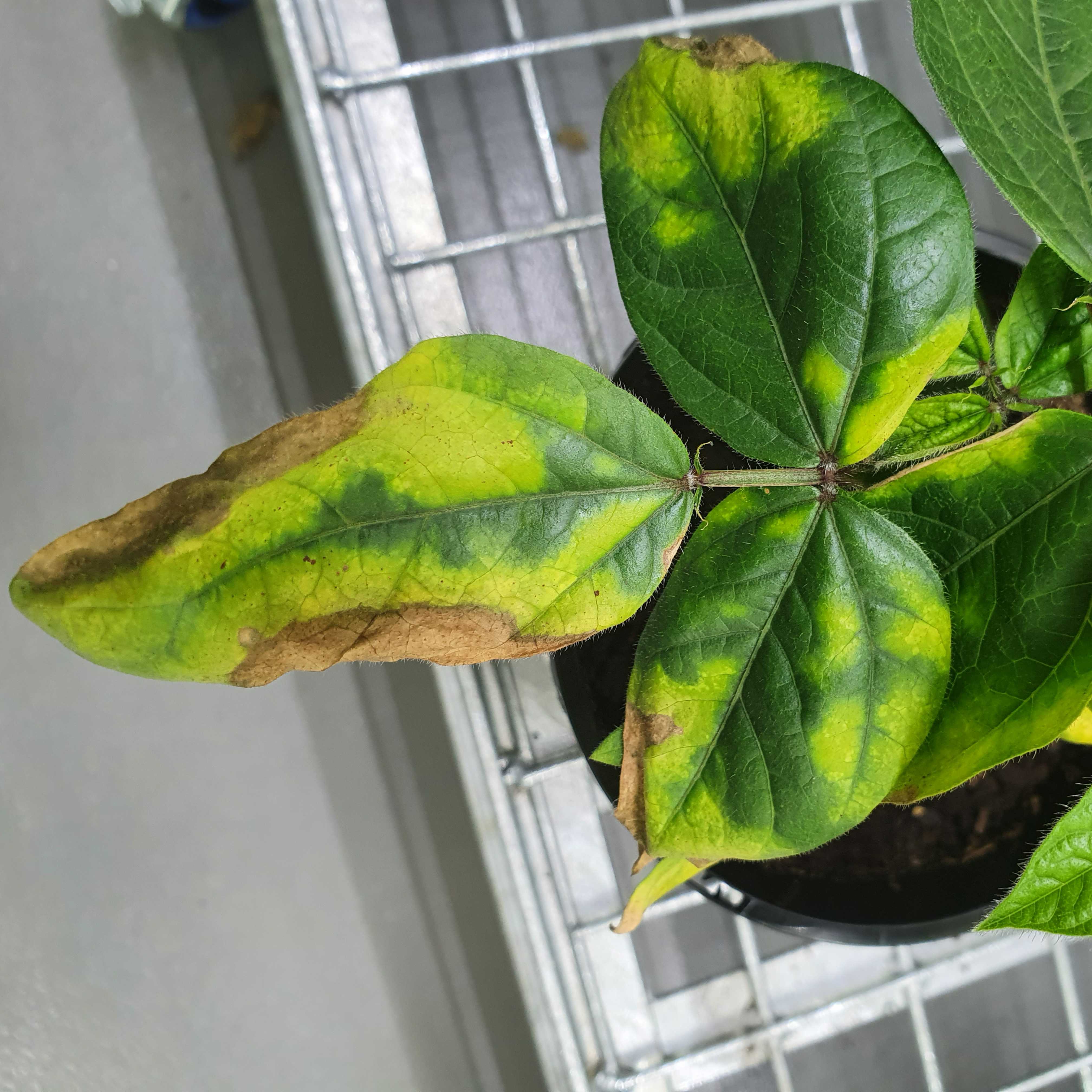
Tan spot symptoms on mungbean cv. Berken at seven days after inoculation with Curtobacterium flaccumfaciens pv. flaccumfaciens through stem injection.
At 25 days after inoculation, harvest all the symptomatic trifoliates, place these on a black background, and take photos.
Import the photos into an Image Analysis (IA) software, for example, Leaf Doctor (Pethybridge and Nelson 2015), and record the percent diseased area.
Estimation of the percent diseased area on mungbean leaves inoculated with Curtobacterium flaccumfaciens pv. flaccumfaciens, using the Image Analysis software Leaf Doctor (Pethybridge and Nelson 2015)
Note: You can also change the thresholds to estimate the chlorotic area only, and subsequently calculate the necrotic area by subtracting the chlorotic area from total diseased area.
Reference:
Pethybridge, S. J., Nelson, S. C. (2015). Leaf Doctor: A new portable application for quantifying plant disease severity. Plant disease, 99 (10): 1310-1316.
Re-isolating Cff from symptomatic plants
Randomly choose a subset of diseased plants for re-isolation of Curtobacterium flaccumfaciens pv. flaccumfaciens, as follows.
Surface-sterilize the symptomatic leaves by spraying with 70% ethanol and wiping with autoclaved paper towels in a laminar flow cabinet.
Excise 1-2 cm pieces of leaf tissue from the margin of leaf lesions and cut in halves.
Place one half of the tissue in a drop of water on a microscope slide to observe under a compound microscope, and place the other half in a microfuge tube containing 100 µl sterile water.
Upon confirmation of bacterial oozing under the microscope, use a sterile loop to streak the bacterial suspension from the microfuge tube onto Nutrient Agar or MSCFF medium (Maringoni et al. 2006b), which is a semi-selective medium for Curtobacterium flaccumfaciens pv. flaccumfaciens.
Note: MSCFF (Maringoni et al. 2006b) is also known as CFFSM (Maringoni et al. 2006a). Detailed recipe for making MSCFF can be found in Maringoni et al. (2006b) and also here: Be-4.3-Curtobacterium-flaccumfaciens-pv.-flaccumfaciens.pdf (seedhealth.org)
Note: The incorporation of several antimicrobial products in the MSCFF semi-selective medium helps with the isolation of Cff by inhibiting growth of saprophytic bacteria. Also, presence of skim milk powder and Congo red in MSCFF facilitates visualization of the Cff colonies because Cff can hydrolyze casein (Holt et al. 1994) and Congo red (Diatloff et al. 1993). Cff on MSCFF forms circular colonies, with a an orange or pink coloration, and casein hydrolysis results in a slight fading of the dye around the colonies.
Note: If you do not have access to the chemicals included in MSCFF, any media containing Congo red results in small, butyrous, raised, glossy, pinkish Cff colonies, which makes it easier to distinguish from the background microflora (Diatloff et al. 1993). Adding skim milk to the medium also helps identifying Cff colonies as slight fading of casein can be noticed around Cff colonies. However, it should be noted that other bacteria may also show the same properties (Maringoni et al. 2006b); therefore, ultimate identification shoudl be done through colony PCR and sequencing, or Cff-specific PCR.
Curtobacterium flaccumfaciens pv. flaccumfaciens colonies on Nutrient Agar (left) and modified* MSCFF medium (right).
*The MSCFF medium was prepared according to Maringoni et al. (2006b), except that sodium azide was not added to the medium.
Reference:
Diatloff, A., Wong, W.C. and Wood, B.A. (1993). Non destructive methods of detecting Curtobacterium flaccumfaciens pv. flaccumfaciens in mungbean seeds. Letters in Applied Microbiology, 16: 269–273.
Holt, G.J., Krieg, N.R., Sneath, P.H.A., Staley, J.T., Williams, S.T. (1994). Bergey’s manual of determinative bacteriology, 9th ed, 787p., Lippincot Williams and Wilkins, Philadelphia.
Maringoni, A. C., Camara, R. D. C. (2006a). Curtobacterium flaccumfaciens pv. flaccumfaciens detection in bean seeds using a semi-selective culture medium. Brazilian Journal of Microbiology, 37: 451-455. (refers to the semi-selective medium as CFFSM)
Maringoni, A. C., Camara, R. C., Souza, V. L. (2006b). Semi-selective culture medium for Curtobacterium flaccumfaciens pv. flaccumfaciens isolation from bean seeds. Seed Science and Technology, 34(1): 117-124. (refers to the semi-selective medium as MSCFF)