Mar 31, 2026
In vitro Infection of SHK-1 Cell Cultures with the Intracellular Pathogen P. salmonis
- Pamela Aravena1,
- Javiera Ortiz Severín1,
- Verónica Cambiazo1
- 1University of Chile


Protocol Citation: Pamela Aravena, Javiera Ortiz Severín, Verónica Cambiazo 2026. In vitro Infection of SHK-1 Cell Cultures with the Intracellular Pathogen P. salmonis. protocols.io https://dx.doi.org/10.17504/protocols.io.j8nlkrqz1v5r/v1
License: This is an open access protocol distributed under the terms of the Creative Commons Attribution License, which permits unrestricted use, distribution, and reproduction in any medium, provided the original author and source are credited
Protocol status: Working
We use this protocol and it's working
Created: June 02, 2025
Last Modified: March 31, 2026
Protocol Integer ID: 219425
Keywords: Piscirickettsia salmonis, SHK-1 cells, Infection, derived cell line, infection, cell, cell line
Funders Acknowledgements:
ANID FONDECYT
Grant ID: 1211893
Millennium Science Initiative Program
Grant ID: ICN2021_044
Abstract
This protocol describes step by step growth and collection
of P. salmonis cells for infecting the SHK-1 salmon-derived cell line in
flasks or 6-well plates.
Image Attribution
Pamela Aravena
Guidelines
Sterility must be maintained when working with P. salmonis and SHK-1 cell cultures. P. salmonis culture medium contains FBS and other nutrients that can be easily contaminated if not careful. We recommend using a biosafety cabinet or a laminar flow cabinet.
Materials
Leibovitz's L-15 Medium, powderThermo FisherCatalog #41300039
Fetal Bovine Serum, Regular, USDA Safety TestedCorningCatalog #35-010-CV
PBS, pH 7.4Gibco - Thermo Fisher ScientificCatalog #10010023
Tryptic Soy Broth, Casein-peptone soymeal-peptone broth, Millipore, Catalog #105459
BD BACTO Agar 100g, Catalog #214050
NaClMerck MilliporeSigma (Sigma-Aldrich)Catalog #S5886
L-CysteineMerck MilliporeSigma (Sigma-Aldrich)Catalog #C7352
Iron(III) chloride hexahydrateMerck MilliporeSigma (Sigma-Aldrich)Catalog #44944
Gentamicin SolutionMerck MilliporeSigma (Sigma-Aldrich)Catalog #G1397
Safety warnings
P. salmonis growth is sensitive to changes in agitation, media composition and (particularly) temperature. If a P. salmonis culture do not reach OD600 = 1.2-1.5, subculturing and infecting with that culture may not work.
P. salmonis Cultivation
1w
P. salmonis CGR02 strain (GCA_001534725.1) was isolated from Salmo salar liver, and it was obtained from ETECMA E.I.R.L (2016, Puerto Montt, Chile).
Bacteria are recovered from frozen stocks in Nutrient Piscirickettsia Broth (NPB; 30 gr/L Tryptic Soy Broth, 256.6 mM NaCl, 8.25 mM L-cysteine, 37 µM FeCl3) and plated into NPB plus 1.5% agar (NPA). After bacterial growth (5-7 days at 18 ºC), plates are used to inoculate 5 mL of NPB.
1w
The strain is grown at 18 ºC with agitation on NPB or in NPA plates. Incubate in a shaking incubator at 180 rpm and 18 ºC for 36 h to obtain exponentially growing bacteria, or 96 h for stationary state bacteria.
To monitor bacterial growth, take an aliquot of the culture to measure the optical density at 600 nm (OD600) and to count the number of bacterial cells using a Neubauer improved cell counting chamber. Use this quantification to estimate the multiplicity of infection (MOI) for the infection.
Before harvesting the cultures for infection experiments, test the purity of P. salmonis cultures by Gram-staining and the PCR-RFLP assay previously described in Mandakovic et al.
Infection of SHK-1 Cells
3d
Cultivate SHK-1 cells (ECACC N° 97111106) in Leibovitz's L-15 Medium (Gibco, USA) supplemented with 5% of inactivated fetal bovine serum (FBS) and 40 μM of 2-mercaptoethanol, at 18 ºC.
Seed the SHK-1 cells at a density of 3.5 x 10⁵ cells per T-25 flask, or 3x10⁵ cells in a 6-well plate, to obtain 80% confluence.
Incubate overnight at 18 °C before infecting with P. salmonis CGR02 at stationary phase, or with bacteria that was heat-inactivated by 60 min at 65 ºC (control), at a MOI of 100:1 (bacteria:cells).
Incubate infected cells at 18 ºC for three days. To eliminate extracellular bacteria, wash the infected cells with cold PBS 1X and then add Leibovitz's L15 medium with gentamicin (100 µg/mL), and incubate for 40 min at 18 ºC. After incubation, wash the cells with PBS 1X and incubate with fresh Leibovitz's L15 medium supplemented with 5% of FBS, without antibiotics.
3d
Monitor the progression of the infection under an inverted optical microscope every day post-infection (dpi).
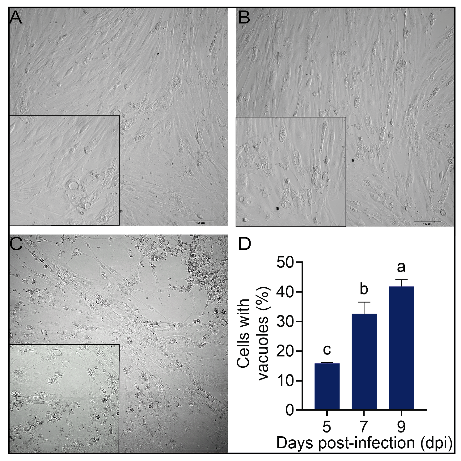
To determine the percentage of P. salmonis containing vacuoles (PCV) during the course of the infection, examine cells by visual inspection from at least six representative bright-field microscopy images (NIS-elements program, Nikon) of two independent infection assays per time point.
Here, a total of 3838 total cells were examined at 5 dpi, 2728 cells at 7 dpi, and 2145 cells at 9 dpi. Then, the number of SHK-1 cells with bacterial vacuoles was determined (Figure 1).
Figure 1.
In vitro infection of SHK-1 cells. (A-C) Representative bright-field microscopy
images of P. salmonis-infected cells. (A) 5 dpi, (B) 7 dpi, (C) 9 dpi. (D)
Percentage of cells with vacuoles in the field of view. Data reflect means ± SD
(N = 6 biological replicates); Images were taken using a Nikon C2+ microscope,
and the images were analyzed with the NIS-elements program (Nikon); letters
above bars indicate significant differences (p value < 0.05). Bar =
100 µm.
Protocol references
Zúñiga A, Aravena P, Pulgar R, Travisany D, Ortiz-Severín J, Chávez FP, Maass A, González M and Cambiazo V (2020) Transcriptomic Changes of Piscirickettsia salmonis During Intracellular Growth in a Salmon Macrophage-Like Cell Line. Front. Cell. Infect. Microbiol. 9:426. doi: 10.3389/fcimb.2019.00426