Jun 30, 2026
Immunoprecipitation (IP) Protocol for MAP2K1 Using MAP2K1 Recombinant Monoclonal Antibody (CSB-RA957619A0HU)
- Rosie Liu1
- 1CUSABIO
- CUSABIO TECHNOLOGY LLC


External link: https://www.cusabio.com/
Protocol Citation: Rosie Liu 2026. Immunoprecipitation (IP) Protocol for MAP2K1 Using MAP2K1 Recombinant Monoclonal Antibody (CSB-RA957619A0HU). protocols.io https://dx.doi.org/10.17504/protocols.io.kxygxrpq4g8j/v1
License: This is an open access protocol distributed under the terms of the Creative Commons Attribution License, which permits unrestricted use, distribution, and reproduction in any medium, provided the original author and source are credited
Protocol status: Working
We use this protocol and it's working
Created: June 30, 2026
Last Modified: June 30, 2026
Protocol Integer ID: 320056
Keywords: MAP2K1 protein; MAP2K1 Recombinant Monoclonal Antibody; Hela whole cell; Immunoprecipitation (IP) validation; Western Blotting (WB) detection, using map2k1 recombinant monoclonal antibody, map2k1 recombinant monoclonal antibody, protocol for map2k1, recombinant monoclonal antibody, map2k1, endogenous map2k1, efficiency of this antibody, antibody, immunoprecipitation, protein from hela, mek1
Abstract
This protocol is designed to specifically immunoprecipitate and enrich endogenous MAP2K1 (MEK1) protein from Hela whole cell lysate using MAP2K1 Recombinant Monoclonal Antibody (CSB-RA957619A0HU) and verify the IP specificity and efficiency of this antibody via subsequent Western Blotting (WB) detection.
Guidelines
Principle
Immunoprecipitation is an affinity enrichment technique based on the specific binding between antigen and antibody. In this assay, the MAP2K1-specific monoclonal antibody CSB-RA957619A0HU is incubated with HeLa cell lysate to bind endogenous MAP2K1 protein and form stable immune complexes. Protein G magnetic beads are then added to capture the immune complexes via specific interaction between Protein G and the Fc region of rabbit IgG. After removing non-specific binding proteins through repeated washing, the target protein is eluted and detected by Western Blotting, achieving specific enrichment and identification of the MAP2K1 protein from complex cell lysate.
Experimental Notes
- Maintain all protein samples and reagents at 4°C or on ice throughout the experiment to prevent protein degradation and reduce non-specific hydrophobic binding.
- The antibody dosage can be optimized in the range of 2–5 µg per 500 µg total protein, or diluted at a ratio of 1:200–1:1000 according to the actual expression level of MAP2K1 in samples.
- Isotype control and input control are indispensable for each experiment to distinguish specific binding from non-specific background and verify the reliability of results.
- Resuspend magnetic beads gently by pipetting or tube inversion; vigorous vortexing may damage the bead surface and increase non-specific adsorption.
- Ensure complete removal of supernatant during each washing step to avoid residual unbound proteins, which will increase background noise.
- For samples with high background, a pre-clearing step (incubating the lysate with control IgG and beads before adding the target antibody) can be added to reduce non-specific binding.
Materials
Cell Line
- Hela human cervical cancer cell lineSample
Antibodies
- MAP2K1 Recombinant Monoclonal Antibody (CUSABIO; Code: CSB-RA957619A0HU, Rabbit IgG isotype, non-conjugated, affinity-chromatography purified): recommended IP dilution: 1:200–1:1000
- Rabbit Control IgG (isotype control for immunoprecipitation)
- HRP-conjugated Protein G Antibody (for WB secondary detection): working dilution 1:2000
Reagents
- RIPA Lysis Buffer: 10 mM Tris-HCl (pH 7.6), 1 mM EDTA, 0.1% SDS, 0.1% sodium deoxycholate, 1% Triton X-100
- Protease Inhibitor Cocktail: including PMSF (0.5–1 mM), leupeptin (10–100 µM), aprotinin (0.01–0.03 µM)
- Ice-cold PBS: 10 mM phosphate buffered saline, pH 7.4, 150 mM sodium chloride
- PBST: PBS supplemented with 0.05% Tween-20
- 150 mM Glycine-HCl Elution Buffer (pH 1.5–2.5): pH 1.5–2.5
- 1 M Tris-HCl Neutralization Buffer (pH 9.4)
- 6×Sample Loading Buffer: Tris-HCl (pH 6.8), 4% SDS, 0.2% bromophenol blue, 20% glycerol, 9% DTT
- Protein G Magnetic Beads
- BCA Protein Quantification Kit
- SDS-PAGE gel preparation reagents: (acrylamide, N, N-methylenebisacrylamide, Tris-HCl, APS, TEMED)
- Tris-Glycine-SDS Electrophoresis Buffer
- Tris-Glycine-Methanol Transfer Buffer
- ECL Chemiluminescent Substrate
- PVDF or NC membrane
- Non-fat milk (blocking reagent)
Equipment & Consumables
- Low-temperature high-speed centrifuge
- Ultrasonic cell disruptor
- Magnetic separator
- Vertical electrophoresis system
- Wet/semi-dry protein transfer system
- Chemiluminescence gel imaging system
- Microplate reader (for BCA assay)
- Pre-chilled 1.5 mL microcentrifuge tubes
- Low-retention pipette tips
- Ice box and ice
Troubleshooting
Problem
No target band in IP lane (Input lane shows clear band) (Possible Causes: Insufficient antibody dosage; insufficient incubation time; protein degradation during sample preparation; incomplete binding of magnetic beads)
Solution
Appropriately increase antibody dosage; extend overnight incubation time; add fresh protease inhibitors and strictly maintain low temperature; extend magnetic bead incubation time or increase bead volume.
Problem
Obvious target band in Control IgG lane (Possible Causes: Non-specific binding of magnetic beads; insufficient washing; excessive total protein input)
Solution
Pre-block magnetic beads with 1% BSA before use; increase washing times or use a higher-stringency washing buffer; reduce total protein input.
Problem
High background in WB result (Possible Causes: Incomplete washing; excessive antibody concentration; non-specific binding of secondary antibody)
Solution
Increase washing times and duration; optimize primary/secondary antibody dilution ratio; replace blocking reagent if necessary.
Problem
Weak target band intensity in IP lane (Possible Causes: Low endogenous expression of MAP2K1; low IP efficiency; protein loss during washing)
Solution
Increase total protein input for IP reaction; optimize antibody dosage and incubation time; reduce washing times or use milder washing buffer.
Problem
Antibody heavy/light chain interferes with target detection (Possible Causes: Thermal denaturation releases antibody chains; target MW is close to 55 kDa (heavy chain) or 25 kDa (light chain))
Solution
Use acid elution instead of thermal denaturation; use light chain-specific secondary antibody or HRP-Protein A/G for WB detection.
Safety warnings
1. Components in protease inhibitor cocktail (such as PMSF) are highly toxic. Avoid direct contact with skin and mucous membranes. Operate with protective gloves and masks in a fume hood.
2. Protease inhibitors are unstable at room temperature and will lose activity quickly. They must be added to lysis buffer immediately before use, and the working solution should be kept on ice at all times.
3. The antibody buffer contains 0.02% sodium azide, which is toxic and can form explosive metal azides when reacting with copper/lead pipelines. Dispose of waste liquid properly and avoid direct contact.
4. Boiling samples may cause liquid splashing and tube lid popping. Use a safety rack for boiling water bath, and ensure tube lids are tightly closed to prevent sample loss and contamination.
5. Repeated freeze-thaw will cause antibody inactivation and protein degradation. Aliquot antibodies and protein samples into small volumes before storage at -80°C.
6. Methanol in transfer buffer is volatile and toxic. Perform transfer operations in a well-ventilated area and avoid prolonged inhalation.
7. Sonication operation may produce bioaerosols containing cell components. It should be performed in a fume hood with proper personal protective equipment.
Before start
All sample preparation procedures must be strictly performed on ice to prevent target protein degradation and denaturation. Harvested cells should be processed immediately to maintain protein integrity. Protease inhibitors must be freshly added to lysis buffer right before use. Repeated freeze-thaw cycles of cell lysates are prohibited. Sonication uses intermittent ice-cooled pulses to avoid heat-induced protein denaturation.
Hela Cell Lysate Preparation
Culture Hela cells until the confluence reaches 80%. Place the culture dishes on ice and wash the cells twice with ice-cold PBS.
Scrape adherent cells with ice-cold PBS and collect the cell suspension into a pre-chilled centrifuge tube. Centrifuge at 2500 rpm for 5 min at 4°C to pellet cells, then discard the supernatant carefully.
Resuspend the cell pellet with ice-cold RIPA lysis buffer supplemented with fresh protease inhibitors (1 mL RIPA buffer per 1×10^7^ cells).
Sonicate the cell suspension on ice with appropriate ultrasonic power to facilitate complete cell lysis.
Incubate the lysate on ice for 20–30 min to ensure sufficient lysis and protein solubilization.
Centrifuge the lysate at 12000 rpm for 20 min at 4°C. Transfer the clear supernatant to a new pre-chilled tube, avoiding the upper lipid layer and cell debris pellet.
Determine the total protein concentration of the supernatant via BCA assay. Fresh lysate is recommended for immediate IP. If storage is required, aliquot the lysate and store at -80°C.
Assay Procedures
Sample Setup:
Aliquot 500 µL of HeLa cell lysate containing 500 µg of total protein into each pre-chilled centrifuge tube. Prepare three groups in parallel:
IP group: for target antibody incubation
Negative control group: replace the target antibody with an equal amount of rabbit control IgG
Input control: reserve 10 µg of cell lysate as the loading reference
Antigen-Antibody Complex Formation:
- Add 2 µg of MAP2K1 Recombinant Monoclonal Antibody (CSB-RA957619A0HU) to the IP group tube.
- Add an equal mass of rabbit control IgG to the negative control tube.
- Gently mix the solutions and incubate at 4°C overnight with gentle end-over-end rotation.
Immunocomplex Capture with Magnetic Beads:
- Pre-wash the Protein A/G magnetic beads with ice-cold PBS 2–3 times before use.
- Add 50 µL of pre-washed magnetic beads to each reaction tube.
- Incubate at 4°C for 2–4 hours with continuous gentle rotation to allow binding between the antibody Fc region and Protein A/G on beads.
Complex Collection and Washing:
- Collect the bead-antibody-antigen complexes via centrifugation at 2500 rpm for 1–2 minutes at 4°C. Carefully remove and discard the supernatant.
- Wash the bead complexes sequentially with the following buffers, with 1 mL of each buffer for 2 repeated washes:
a. RIPA lysis buffer
b. PBST buffer
c. PBS buffer
- For each wash, fully resuspend the beads, incubate on ice for 5 minutes, then centrifuge at 2500 rpm, 4°C to re-collect the beads and discard the supernatant.
Elution and Denaturation (Two elution methods are available):
Thermal denaturation (routine use):
- Add an appropriate volume of 1× SDS sample loading buffer to resuspend the washed bead pellet.
- Boil the sample at 100°C for 10 minutes to denature proteins and dissociate immunocomplexes.
- Briefly centrifuge the tube, then transfer the supernatant to a new, clean tube for western blot detection.
- Note: Thermal denaturation may cause Protein A/G to detach from beads and enter the supernatant, which may interfere with detection results.
Acid elution (lower background, suitable for all protein sizes):
- Add 50 µL of 150 mM glycine-HCl (pH 1.5–2.5) to the bead pellet, gently resuspend, and incubate for several minutes.
- Centrifuge to collect the supernatant and repeat elution once. Combine the two eluates.
- Add 2 µL of 1 M Tris-HCl (pH 9.4) to neutralize the eluate.
- Add 20 µL of 6× sample loading buffer and boil the solution for subsequent WB detection.
Western Blot (WB) Detection:
Load the eluted samples and input control onto an SDS-PAGE gel for electrophoresis separation, followed by protein transfer and immunoblotting. Use HRP-conjugated Protein G (1:2000 dilution) as the secondary detection reagent.
Result Analysis
Specificity Verification:
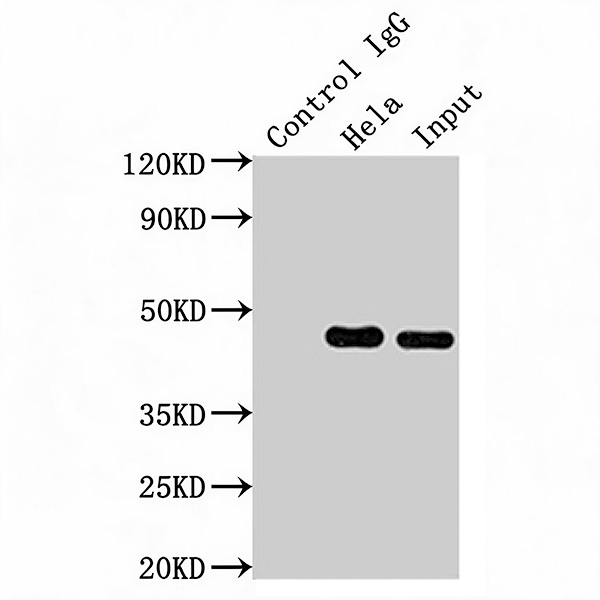
Immunoprecipitation (IP) validation of MAP2K1 Recombinant Monoclonal Antibody
- The Input lane (HeLa whole cell lysate, 10 µg) should present a clear target band at approximately 45 kDa (the molecular weight of MAP2K1/MEK1), confirming target protein expression and a valid detection system.
- The Control IgG lane (isotype control) should show no obvious target band, indicating no non-specific binding of IgG or magnetic beads to MAP2K1, and verifying the specificity of the IP reaction.
- The experimental IP lane (CSB-RA957619A0HU + HeLa lysate) should show a distinct target band at the same molecular weight as the Input lane, confirming successful immunoprecipitation of endogenous MAP2K1 by the antibody.
Performance Evaluation:
Compare the band intensity of the IP group with the input group to estimate the enrichment efficiency of the antibody. A bright, specific band in the IP lane with no obvious non-specific bands demonstrates good IP specificity and efficiency of CSB-RA957619A0HU.
Interference Assessment:
Since the molecular weight of MAP2K1 (~45 kDa) is close to the IgG heavy chain (~55 kDa), heavy chain interference may occur with thermal denaturation. The use of HRP-conjugated Protein G can effectively reduce such interference. If severe overlap is observed, the acid elution method is recommended to further reduce antibody chain contamination.
Protocol references
○ MAP2K1 Recombinant Monoclonal Antibody (CSB-RA957619A0HU)
○ Hela whole cell lysate
○ Target Analyte: MAP2K1 (MEK1)