Jan 07, 2026
Immunolabelling and clearing of large samples of peripheral nerve tissue
Forked from a private protocol
- 1University of Melbourne
- SPARCTech. support email: [email protected]



Protocol Citation: John-Paul Fuller-Jackson, Peregrine B Osborne, Janet R Keast 2026. Immunolabelling and clearing of large samples of peripheral nerve tissue. protocols.io https://dx.doi.org/10.17504/protocols.io.14egn67nql5d/v1
License: This is an open access protocol distributed under the terms of the Creative Commons Attribution License, which permits unrestricted use, distribution, and reproduction in any medium, provided the original author and source are credited
Protocol status: Working
We use this protocol and it's working
Created: May 07, 2024
Last Modified: January 07, 2026
Protocol Integer ID: 99403
Keywords: idisco, light sheet microscopy, tissue clearing, nerve tract, ganglia, large regions of adipose tissue, peripheral nerve tissue, adipose tissue, noradrenergic ganglion neuron, peripheral nerve tract, adjacent neural tissue, whole mount immunolabeling, antibody, clearing method, clearing of large sample, adipo, examples of antibody, ganglia, immunolabeling
Funders Acknowledgements:
NIH SPARC
Grant ID: 3OT2OD023872
Abstract
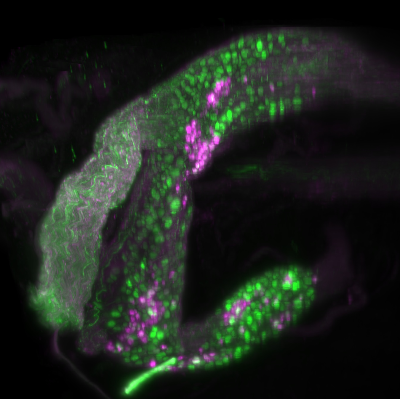
A whole mount immunolabeling and clearing method (iDISCO) combined with the pre-treatment steps in ADIPO-CLEAR is used to visualize neural structures in large samples of peripheral nerve tracts or ganglia. These tissues can be associated with large regions of adipose tissue that are difficult to remove without damaging the adjacent neural tissue. Examples of antibodies used for this type of study are also provided (tyrosine hydroxylase for noradrenergic ganglion neurons and their axons; Beta-tubulin as a pan-neural marker).
Materials
Materials
MethanolMerck MilliporeSigma (Sigma-Aldrich)Catalog #M3641 GlycineMerck MilliporeSigma (Sigma-Aldrich)Catalog #G8790-1KG Sodium azideChemSupplyAustraliaCatalog #SA189-100G
DichloromethaneMerck MilliporeSigma (Sigma-Aldrich)Catalog #320269
Dibenzyl etherMerck MilliporeSigma (Sigma-Aldrich)Catalog #108014
Ethyl cinnamateMerck MilliporeSigma (Sigma-Aldrich)Catalog #112372
1X Dulbecco’s Phosphate Buffered Saline (DPBS) Thermo Fisher ScientificCatalog #14190094
Gelatin from porcine skinMerck MilliporeSigma (Sigma-Aldrich)Catalog #G1890
Hydrogen peroxide 30%Merck Millipore (EMD Millipore)Catalog #822287.1000
ThimerosalMerck MilliporeSigma (Sigma-Aldrich)Catalog #T5125
Triton X-100Merck MilliporeSigma (Sigma-Aldrich)Catalog #T8787-50ML
Anti-class III beta tubulin antibody (rabbit)BioLegendCatalog #802001 Anti-tyrosine hydroxylase antibody (sheep)Merck Millipore (EMD Millipore)Catalog #AB1542 AF647 donkey anti-sheep antibodyInvitrogenCatalog #10729004 Cy3 donkey anti-rabbit antibodyJackson ImmunoResearch Laboratories, Inc.Catalog #711-165-152
Equipment
Equipment
Ultramicroscope Blaze
NAME
Light sheet microscope
TYPE
Ultramicroscope
BRAND
140-006-739.04
SKU
LINK
Solutions
PBS: phosphate-buffered saline, 0.1 M, pH 7.2
B1n buffer: 0.3 M glycine, 0.1% Triton X100, 0.01% sodium azide in water
DPBS: 1x Dulbecco's phosphate-buffered saline
DPBSG-T: 1x Dulbecco's phosphate-buffered saline containing 0.2% gelatin, 0.5% Triton X-100 and 0.01% thimerosal
Primary antibodies
| A | B | C | D | E | |
| Abbreviation | Synonym | RRID | Host species | Dilution | |
| TH | Tyrosine hydroxylase | AB_90755 | Sheep | 1:1000 | |
| Tuj1 | Class III beta-tubulin | AB_2564645 | Rabbit | 1:2000 |
Secondary antibodies
| A | B | C | D | |
| Tag-antibody | Host species | RRID | Dilution | |
| AF647 anti-sheep | Donkey | AB_2535865 | 1:500 | |
| Cy3 anti-rabbit | Donkey | AB_2307443 | 1:2000 |
Large peripheral nerve tissue preparation
1h
While immersed in phosphate buffered saline (PBS), pH 7.2, use a scalpel blade to sub-dissect large sample of peripheral nerve tissue into smaller samples, generally no larger than 10 mm x 10 mm x 10 mm.
Dehydration and Delipidation
1d
Wash samples in B1n buffer (1 h) on rotation 12 rpm at 4 ºC.
Dehydrate samples in a series of methanol in B1n buffer dilutions on rotation 12 rpm at 4 ºC.
- 20% methanol in B1n buffer (1 h)
- 40% methanol in B1n buffer (1 h)
- 60% methanol in B1n buffer (1 h)
- 80% methanol in B1n buffer (1 h)
- 100% methanol (1 h)
Incubate samples in 100% dichloromethane (1 h) on rotation 12 rpm at 4 ºC.
Incubate samples overnight in 100% dichloromethane on rotation 12 rpm at 4 ºC.
Incubate samples in 100% dichloromethane (1 h) on rotation 12 rpm at 4 ºC. If sample has not sunk to the bottom of the vessel continue washes with dichloromethane until it has.
Bleaching
1d
Wash samples twice in 100% methanol (1 h) on rotation 12 rpm at 4 ºC.
Bleach samples overnight in 6% hydrogen peroxide in methanol at 4°C, protected from light.
Blocking
5d
Rehydrate samples in a series of methanol in B1n buffer dilutions while on rotation at 12 rpm at 4°C:
- 100% methanol (1 h)
- 80% methanol in B1n buffer (1 h)
- 60% methanol in B1n buffer (1 h)
- 40% methanol in B1n buffer (1 h)
- 20% methanol in B1n buffer (1 h)
- B1n buffer (1 h)
Incubate samples in DPBS containing 0.2% gelatin, 0.5% Triton X-100 and 0.01% thimerosal (DPBSG-T) for 4 days while on rotation at 12 rpm
Primary antibody incubation
2w
Incubate samples in primary antibody solution containing DPBSG-T for 14 days at 37°C with agitation. Volume of solution need only be sufficient to cover the sample.
Secondary antibody incubation
2d
Wash samples in DPBSG-T (6 x 1 h).
Incubate samples in secondary antibody solution containing DPBSG-T for 2 days at 37°C with agitation. Volume of solution need only be sufficient to cover the sample.
Dehydration and delipidation
1d
Wash samples in DPBSG-T (6 x 1 h).
Dehydrate samples in a series of methanol in DPBS dilutions while on rotation at 12 rpm:
- 20% methanol in DPBS (1 h)
- 40% methanol in DPBS (1 h)
- 60% methanol in DPBS (1 h)
- 80% methanol in DPBS (1h)
- 100% methanol (2 x 1 h)
Incubate samples in a solution of 2/3 dichloromethane and 1/3 methanol overnight on rotation. Ensure that samples sink to the bottom of the vial at the end of this step, otherwise continue incubation in freshly made solution.
Incubate samples in 100% dichloromethane for 30 mins while on rotation at 12 rpm. Repeat this step until samples sink.
Clearing
2h
Incubate samples in dibenzyl ether until the samples have become clear. Ensure each vial is completely filled with dibenzyl ether to minimize sample oxidation as a result of large amounts of air in the vial. This process should not take longer than 2 h.
Storage
Store cleared samples in fresh dibenzyl ether. Keep away from light (wrap in foil and store in an opaque container).
Light sheet microscopy
Prior to visualization on a light sheet microscope, samples should be transferred into ethyl cinnamate, at least 3 h prior.
Remove sample from ethyl cinnamate and gently dry on a tissue. Affix sample to plastic mount using the minimum amount of super glue required. Tips:
- Avoid adhering the sample to the base via any region of the sample that is of interest; both the super glue and proximity to the plastic will reduce imaging quality in that area.
- Some sample holders are available that can hold the sample in place without glue, try these if they are suitable to the orientation required.
- Orientate the sample such that the thinnest plane of the sample is perpendicular to the light sheet beams. The further light has to travel through a sample, the poorer the image quality.
- Orientate the sample so that the region of interest is facing as close to the lens as possible.
- Orientate the sample so that the region of interest is flat, the best resolution of the light sheet microscope is in XY.
For viewing sub-dissections of the large peripheral nerve tissue, it is best to begin with lower magnification to view the whole sample at once and then identify regions of interest such as clusters of neurons. These can then be inspected at higher magnification. Sometimes it is necessary to reorient the sample as described above for better illumination and thus imaging.
Protocol references
Renier, N., Wu, Z., Simon, D.J., Yang, J., Ariel, P., Tessier-Lavigne, M., 2014. iDISCO: a simple, rapid method to immunolabel large tissue samples for volume imaging. Cell 159, 896–910. https://doi.org/10.1016/j.cell.2014.10.010
Belle, M., Godefroy, D., Couly, G., Malone, S.A., Collier, F., Giacobini, P., Chédotal, A., 2017. Tridimensional Visualization and Analysis of Early Human Development. Cell 169, 161-173.e12. https://doi.org/10.1016/j.cell.2017.03.008
Fuller-Jackson, J.P., Osborne, P.B., Keast, J.R., 2021. Regional Targeting of Bladder and Urethra Afferents in the Lumbosacral Spinal Cord of Male and Female Rats: A Multiscale Analysis. eNeuro 8. https://doi.org/10.1523/ENEURO.0364-21.2021
Chi, J., Crane, A., Wu, Z., Cohen, P., 2018. Adipo-Clear: A Tissue Clearing Method for Three-Dimensional Imaging of Adipose Tissue. J Vis Exp 58271. https://doi.org/10.3791/58271