May 27, 2025
Immunolabeling and Quantitative Analysis of Synapses in Human iPSC-Derived Dopaminergic Neurons
- 1Montreal Neurological Institute - McGill University



Protocol Citation: Roxanne Larivière, Edward A. Fon 2025. Immunolabeling and Quantitative Analysis of Synapses in Human iPSC-Derived Dopaminergic Neurons. protocols.io https://dx.doi.org/10.17504/protocols.io.eq2lyq7opvx9/v1
License: This is an open access protocol distributed under the terms of the Creative Commons Attribution License, which permits unrestricted use, distribution, and reproduction in any medium, provided the original author and source are credited
Protocol status: Working
We use this protocol and it's working
Created: May 02, 2025
Last Modified: May 27, 2025
Protocol Integer ID: 210771
Keywords: iPSC , Dopaminergic neurons, Synapse, Immunofluorescence, Synaptophysin, PSD-95, cellular assay, labeling synapse, derived dopaminergic neurons this protocol, quantitative analysis of synapse, derived dopaminergic neuron, synaptophysin, dopaminergic neuron, synapse, induced pluripotent stem cell, pluripotent stem cell, immunofluorescence, using immunofluorescence, neuron
Funders Acknowledgements:
Michael J. Fox Foundation for Parkinson's research
Grant ID: MJFF-025378
Abstract
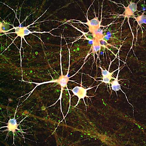
This protocol outlines the procedure for labeling synapses with pre-synaptic and post-synaptic markers, synaptophysin and PSD-95 respectively, and microtubule-associated protein 2 (MAP2) in induced pluripotent stem cell (iPSC)-derived dopaminergic neurons using immunofluorescence.
Materials
Before start
iPSC-derived dopaminergic neurons are cultured on 96-well plates for up to 4 to 6 weeks of differentiation. We use black Costar 96-well assay plates (Corning cat. no. 3904), which are compatible with imaging using the Opera Phenix high-content screening system.
Be extremely gentle when dispensing into the wells, as dopaminergic neurons are prone to detaching with forceful pipetting.
Fixation of Dopaminergic Neurons in 96-well plates
35m
- Carefully aspirate the culture media, leaving 50 µL in each well.
- Gently add50 µL of 8% paraformaldehyde (PFA) to each well to reach a final concentration of 4 % (v/v) PFA.
- Incubate at room temperature for 00:20:00 .
- Remove the fixative and wash each well three times with 75 µL of 1X PBS. Allow each wash to incubate for 00:05:00 .
35m
Permeabilization
25m
- Prepare the permeabilization solution by diluting Triton X-100 to a final concentration of 0.5 % (v/v) in 1X PBS.
- Add 60 µL of the permeabilization solution to each well and incubate for 00:10:00 at room temperature.
- Remove the solution and wash wells three times with 75 µL of 1X PBS, allowing each wash to incubate for00:05:00 .
25m
Blocking
1h
- Prepare the blocking buffer by mixing 5 % (v/v) normal goat serum and 0.02 % (v/v) Triton X-100 in 1X PBS.
- Add60 µL of the blocking buffer to each well and incubate for 01:00:00 atRoom temperature .
1h
Primary Antibody Incubation
16h
1. Prepare the primary antibody solution in blocking buffer with the following dilutions:
- Rabbit anti-Synaptophysin: 1:500
- Mouse anti-PSD-95: 1:250
- Chicken anti-MAP2: 1:1500
2. Add 60 µL of the antibody mix to each well.
3. Incubate Overnight at 4 °C with gentle shaking.
4. Remove antibody mix and wash four times with 75 µL 1X PBS, allowing each wash to incubate for 00:05:00 .
16h
Secondary Antibody Incubation
2h 15m
1. Prepare the secondary antibody solution in 1 % (v/v) normal goat serum diluted in 1X PBS with the following components:
- Alexa Fluor 488 anti-Rabbit: 1:1000
- Alexa Fluor 647 anti-Mouse: 1:1000
- Alexa Fluor 555 anti-Chicken: 1:1000
- Hoechst 33342: 1:5000
2. Add 60 µL of the antibody mix to each well.
3. Incubate for02:00:00 at Room temperature with gentle shaking, protected from light.
4. Remove secondary antibody mix and wash four times with 75 µL 1X PBS, allowing each wash to incubate for 00:05:00 .
2h 15m
Imaging using the Opera Phenix high-content imaging system
1. Insert the prepared plate into the Opera Phenix chamber.
2. Utilize a 40× water immersion objective. Capture images using the Hoechst, 488 nm, 555 nm and 647 nm laser channels. Acquire 15-18 fields of view per well
Synapse Detection and Data analysis using Cell Profiler software
Images are analyzed using a Cell Profiler automated synaptic quantification pipeline, previously published by Berryer et al., eLife, 2023. https://doi.org/10.7554/eLife.80168
Protocol references
1. Martin H Berryer, Gizem Rizki, Anna Nathanson, Jenny A Klein, Darina Trendafilova, Sara G Susco, Daisy Lam, Angelica Messana, Kristina M Holton, Kyle W Karhohs, Beth A Cimini, Kathleen Pfaff, Anne E Carpenter, Lee L Rubin, Lindy E Barrett (2023) High-content synaptic phenotyping in human cellular models reveals a role for BET proteins in synapse assembly eLife 12:e80168 https://doi.org/10.7554/eLife.80168