Aug 26, 2023
Immunolabeling and PI staining Drosophila melanogaster germaria
- shelbilrussell 1
- 1Assistant Professor, Department of Biomolecular Engineering, University of California Santa Cruz


Protocol Citation: shelbilrussell 2023. Immunolabeling and PI staining Drosophila melanogaster germaria. protocols.io https://dx.doi.org/10.17504/protocols.io.5jyl8p2n9g2w/v1
License: This is an open access protocol distributed under the terms of the Creative Commons Attribution License, which permits unrestricted use, distribution, and reproduction in any medium, provided the original author and source are credited
Protocol status: Working
We use this protocol and it's working
Created: August 26, 2023
Last Modified: August 26, 2023
Protocol Integer ID: 87004
Keywords: staining drosophila melanogaster germaria protocol, drosophila melanogaster germaria protocol, drosophila ovary, immunolabeling germarium protein, germarium protein, immunolabeling, protein
Funders Acknowledgements:
National Institutes of Health
Grant ID: R00GM135583
Abstract
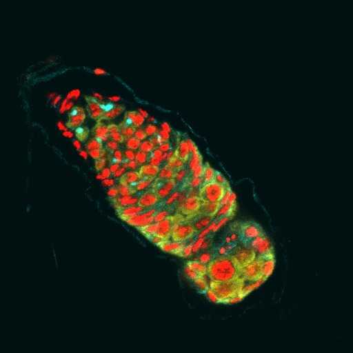
Protocol for staining (Wolbachia-infected) Drosophila ovaries for immunolabeling germarium proteins.
Make devitellinizing (DV) solution
a. Measure and add to a 1.5 ml eppendorf tube in the fume hood:
10x PBS 100 ul
dH20 400 ul
32% EM-grade paraformaldehyde 500 ul **opened < 10 days ago**
NP40 detergent 5.0 ul
____________________
~ 1 mL DV solution
b. Vortex this until NP40 is no longer visible as a blob on the side of the tube.
Dissect ovaries
a. Set up dissecting dish, filling each well with 1xPBS
b. Label 1.5 ml eppendorf tube for each sample and fill with ~1 ml of 1x PBS
c. Knock out flies on CO2 pad
d. Sort flies for target genotypes (if needed)
e. Dissect ovaries in PBS in 3 minutes or less to prevent “aggregation effects”.
Note: This amounts to only 5-10 flies of one genotype.
f. Gently separate ovarioles with pins.
g. Transfer ovaries to tube of 1x PBS
Fix ovaries
a. Briefly spin down ovaries (at ~2500xg) and remove 1x PBS
Note: ovaries may be on the side of tube - be careful to not disturb
b. In the fume hood, add 600 uL heptane to each tube and start timer
c. Add 200 uL of DV solution
d. Mix vigorously by hand for 1 min, or until emulsion forms between two phases.
e. Place on rotator for remaining 19 min
Rinse and treat with RNAseA (to degrade RNA for PI-DNA staining)
a. When ~30 seconds remain on the time, centrifuge ovaries briefly (at ~2500xg) to pellet
b. In the fume hood, remove and dispose the gunky top phase first, being careful to not disturb the pellet. When in doubt, remove less and add an extra wash.
c. Add ~1.2 mL 1x PBS-T (1% Triton X-100), centrifuge, pipette, and dispose residual heptane and fixative.
d. Repeat rinse once more quickly.
e. On third addition of fresh 1x PBS-T, incubate for 10 minutes prior to centrifuging and removal of buffer. Repeat two more times for a total of 5 washes.
Thaw frozen RNAseA (10mg/ml) at room temperature
f. After removing the final 1x PBS-T, add RNAseA
g. Incubate overnight at room temperature
Remove RNAse A
a. Centrifuge and remove and store RNAseA at -20C, marking use
Note: RNAseA aliquots are good for up to 10 uses, with freezes in between
b. Rinse 2x briefly and then wash 4x for 10 min each in PBS-T
Block tissue
Block in PBS-T with 1% (w/v) bovine serum albumin for 1 hr at room temp
Bind primary antibody
a. Suspend in PBS-T containing primary antibody at proper dilution (__:_____).
b. Incubate overnight at 4C.
Remove primary antibody
a. Centrifuge and remove primary antibody, discarding used solution
b. Rinse 2x briefly and then wash 4x for 10 min each in PBS-T
Bind secondary antibody
a. Resuspend in secondary antibody solution at proper dilution (__:_____)
b. Incubate overnight at 4C.
Remove secondary antibody
a. Centrifuge to remove secondary antibody, discarding used solution.
b. Rinse 2x briefly and then wash 4x for 10 min each in PBS-T.
Stain DNA with propidium iodide (PI)
After the removal of the last wash, add 60-100 uL of PI mounting medium to each tube.
Flick to coat all oocytes in medium and spin down with the microcentrifuge to pull solution to bottom.
Incubate overnight at room temperature or at 4C over the weekend.
Mount oocytes
Spin down oocytes and remove as much PI-containing mounting medium as possible.
Resuspend in 30 uL 70% glycerol in 1xPBS.
Using a p1000 tip, transfer each set of ovaries to a clean microscope slide.
Use pins to separate ovaries into ovarioles and eggs - position mature eggs around perimeter, in a circle.
Carefully lay coverslip over samples *do not move coverslip after placement (to avoid distorting oocytes).
Manage the amount of mounting media under slide:
If too much, wick excess with a kimwipe
If too little, add carefully with a tip to edge (allowing only what is required to fill area to wick out)
Paint edges of coverslip with nail polish to seal sample. Allow to fully dry in a dark drawer.
Store at -20C prior to imaging. Image within a month.