Feb 08, 2022
Immunohistochemistry of porcine enteric neurons
- Gemma Mazzuoli-Weber1,
- Michael Schemann2,
- Kristin Elfers1,
- Birgit Kuch3,
- Susanne Hoppe4
- 1University of Veterinary Medicine Hannover, Foundation, Germany;
- 2Techical Univeristy of Munich, Germany;
- 3Technical University of Munich, Germany;
- 4Institute for Physiology and Cell Biology University of Veterinary Medicine Hannover, Foundation
- SPARCTech. support email: [email protected]


Protocol Citation: Gemma Mazzuoli-Weber, Michael Schemann, Kristin Elfers, Birgit Kuch, Susanne Hoppe 2022. Immunohistochemistry of porcine enteric neurons. protocols.io https://dx.doi.org/10.17504/protocols.io.b4qrqvv6
License: This is an open access protocol distributed under the terms of the Creative Commons Attribution License, which permits unrestricted use, distribution, and reproduction in any medium, provided the original author and source are credited
Protocol status: Working
We use this protocol and it’s working
Created: February 04, 2022
Last Modified: February 08, 2022
Protocol Integer ID: 57841
Keywords: immunohistochemistry of porcine enteric neuron, porcine enteric neuron, myenteric plexus of the porcine colon, porcine colon, localization of neurochemical marker, neurochemical marker, immunohistochemistry, myenteric plexus, neuron
Abstract
This protocol is to investigate the presence and localization of neurochemical markers in the submucosal and myenteric plexus of the porcine colon using immunhistochemical staining techniques.
Materials
- samples of pocine colon
- Krebs solution for preparation containing in (mM): 117 NaCl, 11 Glucose, 4.7 KCl, 1.2 MgCl2, 1.2 NaH2PO4, 25 NaHCO3, 2.5 CaCl2; Carl Roth GmbH & Co. KG (Karlsruhe, Germany)
- Sylgard® 184; World Precision Instruments (Sarasota, FL, USA)
- 4% paraformaldehyde; Sigma-Aldrich Chemie GmbH (Schnelldorf, Germany)
- 0.002% picric acid; Sigma-Aldrich Chemie GmbH (Schnelldorf, Germany)
- 0.1 mol/l phosphate buffer; Sigma-Aldrich Chemie GmbH (Schnelldorf, Germany)
- phosphate-buffered saline (PBS): H2O, Sodium Phosphate Monobasic, Sodium Phosphate Dibasic, NaCl; Sigma-Aldrich Chemie GmbH (Schnelldorf, Germany)
- 0.1% NaN3; Carl Roth GmbH & Co. KG (Karlsruhe, Germany)
- 4% horse serum; Sigma-Aldrich Chemie GmbH (Schnelldorf, Germany)
- Triton X-100; Sigma-Aldrich Chemie GmbH (Schnelldorf, Germany)
- object slides; Gerhard Menzel B. V. & Co. KG (Braunschweig, Germany)
- glycerol; Sigma-Aldrich Chemie GmbH (Schnelldorf, Germany)
- cover slips; Omnilab-Laborzentrum GmbH & Co. KG (Gehrden, Germany)
- all antibodies from table 1 and table 2
- epifluorescence microscope; Olympus Corporation (Hamburg, Germany)
- Olympus cellSens Standard Software, Olympus Corporation (Hamburg, Germany)
Troubleshooting
Samples of colon were taken from apparently healthy pigs, placed in ice-cold oxygenated Krebs solution for preparation and immediately transferred to the laboratory. Tissues were then dissected in the ice-cold oxygenated Krebs solution for preparation to obtain whole-mount inner submucosal plexus or myenteric plexus preparations.
Tissue specimens were fixed overnight in a solution containing 4% paraformaldehyde and 0.002% picric acid in 0.1 mol/l phosphate buffer and then washed (3 x 10 min) in PBS.
The preparations were then incubated in PBS/NaN33/horse serum for 1h at room temperature followed by 12h and 2h incubation with the primary and secondary antibodies, respectively.
Specimens were washed in PBS, mounted on object slides and cover slipped with a solution of PBS (pH 7.0)/NaN3 containing 65% glycerol.
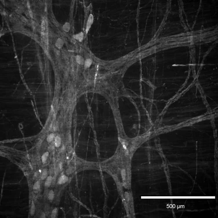
The preparations were examined with an epifluorescence microscope equipped with appropriate filter blocks. Pictures were acquired with a camera connected to a computer and controlled by Olympus cellSens Standard Software.
Pictures of the stained ganglia were acquired.