Jul 23, 2024
Immunohistochemical labelling of spinal cord sections for chemoarchitectural analysis of segments
- 1University of Melbourne
- SPARCTech. support email: [email protected]


Protocol Citation: John-Paul Fuller-Jackson, Peregrine B Osborne, Janet R Keast 2024. Immunohistochemical labelling of spinal cord sections for chemoarchitectural analysis of segments. protocols.io https://dx.doi.org/10.17504/protocols.io.14egn6376l5d/v1
License: This is an open access protocol distributed under the terms of the Creative Commons Attribution License, which permits unrestricted use, distribution, and reproduction in any medium, provided the original author and source are credited
Protocol status: Working
We use this protocol and it's working
Created: May 23, 2024
Last Modified: July 24, 2024
Protocol Integer ID: 100327
Keywords: neuroanatomy, immunohistochemistry, chemoarchitecture, spinal cord, sensory, motor, visceral, autonomic, immunohistochemical labelling of spinal cord section, adult rat lumbosacral spinal cord, spinal cord section, immunohistochemical visualisation of the chemoarchitecture, lumbosacral spinal cord, spinal cord, chemoarchitectural analysis of segment, immunohistochemical labelling, immunohistochemical visualisation, neuronal nitric oxide synthase, chemoarchitectural analysis, chemoarchitecture, vesicular glutamate transporter, rostrocaudal axis of each segment, choline acetyltransferase
Funders Acknowledgements:
NIH SPARC
Grant ID: 3OT2OD023872
Disclaimer
ETHICS DISCLAIMER
The protocols.io team notes that research involving animals and humans must be conducted according to internationally-accepted standards and should always have prior approval from an Institutional Ethics Committee or Board.
Abstract
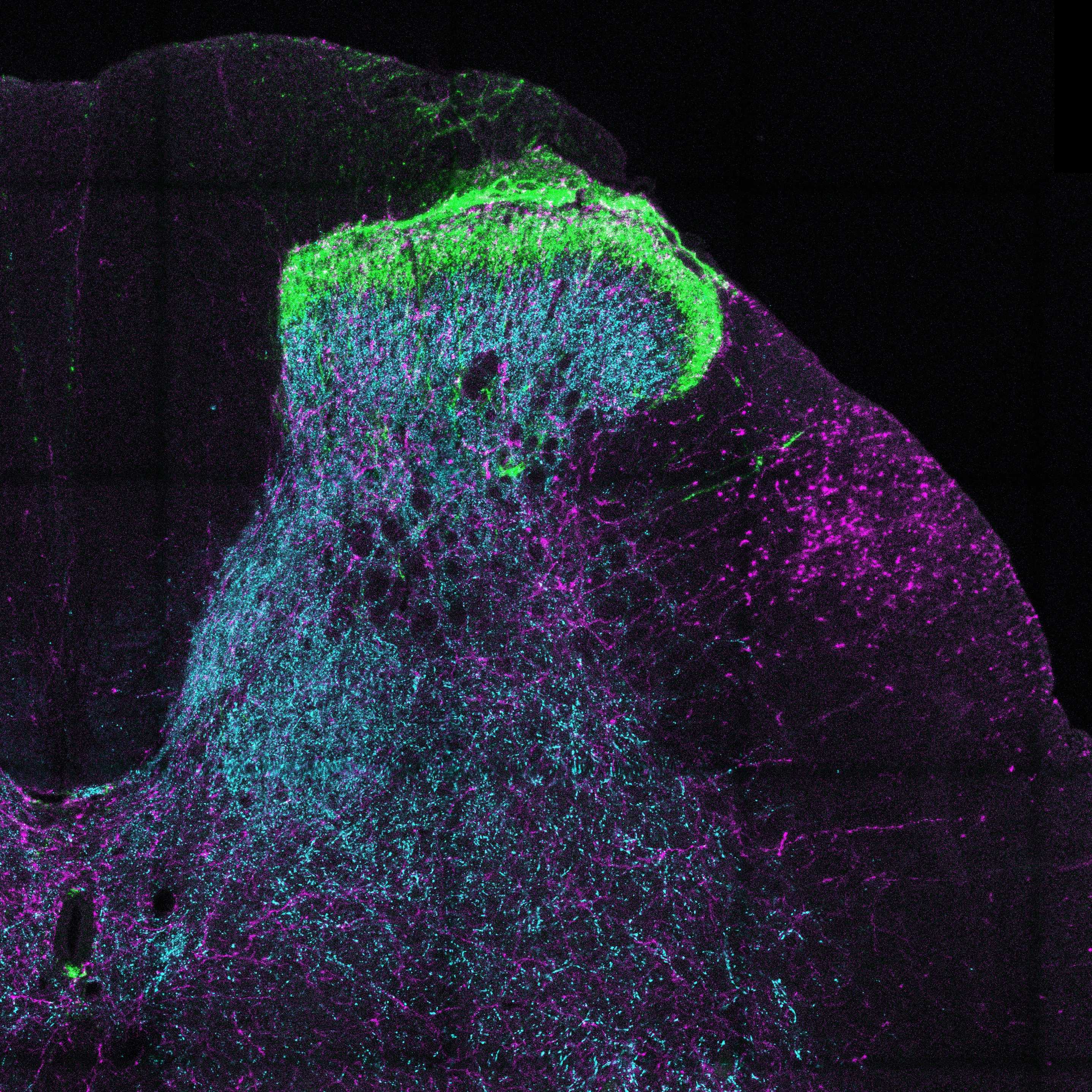
This protocol is used for immunohistochemical visualisation of the chemoarchitecture of the adult rat lumbosacral spinal cord (and other segments for comparison). Spinal cords were sub-dissected into segments, and transverse sections were obtained from across the rostrocaudal axis of each segment. Two combinations of antibodies were used:
- Combination 1: neuronal nitric oxide synthase (nNOS), choline acetyltransferase (ChAT) and NeuN
- Combination 2: calcitonin gene-related peptide (CGRP), vesicular glutamate transporter 1 (VGlut1) and tyrosine hydroxylase (TH)
Materials
MATERIALS
Horse serumMerck MilliporeSigma (Sigma-Aldrich)Catalog #12449C
OCT (Optimal Cutting Temperature compound)Sakura FinetekCatalog #4583
Goat anti-ChAT antibodyMerck Millipore (EMD Millipore)Catalog #AB144P
Anti-NeuN Antibody, clone A60Merck Millipore (EMD Millipore)Catalog #MAB377
Anti-neuronal nitric oxide synthase antibody (rabbit)InvitrogenCatalog #61-7000
Goat anti-CGRP antibody; AB_2290729Bio-Rad LaboratoriesCatalog #1720-9007
Anti-VGlut1 antibody (guinea pig)Merck Millipore (EMD Millipore)Catalog #AB5905
Mouse anti-TH (tyrosine hydroxylase) antibodyCatalog #22941
AF488 Donkey anti-goat IgGJackson ImmunoResearch Laboratories, Inc.Catalog #705-545-147 AF488 Donkey anti-mouse IgGJackson ImmunoResearch Laboratories, Inc.Catalog #715-545-150 Cy3 Donkey anti-rabbit IgGJackson ImmunoResearch Laboratories, Inc.Catalog #711-165-152 Cy3 Donkey anti-guinea pig IgGJackson ImmunoResearch Laboratories, Inc.Catalog #706-165-148 AF647 Donkey anti-sheep IgGMolecular ProbesCatalog #A21448 AF647 Donkey anti-mouse IgGInvitrogenCatalog #A31571
Solutions:
- PBS: phosphate-buffered saline, 0.1 M, pH 7,2
- PBS containing 0.1% sodium azide
- PBS containing 30% sucrose (w/v)
- Blocking solution: PBS containing 10% normal horse serum and 0.5% triton X-100
- PBS containing 0.1% sodium azide, 2% normal horse serum and 0.5% triton X-100
Primary Antibodies:
| A | B | C | D | E | F | |
| Abbreviation | Gene name | Synonym | RRID | Host Species | Dilution | |
| NeuN | FOX3 | NeuN | AB_2298772 | Mouse | 1:2000 | |
| ChAT | chat | Choline acetyltransferase | AB_2079751 | Goat | 1:500 | |
| nNOS | nnos | Neuronal nitric oxide synthase | AB_2313734 | Rabbit | 1:2000 | |
| CGRP | calca | Calcitonin gene-related peptide | AB_2290729 | Goat | 1:2000 | |
| TH | th | Tyrosine hydroxylase | AB_572268 | Mouse | 1:2000 | |
| VGlut1 | slc17a7 | vesicular glutamate transporter 1 | AB_2301751 | Guinea pig | 1:5000 |
Secondary Antibodies:
| A | B | C | D | |
| Tag-antibody | Host species | RRID | Dilution | |
| Anti-mouse AF488 | Donkey | AB_2340846 | 1:2000 | |
| Anti-goat AF488 | Donkey | AB_2336933 | 1:1000 | |
| Anti-rabbit Cy3 | Donkey | AB_2307443 | 1:3000 | |
| Anti-guinea pig Cy3 | Donkey | AB_2340460 | 1:2000 | |
| Anti-sheep AF647 | Donkey | AB_2535865 | 1:500 | |
| Anti-mouse AF647 | Donkey | AB_162542 | 1:1000 |
Sub-dissection of spinal cord into segments
In a silicone gel-lined petri dish, immerse the fixed spinal cord in phosphate-buffered saline (PBS; 0.1 M, pH7.2).
If still present on the spinal cord, carefully remove the dura mater from the outside of the spinal cord using fine forceps and iris scissors. Take care not to damage the spinal cord or remove the spinal roots as these will be needed as landmarks.
Pin the spinal cord flat by laying the spinal cord dorsal surface facing the gel, and individually pinning all of the ventral spinal roots out perpendicularly.
Identify each of the spinal cord segments using the following landmarks:
- Each segment is defined by a ventral root, with the boundaries between segments where one set of rootlets ends, and another begins.
- In the lumbar spinal cord, the lumbar enlargement is the widest portion, containing segments L3-L5.
- The ventral roots of the sacral segments are much thinner than those of the lumbar segments.
Starting with the most caudal segments, use a scalpel blade to sub-dissect each segment, cutting at the exact point between two sets of rootlets. Store the segments in separate tubes of PBS containing 0.1% sodium azide, labelled appropriately until further use.
Preparation of cryosections
Cryoprotect fixed spinal cord segments in PBS containing 30% sucrose. This should be performed at 4 ºC, 24-72h prior to cutting.
Embed tissue in cryomold using OCT, freeze in cryostat and cut sections (40 µm), collecting sections progressively across sets of 4 wells to collect 160 µm spaced series.
Immunostaining
Wash sections in PBS (3 x 10 min)
Incubate sections in blocking solution at room temperature for 2 h
Incubate sections in appropriate dilutions of primary antibodies (or combinations of primary antibodies) for 48-72h. Antibodies are diluted in PBS containing 0.1% sodium azide, 2% horse serum, and 0.5% triton-X.
Wash sections in PBS (3 x 10 min)
Incubate sections in appropriate dilutions of secondary antibodies (or combinations of secondary antibodies) 4 h in the dark. Antibodies are diluted in PBS containing 2% horse serum, and 0.5% triton-X.
Wash sections in PBS (3 x 10 min)
Mount sections onto glass slides and coverslip in preferred anti-fade mountant.
Microscope
Labeled neurons are counted and classified according to their immunoreactivity, including only nucleated neuronal profiles in the analysis.
Note
For digital analysis, tile-scanning of complete spinal cord sections is recommended, ensuring that the order of sections (rostral to caudal) is noted.