Apr 28, 2025
Version 1
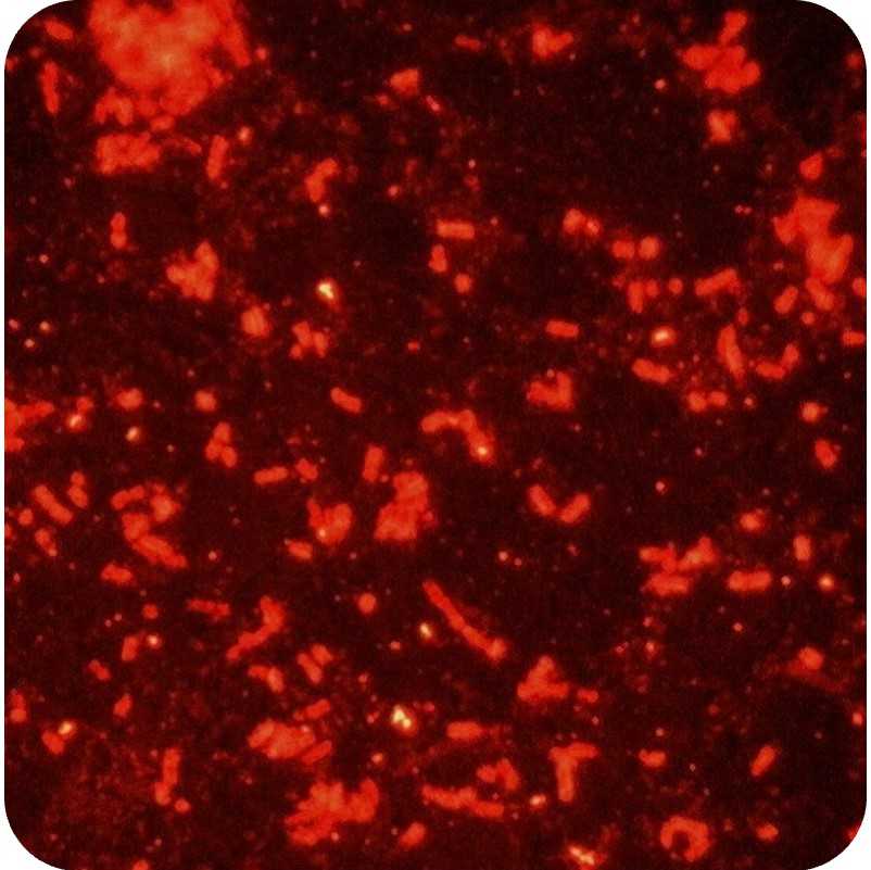
Immunofluorescence Staining of Bacterial Samples V.1
- Pedro Fonseca1,2,3,4
- 1INESC-MN;
- 2Instituto Superior Técnico, Universidade de Lisboa;
- 3Unidade Militar Laboratorial de Defesa Biológica e Química, Exército Português;
- 4Instituto Nacional de Saúde Doutor Ricardo Jorge, I.P.
- Advanced Integrated Microsystems Doctoral Program


Protocol Citation: Pedro Fonseca 2025. Immunofluorescence Staining of Bacterial Samples. protocols.io https://dx.doi.org/10.17504/protocols.io.4r3l2eknjl1y/v1
License: This is an open access protocol distributed under the terms of the Creative Commons Attribution License, which permits unrestricted use, distribution, and reproduction in any medium, provided the original author and source are credited
Protocol status: Working
We use this protocol and it's working
Created: April 27, 2025
Last Modified: April 28, 2025
Protocol Integer ID: 177667
Keywords: Immunofluorescence, Bacterial Staining, Fluorescence Microscopy, Antibody Labeling, Monolayer Preparation, Slide Coating, immunofluorescence staining of bacterial sample, immunostaining of bacterial sample, immunofluorescence staining, nanosight lm10 device for nanoparticle, nanoparticle tracking analysis, bacterial sample, fluorescence microscopy, analyzing nanoparticle, immunofluorescence, nanosight lm10 device, antibody incubation for improved reproducibility, nanoparticle, antibody, antibody incubation, secondary antibody incubation, concentration measurement
Disclaimer
DISCLAIMER – FOR INFORMATIONAL PURPOSES ONLY; USE AT YOUR OWN RISK
The protocol content here is for informational purposes only and does not constitute legal, medical, clinical, or safety advice, or otherwise; content added to protocols.io is not peer reviewed and may not have undergone a formal approval of any kind. Information presented in this protocol should not substitute for independent professional judgment, advice, diagnosis, or treatment. Any action you take or refrain from taking using or relying upon the information presented here is strictly at your own risk. You agree that neither the Company nor any of the authors, contributors, administrators, or anyone else associated with protocols.io, can be held responsible for your use of the information contained in or linked to this protocol or any of our Sites/Apps and Services.
Abstract
This protocol describes a step-by-step method for preparing, loading, and analyzing nanoparticle-containing samples using the NanoSight LM10 device for nanoparticle tracking analysis (NTA). Emphasis is placed on safe device assembly, optimized sample preparation, and correct software settings to ensure reliable size distribution and concentration measurements. Proper handling techniques to prevent instrument damage and achieve high-quality imaging are also detailed.This protocol describes the preparation, fixation and immunostaining of bacterial samples for fluorescence microscopy. It includes detailed steps for monolayer preparation, blocking, primary and secondary antibody incubations, and mounting, with options to optimize slide preparation and antibody incubation for improved reproducibility and signal quality.
Guidelines
- Use aqueous solvents compatible with the standard LM10 configuration. For non-aqueous media, contact the manufacturer.
- Avoid bubbles by de-gassing samples when necessary to maintain imaging quality.
- Remove large aggregates (>10 µm) via centrifugation or filtration to prevent clogs and light scatter interference.
- Dilute samples to a particle concentration of 108–109 particles/mL for optimal visualization.
- Observe around 10–100 particles in the field of view for accurate measurement.Always keep fluorescent antibodies and stained slides protected from light.
- Keep antibodies on ice during preparation.
- Use clean, particle-free solutions and sterile pipette tips.
- For reproducibility, prefer pre-coated Poly-L-lysine slides when possible.
Pre-test antibody concentrations if using new lots or targets.
Materials
- 70% Ethanol
- Poly-L-lysine solution (1 mg/mL) or Pre-coated Poly-L-lysine slides
- Phosphate Buffered Saline (PBS), pH 7.4
- PBS with 0.5% Triton X-100 (PBS-Tx)
- Fish Skin Gelatin 0.2% PBS solution (blocking agent)
- Ultrapure water (preferably Milli-Q)
- Primary antibodies (1:100 working solution; on ice)
- Fluorescent secondary antibodies (1:100 working solution; on ice, protected from light)
- 1.5 mL Eppendorf tubes
- Microscope slides
- P200 micropipette and sterile tips
- Humid chamber
- Slide holders for washing
- DAKO pen
- Filter paper
- Coverslips
- Mounting media (e.g., VectaShield® or Fluoromount™)
- Clear nail varnish (optional)
Safety warnings
- Device Integrity: Ensure the 'O'-ring is properly sealed before sample injection. Leakage can result in irreparable damage to the laser.
- Pressure Control: Inject samples slowly to avoid pressure build-up that could bypass seals or damage the optical window.
- Microscope Safety: Exercise caution when adjusting the microscope stage to avoid contact between the objective and the LM10 window.Handle all fluorescent reagents in low light to prevent signal degradation.
- Avoid letting slides dry during staining steps.
- Ensure thorough but gentle washes to prevent detaching bacterial monolayers.
Carefully apply and seal coverslips to prevent sample loss and preserve fluorescence.
Before start
- Turn off and unplug the NanoSight LM10 before cleaning or assembling.
- Prepare all reagents and ensure syringes and beakers are clean and particle-free.
- Degas samples when applicable.
- Verify that all software (NTA 2.1) is installed and functioning on the connected computer.Turn on the humid chamber to equilibrate at desired temperature.
- Prepare all antibody working solutions fresh.
- Clean glassware and ensure the workspace is free of dust and particles.
Have all washing buffers (PBS, PBS-Tx) ready and at room temperature.
Bacterial Preparation
Transfer 1 mL of 24-hour bacterial culture into a 1.5 mL Eppendorf tube.
Centrifuge at 4000 × g for 4 minutes.
Discard supernatant and resuspend pellet in 1 mL PBS.
Repeat centrifugation and resuspension two more times (total three washes).
After final wash, resuspend pellet in 100 µL PBS.
Slide Preparation
Option 1: Manual Poly-L-lysine Coating
Clean slides with 70% ethanol.
Pipette 100 µL Poly-L-lysine solution onto filter paper.
Firmly swipe the filter paper once across each slide.
Option 2: Pre-coated Slides
Use pre-coated Poly-L-lysine slides directly.
Draw hydrophobic barriers using the DAKO pen.
Pipette 100 µL bacterial suspension into compartments.
Incubate in a humid chamber at room temperature for 10–15 minutes.
Gently blot excess with a paper towel.
Check under microscope for monolayer formation.
Blocking
Pipette 100 µL 0.2% Fish Skin Gelatin into each compartment.
Incubate in a humid chamber for 30 minutes at room temperature.
Wash slides:
- Brief rinse with PBS-Tx
- 5-minute wash with PBS
Primary Antibody Incubation
Pipette 30 µL primary antibody working solution into experimental compartments.
Pipette 30 µL Fish Skin Gelatin into control compartments.
Incubation options:
- 1 hour at 37°C in humid chamber, or
- Overnight at 4°C in humid chamber (recommended for improved sensitivity)
Wash slides:
- Brief rinse with PBS-Tx
- 10-minute PBS-Tx wash at room temperature
- 5-minute PBS wash
Mounting
Apply mounting medium (e.g., VectaShield® or Fluoromount™).
Gently place coverslip, avoiding air bubbles.
Optionally seal coverslip edges with clear nail varnish.
Store slides in the dark until imaging.
Visualize using a fluorescence microscope.
Protocol references
1. Skerker JM, Berg HC. Direct observation of extension and retraction of type IV pili. Proc Natl Acad Sci U S A. 2001;98(12):6901–4.
2. Kuru E, Hughes HV, Brown PJ, Hall E, Tekkam S, Cava F, et al. In situ probing of newly synthesized peptidoglycan in live bacteria with fluorescent D-amino acids. Angew Chemie - Int Ed. 2012;51(50):12519–23.
3. Im K, Mareninov S, Diaz MFP, Yong WH. An Introduction to Performing Immunofluorescence Staining. In: Yong WH, editor. Physiology & behavior [Internet]. New York, NY: Springer New York; 2019. p. 299–311. (Methods in Molecular Biology; vol. 1897). Available from: http://link.springer.com/10.1007/978-1-4939-8935-5