Apr 30, 2025
Immunofluorescence Labeling of TH, Alpha-Synuclein, and MAP2 in iPSC-Derived Dopaminergic Neurons
- 1Montreal Neurological Institute - McGill University


Protocol Citation: Roxanne Larivière, Edward A. Fon 2025. Immunofluorescence Labeling of TH, Alpha-Synuclein, and MAP2 in iPSC-Derived Dopaminergic Neurons . protocols.io https://dx.doi.org/10.17504/protocols.io.36wgqdo9yvk5/v1
License: This is an open access protocol distributed under the terms of the Creative Commons Attribution License, which permits unrestricted use, distribution, and reproduction in any medium, provided the original author and source are credited
Protocol status: Working
We use this protocol and it is working
Created: December 19, 2024
Last Modified: April 30, 2025
Protocol Integer ID: 116390
Keywords: IF, immunofluorescence, TH, alpha-synuclein, MAP2 , iPSC, dopaminergic neurons, synuclein, derived dopaminergic neuron, dopaminergic neuron, induced pluripotent stem cell, map2 in ipsc, labeling tyrosine hydroxylase, immunofluorescence labeling, pluripotent stem cell, immunofluorescence labeling of th, tyrosine hydroxylase, using immunofluorescence
Funders Acknowledgements:
Michael J. Fox Foundation for Parkinson's research
Grant ID: MJFF-020696
Abstract
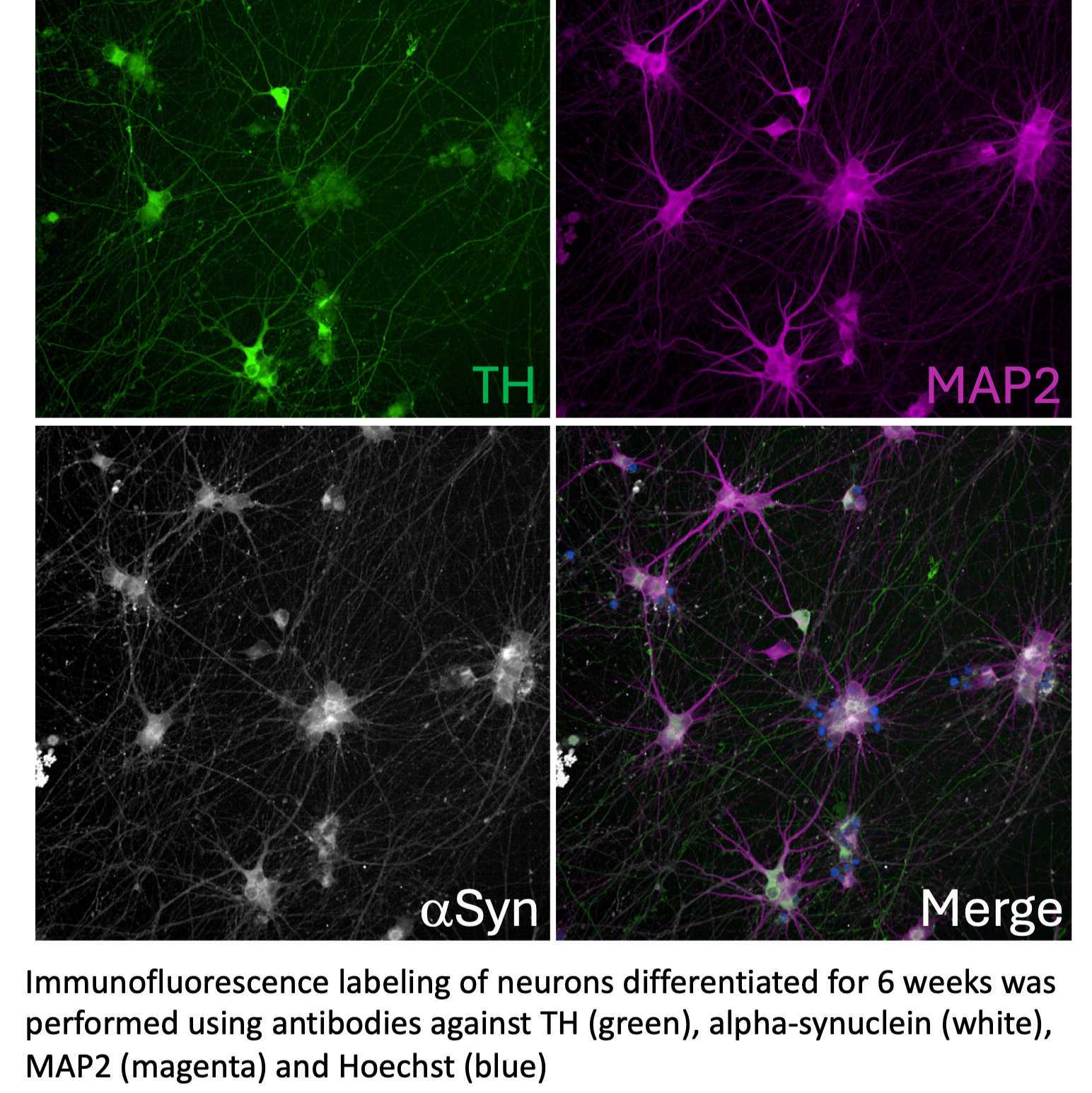
This protocol outlines the procedure for labeling tyrosine hydroxylase (TH), alpha-synuclein (α-syn), and microtubule-associated protein 2 (MAP2) in induced pluripotent stem cell (iPSC)-derived dopaminergic neurons using immunofluorescence.
Materials
Before start
iPSC-derived dopaminergic neurons are cultured on 96-well plates for up to 4 to 6 weeks of differentiation. We use black Costar 96-well assay plates (Corning cat. no. 3904), which are compatible with imaging using the Opera Phenix high-content screening system.
Be extremely gentle when dispensing into the wells, as dopaminergic neurons are prone to detaching with forceful pipetting.
Fixation of dopaminergic neurons in 96 well plates
40m
- Leave 50 µL of media and gently add 50 µL of 8% PFA per well.
- Incubate at Room temperature 00:20:00 .
- Remove PFA and wash 3 times with 1X PBS, 75 µL per well, 00:05:00 each
40m
Permeabilization
25m
- Prepare permeabilization solution: 0.2% TX-100; 1X PBS.
- Incubate in permeabilization solution 00:10:00 , 60 µL per well
- Remove permeabilization solution and wash 3 times with 1X PBS, 75 µL per well, 00:05:00 each
25m
Blocking
1h
- Prepare blocking buffer: 5% normal goat serum; 0.02% TX-100; 1X PBS
- Incubate in blocking solution 01:00:00 , 60 µL per well
1h
Incubation with primary antibodies
16h
- Prepare primary antibody mix in blocking buffer, 60 µL per well
- rabbit TH antibody 1:500
- mouse alpha-synuclein antibody 1:500
- chicken MAP2 antibody 1:1500
- Incubate Overnight at 4 °C with gentle shaking
16h
Washes
20m
- Wash 4 times with 75 µL 1X PBS, 00:05:00 each
20m
Incubation with secondary antibodies
2h
- Prepare secondary antibody mix in 1% normal goat serum; 1X PBS. Dispense 60 µL per well.
- anti-Rabbit Alexa Fluor 488 1:1000
- anti-mouse Alexa Fluor 647 1:1000
- anti-Chicken Alexa Fluor 555 1:1000
- Hoechst 33342 1:5000
- Incubate 02:00:00 at Room temperature with gentle shaking
2h
Washes
20m
- Wash 4 times with 75 µL 1X PBS, 00:05:00 each
- Leave 100 µL of 1X PBS into wells
20m
Imaging
- Samples are ready for imaging