Feb 01, 2023
Immunofluorescence-based assay to assess LRRK2 recruitment by Rab29
- 1Medical Research Council Protein Phosphorylation and Ubiquitylation Unit, School of Life Sciences, University of Dundee, Dow Street, Dundee DD1 5EH, UK


Protocol Citation: Francesca Tonelli 2023. Immunofluorescence-based assay to assess LRRK2 recruitment by Rab29. protocols.io https://dx.doi.org/10.17504/protocols.io.bp2l69r8klqe/v1
License: This is an open access protocol distributed under the terms of the Creative Commons Attribution License, which permits unrestricted use, distribution, and reproduction in any medium, provided the original author and source are credited
Protocol status: Working
We use this protocol and it's working
Created: February 01, 2023
Last Modified: May 31, 2024
Protocol Integer ID: 76248
Keywords: ASAPCRN, LRRK2, Rab29, Golgi, immunofluorescence, bulk of cellular lrrk2, lrrk2 mutation, cellular lrrk2, induced lrrk2 recruitment, lrrk2 recruitment by rab29, lrrk2 recruitment to the golgi, hek293 cell, rab29 mutation, immunofluorescence microscopy, lrrk2 recruitment, samples for immunofluorescence microscopy, confocal immunofluorescence microscopy method, confocal immunofluorescence, fluorescence microscopy, using confocal fluorescence microscopy, confocal fluorescence microscopy, immunoblotting analysis, lrrk2, mutation, expression of rab29, cell, hela cell
Funders Acknowledgements:
ASAP
Grant ID: ASAP-000463
Abstract
Previous studies using confocal fluorescence microscopy showed that transient over-expression of Rab29 recruits the bulk of cellular LRRK2 to the Golgi, which results in its activation (PMID: 29212815). Here we describe our confocal immunofluorescence microscopy method for measuring the co-localization of LRRK2 and Rab29 in a cell-based assay. This method can be used to screen the impact that LRRK2 mutations or Rab29 mutations have on LRRK2 recruitment to the Golgi, as well as the effect of any compound on Rab29-induced LRRK2 recruitment to the Golgi.
Note: This protocol can be used for HEK293 cells and HeLa cells. For other cell types, experimental conditions may need optimising.
In parallel with the preparation of samples for immunofluorescence microscopy, we recommend preparing samples for quantitative immunoblotting analysis (as described in dx.doi.org/10.17504/protocols.io.bsgrnbv6) to confirm successful transfection.
Guidelines
This protocol can be used for HEK293 cells and HeLa cells. For other cell types, experimental conditions may need optimising.
In parallel with the preparation of samples for immunofluorescence microscopy, we recommend preparing samples for quantitative immunoblotting analysis (as described in dx.doi.org/10.17504/protocols.io.bsgrnbv6) to confirm successful transfection.
Materials
Materials:
- HEK293 cells (ATCC #CRL-1573) cultured in complete growth medium: DMEM (Thermofisher Scientific #11960-044) supplemented with 10% Fetal Calf Serum, qualified, Brazil (Thermofisher Scientific #10270-106), penicillin/streptomycin (Thermofisher Scientific #15140-122) and L-glutamine (Thermofisher Scientific #25030-024)
- N-terminus GFP-tagged LRRK2 (wild-type or mutant) cDNA in a pCMV5 vector; HA-empty vector and HA-tagged Rab29 (wild-type or mutant) cDNA in a pCMV5 vector. All plasmids used for our studies are available from the MRC PPU Reagents and Services (https://mrcppureagents.dundee.ac.uk).
- Polyethylenimine (PEI) “Max” (Linear, Mw 40,000) (Polysciences, Inc., #24765); 1 mg/ml (w/v) solution in milliQ water, pH 7.4; sterile filtered.
- Opti-MEM Reduced Serum Medium (ThermoFisher Scientific #31985062)
- Glass coverslips (22mm x 22mm squared glass coverslips)
- Tissue culture-treated flat bottom cell culture 6-well plates (Thermo Scientific Nunc #142475)
- Optional: Poly-L-Lysine solution, mol wt 150,000-300,000, sterile-filtered (Sigma, P4832)
- 4% (w/v) paraformaldehyde in PBS, pH 7.4 (Alfa Aesar by Thermo Fisher Scientific, J61899). Note: This must be methanol-free.
- Phosphate buffered saline (PBS), pH 7.4 (ThermoFisher Scientific #10728775)
- NP-40 alternative (Merck #492016)
- Bovine Serum Albumin (BSA) (Sigma-Aldrich #A7906)
- Primary antibodies: Anti-HA tag mouse monoclonal antibody [HA.C5] (Abcam, ab18181) and anti-ACBD3 rabbit polyclonal antibody (Sigma-Aldrich, HPA015594). Optional: Anti-GFP chicken polyclonal antibody (Abcam, ab13970)
- Secondary antibodies: Goat anti-Mouse IgG (H+L) Highly Cross-Adsorbed Alexa Fluor™ 568 (Invitrogen A11031) and Goat anti-Rabbit IgG (H+L) Highly Cross-Adsorbed Alexa Fluor™ Plus 647 (Invitrogen A32733). Optional: Goat anti-Chicken IgY (H+L) Secondary Antibody, Alexa Fluor™ 488 (Invitrogen A-11039)
- DAPI (bisBenzimide H 33342 trihydrochloride) (Sigma Aldrich #B2261)
- Vectashield Antifade mounting medium (Vector Laboratories, H-1000)
- Glass microscope slides
Equipment:
- CO2incubator for growing cells
- Laminar flow hood for cell culture
- Zeiss confocal laser scanning microscope
Preparing coverslips
Sterilise the glass coverslips by immersing in 100% ethanol for at least 30 minutes.
Leave the coverslips to air dry completely in the laminar flow hood.
Using sterilized tweezers, transfer the sterilized coverslips to the 6 well plates to be used to seed cells for transfection as described below (one coverslip in each well).
Optional: Coat the glass coverslips with Poly-L-Lysine:
The day before plating cells, completely submerge the coverslips from step 1.3 with Poly-L-Lysine solution (1-2 mL per well). Incubate for one hour at room temperature. Wash the cover slips 2/3 times with DMEM and incubate overnight in complete growth media. The next day, aspirate off the media and seed the cells directly onto the cover slips (as described below).
Plating cells
In a laminar flow hood for cell culture, remove culture medium from one flask of HEK293 cells.
Briefly rinse the cell layer with 0.25% (w/v) Trypsin- 0.53 mM EDTA solution to remove all traces of serum.
Add 2 mL of Trypsin-EDTA solution to the flask and incubate at 37°C until the cell layer is dispersed (5-10 minutes).
Add 8 mL of complete growth medium and resuspend cells by gently pipetting.
Dilute the cell suspension with complete growth medium to achieve 50-60% confluency on the day of transfection.
Seed cells in 6 well plated by adding 2 mL of cell suspension from step 9 into each well (containing the sterilized coverslip).
Transfer the plates to a humidified incubator maintaining 37°C and 5% (v/v) CO2 until transfection (18-24 hours after plating).
Transient transfection
In a laminar flow hood for cell culture, prepare a transfection mix by adding 1.6 μg of GFP-LRRK2 cDNA, 0.4 μg of HA-Rab29 (or HA-empty) cDNA and 6 μl of 1 mg/ml PEI solution into 500 μl of Opti-MEM for each well. Vortex for 20/30 seconds.
Incubate the transfection mix for 20 min at RT to allow the DNA/PEI complex to form.
Add the transfection mix to each well drop by drop.
Transfer the plates to a humidified incubator maintaining 37°C and 5% (v/v) CO2 for 24 hours.
Sample preparation for immunofluorescence microscopy
Remove culture medium completely from each well using an aspirator.
Fix cells by adding 4% PFA in PBS. Incubate for 10 min at RT.
Remove PFA completely using a pipette.
Wash the coverslips 3 times with 0.2% (w/v) BSA in PBS.
Permeabilise cells by incubating with 0.1% (v/v) NP-40 alternative in PBS for 10 min at RT.
Remove the solution completely using an aspirator.
Wash the coverslips 3 times with 0.2% (w/v) BSA in PBS.
Block with 1% (w/v) BSA in PBS for 1 h at RT.
Prepare the primary antibody solution by diluting anti-HA tag mouse monoclonal antibody and anti-ACBD3 rabbit polyclonal antibody in 0.2% (w/v) BSA in PBS (1:1000 and 1:200 dilution, respectively).
Optional: If necessary, the GFP signal can be boosted by incubation with an anti-GFP antibody (1:1000 dilution in 0.2% (w/v) BSA in PBS).
Incubate the samples with primary antibodies for 1 h at RT in a humidified chamber.
Wash the coverslips 3 times (5-10 min per wash) with 0.2% BSA in PBS.
Prepare the secondary antibody solution by diluting the secondary antibodies in 0.2% BSA in PBS (1:500 dilution). Add DAPI at 1 μg/ml final concentration to the secondary antibody solution.
Note: If using an anti-GFP antibody in step 4.9, add the appropriate secondary antibody during this step.
Incubate the samples with the secondary antibodies and DAPI for 1 h at RT in the dark in a humidified chamber.
Wash the coverslips 4 times (10 min per wash) with 0.2% BSA in PBS.
Mount the coverslips on glass microscope slides using Vectashield mounting medium (15-20 µl per coverslip).
Store in darkness at 4°C until imaging.
Laser confocal imaging and image analysis
Image cells using a Zeiss LSM 880 laser scanning microscopes using the Plan Apochromat ×63 objective (NA 1.4) objective with a zoom of 0.8 and optical section thickness of 0.8µm (image size 1912 × 1912 pixels, pixel size 0.071 µm).
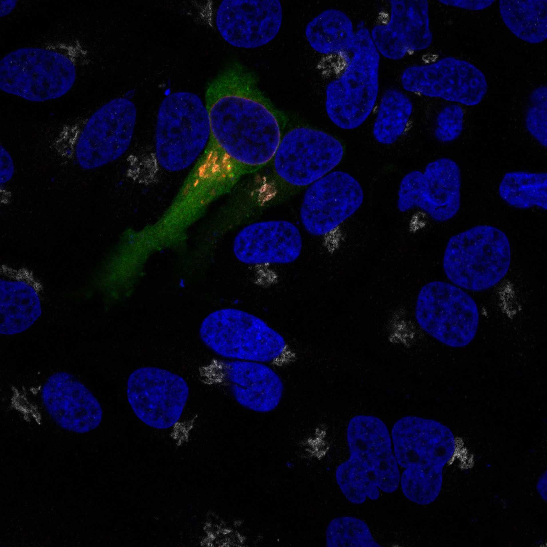
For each sample, collect ten randomly selected fields with GFP-positive cells (i.e. cells that are successfully transfected with GFP-LRRK2). Note: This should be performed blinded to LRRK2 or Rab29 variant.
Cells that are identified in both the LRRK2 (green) channel and Rab29 (red) channel (i.e. cells transfected with both LRRK2 and Rab29) are analysed for colocalization of red and green channels. Cells not expressing both red and green are removed from analysis. The Mander’s coefficient is calculated indicating the proportion of LRRK2 overlapping with Rab29 above a specific threshold (set at 450).
Evaluate statistical significance between different experimental conditions by applying an appropriate statistical method.