May 18, 2026
IHC Validation of TP53 Expression in Paraffin-Embedded Human Glioma Cancer Using TP53 Recombinant Monoclonal Antibody
- Rosie Liu1
- 1CUSABIO
- CUSABIO TECHNOLOGY LLC


Protocol Citation: Rosie Liu 2026. IHC Validation of TP53 Expression in Paraffin-Embedded Human Glioma Cancer Using TP53 Recombinant Monoclonal Antibody. protocols.io https://dx.doi.org/10.17504/protocols.io.x54v9qq4zl3e/v1
License: This is an open access protocol distributed under the terms of the Creative Commons Attribution License, which permits unrestricted use, distribution, and reproduction in any medium, provided the original author and source are credited
Protocol status: Working
We use this protocol and it's working
Created: May 17, 2026
Last Modified: May 18, 2026
Protocol Integer ID: 317246
Keywords: TP53; TP53 Recombinant monoclonal antibody; IHC validation, using tp53 recombinant monoclonal antibody, tp53 recombinant monoclonal antibody this protocol, using cusabio tp53 recombinant monoclonal antibody, cusabio tp53 recombinant monoclonal antibody, embedded human glioma cancer, ihc validation of tp53 expression, visualization of tp53 protein, tp53 protein, human glioma cancer, human glioma cancer tissue, tp53 expression, prognostic biomarker screening, cancer biology research, critical for cancer biology research, cellular context of the tumor tissue, tumor tissue, ihc validation, tumorigenesis mechanism
Abstract
This protocol aims to detect the expression, subcellular localization, and semi-quantitative level of TP53 protein in formalin-fixed paraffin-embedded (FFPE) human glioma cancer tissues using CUSABIO TP53 Recombinant Monoclonal Antibody (CSB-RA825742A0HU) via the IHC technique. This assay allows for the visualization of TP53 protein within the cellular context of the tumor tissue, which is critical for cancer biology research, such as tumorigenesis mechanisms or prognostic biomarker screening.
Guidelines
Principle
IHC is a technique that combines immunology and histochemistry to detect specific antigens in tissue sections. The principle is based on the specific binding between antibodies and antigens. In this experiment:
1. The primary antibody (TP53 Recombinant Monoclonal Antibody) specifically binds to the TP53 protein antigen present in the glioma tissue sections.
2. The HRP-labeled secondary antibody (Goat anti-rabbit IgG polymer) then binds to the primary antibody, forming an antigen-primary antibody-secondary antibody complex.
3. Horseradish peroxidase (HRP) catalyzes the oxidation of the chromogenic substrate DAB in the presence of hydrogen peroxide, producing an insoluble brown precipitate at the antigen site.
4. Hematoxylin counterstaining provides contrast by staining cell nuclei blue, allowing clear visualization of tissue architecture and subcellular localization of TP53 protein expression in the tissue.
Note
1. Tissue Handling: Fresh tissues should be fixed immediately after resection to prevent protein degradation and loss of antigenicity. Over-fixation may mask antigenic epitopes and reduce staining intensity. Under-fixation can cause heavy edge staining with little to no positive signal.
2. Deparaffinization/rehydration: Complete dewaxing is critical for successful staining. Ensure slides are fully immersed in the dewaxing agent, and replace the agent regularly to maintain effectiveness (avoid slides to drying). Pre-baking slides at 60°C for 30-60 minutes greatly enhances paraffin removal.
3. Antigen Retrieval: This is a critical step for IHC staining of FFPE tissues. The high-pressure antigen retrieval method using citric acid buffer (pH 6.0) is optimal for TP53 antigen retrieval. Ensure the buffer does not boil dry during the process. Over-retrieval can destroy antigen epitopes and cause tissue detachment, while under-retrieval leads to weak or no staining. Allow the buffer to cool naturally; rapid cooling may result in poor antigen recovery.
4. Antibody Handling: Aliquot the primary antibody and store at -20°C to avoid repeated freeze-thaw cycles. The diluted primary antibody should be prepared fresh before use.
5. Incubation Conditions: All incubation steps should be performed in a humidified chamber to prevent the sections from drying out, which can cause high background staining.
6. DAB Development: DAB is a light-sensitive reagent and should be prepared fresh and protected from light. Monitor the color development closely under a microscope to avoid over-staining.
7. Slide Storage: Stained slides should be stored in the dark at room temperature. Long-term storage may cause fading of the DAB staining.
Materials
Instruments:
- Leica Bond™ IHC staining system
- Microtome
- Pressure cooker
- Water bath (45°C)
- Incubator (37°C, 60°C)
- Refrigerator (4°C)
- Bright-field microscope with digital camera system
- Humidified incubation chambers
- Pipettes and pipette tips
- Glass slides and coverslips
- Staining jars and racks
Reagents:
- Primary antibody: TP53 Recombinant Monoclonal Antibody (CSB-RA825742A0HU, CUSABIO)
- Secondary antibody: HRP-labeled Goat anti-rabbit IgG polymer
- 10% neutral buffered formalin
- Paraffin wax
- APES (3-Aminopropyltriethoxysilane)
- Acetone
- KCl, Na2HPO4·2H2O, KH2PO4, NaOH, NaCl
- Tween-20
- Trisodium citrate dihydrate
- Citric acid monohydrate
- 3% H2O2 solution
- Normal goat serum
- Bovine serum albumin (BSA)
- DAB (3,3'-Diaminobenzidine) chromogenic substrate
- Hematoxylin
- Graded ethanol series
- Xylene
- Neutral balsam
- Deionized water (ddH2O)
Troubleshooting
Problem
No staining or Extremely weak staining (Possible Causes: 1. Inadequate antigen retrieval 2. Primary antibody concentration too low 3. Incubation time too short 4. Antibody expired or improperly stored 5. Over-fixation of tissue)
Solution
1. Extend antigen retrieval time or try different retrieval buffers 2. Increase primary antibody concentration (e.g., 1:50) 3. Extend primary antibody incubation time 4. Use a fresh aliquot of antibody 5. Ensure proper fixation time (24-48 hours)
Problem
High background staining (Possible Causes: 1. Inadequate blocking 2. Primary antibody concentration too high 3. Sections dried out during incubation 4. Endogenous peroxidase not completely blocked 5. Non-specific binding of secondary antibody)
Solution
1. Extend blocking time or use a different blocking serum 2. Decrease primary antibody concentration 3. Ensure all incubations are performed in a humidified chamber 4. Extend the endogenous peroxidase blocking time 5. Increase the number of PBST washes after secondary antibody incubation
Problem
Non-specific staining (Possible Causes: 1. Primary antibody cross-reactivity 2. Secondary antibody non-specific binding 3. Tissue autofluorescence/autostaining)
Solution
1. Include a negative control (no primary antibody) to confirm specificity 2. Use a pre-adsorbed secondary antibody 3. Ensure proper tissue processing and fixation
Problem
Uneven staining (Possible Causes: 1. Sections not completely covered with reagents 2. Bubbles under the coverslip during mounting 3. Inconsistent antigen retrieval)
Solution
1. Ensure reagents completely cover the tissue sections 2. Mount slides carefully to avoid bubbles 3. Ensure all slides are submerged in antigen retrieval buffer and treated uniformly
Problem
Nuclear staining is weak or absent (Possible Causes: 1. Inadequate antigen retrieval for nuclear antigens 2. Primary antibody does not recognize the nuclear epitope 3. Nuclear proteins degraded during tissue processing)
Solution
1. Optimize antigen retrieval conditions (higher temperature, longer time) 2. Verify the antibody recognizes the nuclear form of TP53 3. Ensure fresh tissues are fixed immediately after resection
Safety warnings
1. Carcinogenic Hazard: DAB is a known carcinogen. Wear gloves, a lab coat, and eye protection when handling DAB solution. All DAB-contaminated materials should be collected and disposed of properly according to institutional safety regulations.
2. Flammable Hazard: Acetone, ethanol, and xylene are highly flammable. Keep them away from open flames and heat sources. Use in a well-ventilated fume hood.
3. Toxic Hazard: Xylene is toxic and may irritate the skin, eyes, and respiratory system. Avoid direct contact and inhalation. Use in a well-ventilated fume hood.
4. Corrosive Hazard: Hydrochloric acid used for differentiation is corrosive. Handle with care and avoid contact with skin and eyes.
5. Biohazard: Human tissue samples may contain infectious agents. Handle all clinical specimens with appropriate biosafety precautions.
6. Toxic Stains: Hematoxylin staining solution contains toxic components (e.g., mercury). Avoid skin contact and ingestion. Dispose of used hematoxylin solution as hazardous waste.
7. Pressure Cooker Safety: Follow the manufacturer's instructions when using the pressure cooker. Do not open the lid until the pressure has completely returned to atmospheric pressure to prevent scalding or explosion.
Ethics statement
This protocol involves the use of animal blood samples. Users must obtain prior approval from their Institutional Animal Care and Use Committee (IACUC) or equivalent ethics committee before performing this protocol. All procedures must comply with applicable institutional and governmental regulations regarding the ethical use of animals.
Before start
Proper pre-experiment sample processing is critical to preserve tissue architecture and antigen integrity, which directly impacts the quality and reliability of subsequent IHC staining results.
Pre-experiment Sample Processing
Tissue Fixation: Fresh human glioma cancer tissues were immediately fixed in 10% neutral buffered formalin for 24-48 hours at room temperature to preserve tissue morphology and antigenicity.
Dehydration and Clearing: The fixed tissues were processed through a graded ethanol series (60%, 75%, 85%, 95%, 100% I, 100% II for dehydration, followed by clearing in xylene (two changes, 15 minutes each).
Paraffin Embedding: The cleared tissues were infiltrated with molten paraffin wax (three changes, 30 minutes each at 60°C) and then embedded in paraffin blocks.
Section Preparation: Paraffin blocks were cut into 4-5 µm thick serial sections using a microtome.
Slide Coating: Glass slides were pre-coated with 2% APES-acetone solution to enhance tissue adhesion. Sections were floated on a 45°C water bath and carefully mounted onto the coated slides.
Drying: Slides were dried at 37°C overnight and then baked at 60°C for 1 hour to ensure complete adhesion of sections to slides.
Reagent Preparation
2% APES-acetone Solution (300ml): Mix 6 mL of APES with 300 mL of acetone thoroughly. Prepare fresh before use.
25× PBS Stock Solution (5000ml): Fully dissolve 25 g KCl, 180 g Na2HPO4·2H2O, 55 g KH2PO4, 7 g NaOH, and 1000 g NaCl in ddH2O. Adjust the final volume to 5000 mL with ddH2O and stir until completely dissolved. Store at room temperature.
1× PBS Working Solution: Dilute 200 mL of 25× PBS stock solution with 4800 mL of ddH2O. Adjust pH to 7.4 if necessary.
PBST Solution (1000ml): Add 5 μL of Tween-20 to 1000 mL of 1× PBS and mix well. Prepare fresh before use.
Citric Acid-Sodium Citrate Antigen Retrieval Buffer (pH 6.0, 500ml)
Trisodium citrate dihydrate stock solution: Dissolve 14.7g trisodium citrate dihydrate in 500ml ddH₂O
Citric acid monohydrate stock solution: Dissolve 4.2g citric acid monohydrate in 200ml ddH₂O
Working solution: Add 9ml citric acid monohydrate solution into 41ml trisodium citrate dihydrate solution, then add ddH₂O to make up to 500ml. Verify pH is 6.0
Blocking Solution: 10% normal goat serum diluted in 1× PBS. Prepare fresh before use.
Primary Antibody Dilution: Dilute TP53 Recombinant Monoclonal Antibody (CSB-RA825742A0HU) at 1:100 in 1% BSA-PBS solution. Prepare fresh before use.
Detection Reagents
- HRP-labeled Goat anti-rabbit IgG polymer
- 0.05% DAB (3,3'-Diaminobenzidine) chromogenic solution (prepare fresh and protect from light)
- Hematoxylin counterstain
- Graded ethanol series (60%, 75%, 85%, 95%, 100%)
- Xylene
- Neutral mounting medium
Assay Procedures
Dewaxing and Hydration
- Immerse slides in xylene forat least 60 minutes.
- Immerse slides in 100% ethanol I for 5 minutes
- Immerse slides in 100% ethanol II for 5 minutes
- Immerse slides in 95% ethanol for 5 minutes
- Immerse slides in 85% ethanol for 5 minutes
- Immerse slides in 75% ethanol for 5 minutes
- Immerse slides in 60% ethanol for 5 minutes
- Rinse slides three times with ddH2O, 5 minutes each
Antigen Retrieval
- Place the hydrated slides in a pressure cooker containing preheated citric acid-sodium citrate buffer (pH 6.0).
- Heat the pressure cooker at 1000 W until the pressure valve rises, then maintain high pressure for 2-3 minutes.
- Turn off the heat and allow the pressure cooker to cool naturally to room temperature (approximately 20-30 minutes).
- Remove slides and rinse three times with 1× PBS, 5 minutes each.
Blocking
Endogenous Peroxidase Blocking:
- Incubate slides with 3% H2O2 solution at room temperature for 10 minutes to quench endogenous peroxidase activity.
- Rinse three times with 1× PBS, 5 minutes each.
Blocking Non-specific Binding:
- Add 10% normal goat serum blocking solution to cover the tissue sections completely.
- Incubate at room temperature for 30 minutes in a humidified chamber.
- Do not rinse; gently tap off excess blocking solution.
Primary Antibody Incubation
- Add diluted TP53 primary antibody (1:100 in 1% BSA-PBS) to cover the tissue sections.
- Incubate overnight at 4°C in a humidified chamber.
- The next day, remove slides from 4°C and allow them to equilibrate to room temperature for 30 minutes.
- Rinse three times with PBST, 5 minutes each.
Secondary Antibody Incubation
- Add HRP-labeled Goat anti-rabbit IgG polymer to cover the tissue sections.
- Incubate at room temperature for 30 minutes in a humidified chamber.
- Rinse three times with PBST, 5 minutes each.
Staining
Chromogenic Reaction:
- Add freshly prepared 0.05% DAB chromogenic solution to cover the tissue sections.
- Monitor the color development under a microscope (usually 1-5 minutes) until the desired staining intensity is achieved.
- Terminate the reaction by immersing slides in CSB-RA825742A0HU.
Counterstaining:
- Counterstain nuclei with hematoxylin for 3-5 minutes.
- Rinse with running tap water for 10 minutes to blue the nuclei.
Detection and Mounting
- Bake the slides at 60°C until completely dry.
- Mount slides with neutral balsam and cover with coverslips.
- Observe the stained slides under a bright-field microscope and capture representative images.
Result Analysis
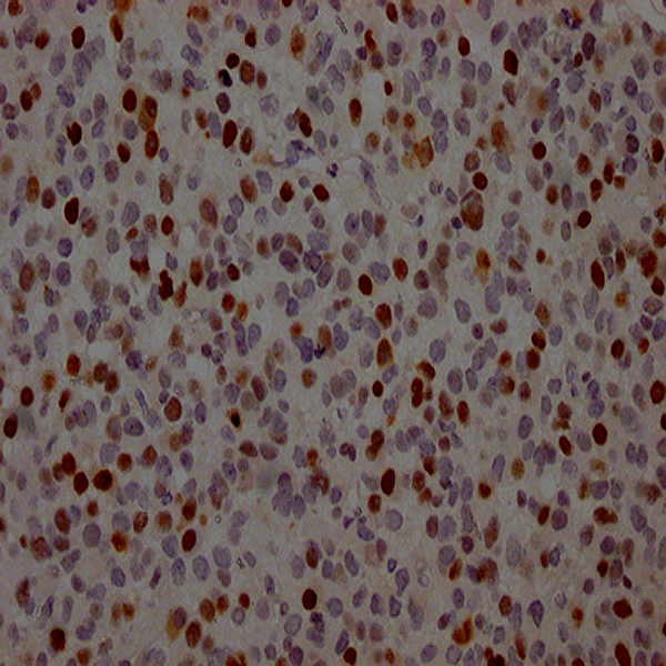
Staining Localization: TP53 is a nuclear transcription factor, so specific positive staining should be localized in the cell nucleus. In the provided IHC image, distinct brown staining is observed in the nuclei of numerous glioma cells, which is consistent with the expected subcellular localization of TP53 protein.
IHC image of CSB-RA825742A0HU diluted at 1:100 and staining in paraffin-embedded human glioma cancer performed on a Leica Bond‱ system.
Staining Intensity: The staining intensity varies among different cells, ranging from weak to strong brown, indicating heterogeneous expression levels of TP53 protein in the glioma tissue sample.
Specificity Assessment: The background staining is very low, and non-specific cytoplasmic or membrane staining is minimal. This demonstrates that the TP53 Recombinant Monoclonal Antibody (CSB-RA825742A0HU) has high specificity for endogenous TP53 protein in human glioma tissues.
Positive Cell Rate: Count the number of TP53-positive cells and total cells in at least 5 random high-power fields (400×) to calculate the positive cell rate. This quantitative data can be used for further statistical analysis of TP53 expression in different glioma subtypes or grades.
Protocol references
○ Product Name: TP53 Recombinant Monoclonal Antibody
○ Catalog Number: CSB-RA825742A0HU
○ Immunogen Species: Homo sapiens (Human)
○ Target Analyte: TP53