Apr 30, 2026
Histone H3.3 Recombinant Monoclonal Antibody (CSB-RA010109A0HU) Immunofluorescence (IF) Protocol
- Rosie Liu1
- 1CUSABIO
- CUSABIO TECHNOLOGY LLC


Protocol Citation: Rosie Liu 2026. Histone H3.3 Recombinant Monoclonal Antibody (CSB-RA010109A0HU) Immunofluorescence (IF) Protocol. protocols.io https://dx.doi.org/10.17504/protocols.io.kxygxjd4kl8j/v1
License: This is an open access protocol distributed under the terms of the Creative Commons Attribution License, which permits unrestricted use, distribution, and reproduction in any medium, provided the original author and source are credited
Protocol status: Working
We use this protocol and it's working
Created: April 30, 2026
Last Modified: April 30, 2026
Protocol Integer ID: 316032
Keywords: Histone H3.3 Recombinant Monoclonal Antibody; Histone H3.3; endogenous Histone H3.3 expression; Immunofluorescence (IF) Protocol, immunofluorescence, recombinant monoclonal primary antibody, primary antibody, hela cell, conjugated secondary antibody, secondary antibody, subcellular localization
Abstract
This standardized IF protocol is specifically developed to detect and visualize the subcellular localization of Histone H3.3 using CUSABIO Histone H3.3 Recombinant Monoclonal Antibody (Cat. No. CSB-RA010109A0HU) in fixed, permeabilized HeLa cells. This technique allows specific detection of the H3.3 protein using a recombinant monoclonal primary antibody, followed by a fluorophore-conjugated secondary antibody, with concurrent nuclear staining with DAPI to delineate cellular architecture.
Guidelines
Principle:
Immunofluorescence uses antigen-antibody specificity to detect target proteins (Histone H3.3) in cells. The primary antibody binds specifically to H3.3, and a fluorophore-conjugated secondary antibody amplifies the signal. DAPI stains DNA (nuclei) to provide cellular context. Fixed/permeabilized cells retain structure and allow antibody access to intracellular antigens.
Key Notes and Precautions:
1. Light Protection Requirement: All operations involving fluorescent reagents (secondary antibody, DAPI) must be performed in the dark. Cover samples with aluminum foil during incubation and washing steps to avoid fluorescence quenching.
2. Cell Density Control: Maintain optimal cell confluence (50-60%) at the time of fixation. Excessively high cell density will lead to cell overlap, insufficient antibody penetration, and non-specific staining; too low cell density will result in insufficient cells for imaging analysis.
3. Fixation and Permeabilization Standardization: Strictly control the fixation time (10-15 min) at room temperature, and permeabilization time (20 min at RT) with 0.2% Triton X-100. Over-fixation may mask antigen epitopes, while insufficient permeabilization will prevent antibody entry into the nucleus, leading to weak or no signal.
4. Blocking: Thorough blocking with 10% normal serum (from the same species as the secondary antibody host, here goat) for 1 hour is essential to reduce non-specific binding of antibodies to the sample.
5. Washing Standardization: Perform all washing steps with gentle shaking to ensure uniform and sufficient washing. Completely aspirate the solution after each wash to avoid dilution of subsequent reagents, which may cause increased background.
6. Antibody Incubation Specification: Ensure the primary and secondary antibody working solutions completely cover the cell monolayer to avoid local drying of the sample, which will cause severe non-specific staining.
7. DAPI Staining Control: Strictly control the DAPI incubation time according to the reagent instructions. Excessively long staining time will lead to high background fluorescence and affect the observation of target protein signals.
8. Negative Control Setting: Always set up a negative control well (incubated with 1× PBS instead of primary antibody) in each experiment to verify the specificity of the staining and rule out non-specific binding of the secondary antibody.
9. Sample Detection Timeliness: After staining is completed, detect and image the sample as soon as possible. Prolonged storage of stained samples, even in the dark, will lead to gradual fluorescence quenching and loss of signal.
10. Humidity Maintenance: During long-term incubation (such as overnight primary antibody incubation), place the 96-well plate in a humidified light-proof box to prevent evaporation of the antibody solution and sample drying.
Materials
Biological Samples
- HeLa human cervical cancer cells in logarithmic growth phase (cell viability �>90%)
Antibodies and Reagents
- Primary antibody: Histone H3.3 Recombinant Monoclonal Antibody (CUSABIO, Cat. No. CSB-RA010109A0HU)
- Secondary antibody: Alexa Fluor 488-conjugated AffiniPure Goat Anti-Rabbit IgG (H+L)
- Normal goat serum
- 4% Methanol-free formaldehyde
- 0.2% Triton X-100
- DAPI nuclear staining reagent
- Sterile 10× PBS and 1× PBS
- Complete cell culture medium
Instruments and Consumables
- Cell culture incubator (37°C, 5% CO₂)
- Biological safety cabinet
- Low-speed benchtop centrifuge
- Inverted microscope (for checking cell density/confluence)
- Fluorescence microscope
- 96-well cell culture plate
- Sterile 15 mL centrifuge tubes
- Sterile pipette tips and pipettes
- Parafilm
- Light-proof incubation box
- Hemocytometer or automated cell counter
Troubleshooting
Problem
No specific fluorescence signal detected (1. Insufficient cell permeabilization, antibody cannot enter the nucleus 2. Primary antibody concentration too low or incubation time insufficient 3. Fluorescence quenching due to light exposure 4. Antigen epitope masked by over-fixation)
Solution
1. Extend permeabilization time to 25-30 min with 0.2% Triton X-100, or optimize concentration to 0.3% for better nuclear penetration 2. Adjust primary antibody dilution to 1:30-1:50, or extend overnight incubation to 16-24 h at 4°C 3. Ensure all fluorescent reagent operations are strictly light-proof, and prepare a fresh secondary antibody working solution 4. Reduce fixation time to 10 min, use methanol-free formaldehyde
Problem
High non-specific background fluorescence (1. Insufficient blocking time or incomplete blocking 2. Secondary antibody concentration too high 3. Insufficient washing steps 4. Primary antibody non-specific binding 5. Sample drying during incubation)
Solution
1. Extend blocking time to 1.5-2 h, or increase normal goat serum concentration to 15% 2. Reduce secondary antibody concentration and optimize dilution ratio 3. Increase washing to 4-5 times, extend each wash to 5 min 4. Optimize primary antibody dilution ratio to reduce non-specific binding 5. Ensure the solution completely covers the sample, and use a humidified incubation box
Problem
Fluorescence signal not co-localized with DAPI nuclear staining (1. Insufficient permeabilization, antibody cannot enter the nucleus 2. Incorrect cell fixation conditions 3. Non-specific binding of the antibody to cytoplasmic proteins 4. Target protein translocation under abnormal cell state)
Solution
1. Optimize permeabilization conditions with 0.2% Triton X-100 to ensure sufficient nuclear membrane penetration 2. Use fresh 4% formaldehyde fixation solution, strictly control the fixation time 3. Increase blocking time, optimize primary antibody dilution ratio, and set up a negative control to verify specificity 4. Use cells in a good logarithmic growth phase with >90% viability
Problem
Severe cell detachment during processing (1. Too vigorous pipetting during washing steps 2. Over-confluent cells before fixation 3. Excessively long permeabilization time 4. Poor cell adherence to the well plate)
Solution
1. Perform all washing steps gently, add solution along the well wall 2. Seed cells at an appropriate density, fix at 50-60% confluence 3. Control permeabilization time within 20 min with 0.2% Triton X-100 4. Use poly-L-lysine coated 96-well plates to improve cell adherence
Problem
Fluorescence signal quenches rapidly during imaging (1. Insufficient light protection during sample preparation 2. No anti-fluorescence quenching mounting medium used 3. Prolonged exposure to excitation light during imaging 4. Expired fluorescent secondary antibody)
Solution
1. Strictly protect samples from light during all steps after secondary antibody incubation 2. Add anti-fluorescence quenching mounting medium after staining 3. Minimize sample exposure time under excitation light, use low-intensity excitation for focusing 4. Use fresh, valid Alexa Fluor 488-conjugated secondary antibody
Problem
Positive fluorescence signal in negative control well (1. Non-specific binding of secondary antibody 2. Insufficient blocking 3. Insufficient washing after secondary antibody incubation 4. Contamination of the secondary antibody)
Solution
1. Increase blocking time, optimize secondary antibody dilution ratio 2. Use normal goat serum (the same species as the secondary antibody host) for blocking 3. Increase washing times and duration after secondary antibody incubation 4. Use a new validated Alexa Fluor 488-conjugated secondary antibody
Safety warnings
1. Chemical Hazard Warning: Formaldehyde is a confirmed carcinogen, toxic, and irritating to the respiratory tract, skin, and eyes. All operations involving formaldehyde must be performed in a chemical fume hood, with nitrile gloves, safety goggles, and a lab coat. Avoid inhalation, skin contact, and ingestion. Dispose of waste according to institutional regulations for hazardous chemicals.
2. Nucleic Acid Dye Safety: DAPI is a known mutagen and potential carcinogen. Wear gloves and goggles when handling, avoid any contact with skin and mucous membranes, and dispose of DAPI-containing waste in accordance with institutional hazardous waste regulations.
3. Biological Safety: All HeLa cell handling must be performed in a biosafety cabinet. Dispose of cell culture waste, contaminated consumables, and liquid waste in accordance with institutional biosafety regulations for human cell lines.
4. Fluorescence Quenching Risk: Fluorescent secondary antibodies and DAPI are extremely sensitive to light. Long-term exposure to ambient light will cause irreversible fluorescence quenching, leading to signal loss or false negative results.
5. Cell Detachment Risk: All washing and reagent replacement steps must be performed gently. Avoid directly pipetting solutions onto the cell monolayer, as this will cause cell detachment from the well plate and loss of experimental samples.
6. Antibody Stability: Aliquot the primary antibody into small volumes upon receipt and store at -20°C. Avoid repeated freeze-thaw cycles, which will significantly reduce the antibody binding activity and lead to staining failure.
Ethics statement
This protocol involves the use of animal blood samples. Users must obtain prior approval from their Institutional Animal Care and Use Committee (IACUC) or equivalent ethics committee before performing this protocol. All procedures must comply with applicable institutional and governmental regulations regarding the ethical use of animals.
Before start
All reagents should be prepared with sterile ultrapure deionized water. Prepare working solutions fresh immediately before use unless otherwise specified. All fluorescent reagent-related operations must be performed in the dark. All cell handling steps must be performed under aseptic conditions in a biosafety cabinet to prevent contamination.
Pre-experiment Sample Processing
Cell Culture: Maintain HeLa cells in appropriate complete cell culture medium at 37°C with 5% CO₂ in a humidified incubator.
Cell Harvest: Select HeLa cells in the logarithmic growth phase with viability greater than 90%. Gently dissociate the cells using a standard method (e.g., trypsinization for adherent cells). Collect HeLa cell suspension into a sterile 15 mL centrifuge tube. Centrifuge at 1000 rpm (approximately 200 × g) for 5 min at room temperature (RT). Carefully aspirate and discard the supernatant.
Cell Washing: Resuspend the cell pellet with 5 mL of pre-warmed complete medium, then centrifuge at 1000 rpm for 5 min. Repeat this wash step twice to remove residual trypsin and cell debris.
Cell Seeding: Resuspend the washed cell pellet in fresh complete medium to an appropriate cell density. Seed the cell suspension into a 96-well cell culture plate, ensuring uniform single-cell dispersion. Incubate the plate overnight at 37°C with 5% CO₂ to allow cells to allow for proper attachment and spreading on the substrate before fixation. Avoid excessive cell confluence; 50-60% confluence at the time of fixation is optimal for IF staining.
Reagent Preparation
10× Phosphate Buffered Saline (PBS, pH 7.4): Dissolve 80 g NaCl, 2 g KCl, 14.4 g Na₂HPO₄, and 2.4 g KH₂PO₄ in 800 mL of water. Adjust pH to 7.4 with HCl or NaOH, then bring the final volume to 1 L. Autoclave for sterilization and store at RT. Dilute to 1× PBS with sterile water before use.
4% Formaldehyde Fixation Solution: Dilute 16% methanol-free formaldehyde stock solution with 1× PBS to a final concentration of 4% (v/v). Always handle in a fume hood. Prepare fresh immediately before use.
0.2% Triton X-100 Permeabilization Solution: Dissolve 0.2 mL of Triton X-100 in 99.8 mL of 1× PBS, mix thoroughly until completely dissolved. Can be prepared as a stock and stored at 4°C.
10% Normal Goat Serum Blocking Buffer: Dilute normal goat serum with 1× PBS to a final concentration of 10% (v/v). Aliquot and store at -20°C. Thaw on ice before use.
Primary Antibody Working Solution: Dilute the Histone H3.3 Recombinant Monoclonal Antibody (CSB-RA010109A0HU) at 1:60 with 1× PBS. Prepare sufficient volume to completely cover the cell sample in each well.
Secondary Fluorescent Antibody Working Solution: Dilute Alexa Fluor 488-conjugated AffiniPure Goat Anti-Rabbit IgG (H+L) to an appropriate concentration (e.g., 1:200–1:500) with 1× PBS. Prepare in the dark and use immediately after preparation.
DAPI Staining Working Solution: Prepare DAPI stock (e.g., 1 mg/mL in water) and dilute to 1–5 μg/mL in PBS (or use a commercial DAPI staining solution).
Assay Procedures
Sample preparation
HeLa cells in logarithmic growth phase (viability greater than 90%) were collected to a sterile 15mL centrifuge tube, centrifuged at 1000 rpm for 5 min, and the supernatant discarded. Cells were washed two times with 5 mL complete medium. Then, they were spread onto a 96-well plate and cultured overnight. Besides, cell density should not be too large, and individual dispersion is appropriate.
Fixation and permeabilization
Wash cells with PBS three times. Fix the cells with 4% formaldehyde for 10-15min at RT. Wash cells with PBS three times. Permeabilize the cells with 0.2% Triton X-100 for 20min at RT. Wash cells with PBS three times.
Blocking
Blocking with 10% normal goat serum for 1h at RT.
Adding antibody
Prepare Histone H3.3 Recombinant Monoclonal Antibody solution at 1:60, and keep the sample completely covered in primary antibody solution. Incubate the primary antibody at 4℃ overnight.
Washing
Wash cells with PBS three times.
Adding detection antibody
Prepare the secondary antibody solution by referring to the recommended dilution ratio in the dark, and keep the sample completely covered in the primary antibody solution. Incubate secondary antibody for 1h at RT. Wash cells with PBS three times.
Washing
Repeat step 5
Nuclear staining
DAPI is often used as a nuclear stain to make the cell structure clearer. Dilute the DAPI reagent according to the instructions and incubate with the sample for a proper time. Dyeing time should not be too long.
Detection
Keep the sample in the dark and detect the fluorescence signal as soon as possible.
Result Analysis
Experimental Outcome
The experiment successfully achieved specific and sensitive detection of endogenous Histone H3.3 in HeLa cells, validating the excellent performance of the CUSABIO Histone H3.3 Recombinant Monoclonal Antibody (Cat. No. CSB-RA010109A0HU) in IF applications.
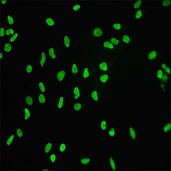
Observed Fluorescence Characteristics
Immunofluorescence staining of HeLa cells with CSB-RA010109A0HU at 1:60,counter-stained with DAPI.
Precise Subcellular Localization: The green fluorescence signal from the target antibody is strictly and exclusively localized in the nucleus of HeLa cells, showing 100% co-localization with the blue DAPI nuclear staining signal. No specific fluorescence signal is detected in the cytoplasmic region, which is completely consistent with the biological function of Histone H3.3 as a core nucleosome protein exclusively expressed in the nucleus.
Signal Intensity and Uniformity: At the 1:60 primary antibody dilution ratio, a strong, uniform, and sharp green fluorescence signal is observed in the nucleus of all healthy adherent HeLa cells, with an extremely high signal-to-noise ratio and no diffuse background signal.
Staining Specificity Confirmation: No non-specific banding or off-target staining is observed in any field of view. The signal is restricted to the nuclear compartment, confirming no cross-reactivity of the antibody with cytoplasmic proteins or other non-target nuclear proteins.
Antibody Performance Validation
Target Specificity: The strict nuclear localization of the fluorescence signal, combined with the absence of non-specific cytoplasmic staining, confirms that Histone H3.3 Recombinant Monoclonal Antibody (Cat. No. CSB-RA010109A0HU) specifically recognizes endogenous Histone H3.3 protein in HeLa cells, with no off-target binding.
Detection Sensitivity: The clear and robust nuclear fluorescence signal detected at a 1:60 primary antibody dilution ratio demonstrates that the antibody has excellent analytical sensitivity for IF applications, enabling reliable detection of endogenous Histone H3.3 expression in routine cell samples.
Assay Reproducibility: Consistent fluorescence signal intensity, localization, and signal-to-noise ratio across multiple replicate wells and different random fields of view indicate that the established IF protocol has high reproducibility and stability.
Biological Interpretation
The uniform nuclear expression of Histone H3.3 in HeLa cells observed in this experiment is fully consistent with its well-documented biological role as a variant histone that is incorporated into nucleosomes independent of DNA replication, regulating chromatin structure and epigenetic gene expression. The precise nuclear localization further validates the specificity of the antibody staining and its suitability for studying the dynamic distribution and function of Histone H3.3 in the nucleus.
Conclusion
The Histone H3.3 Recombinant Monoclonal Antibody (Cat. No. CSB-RA010109A0HU) exhibits exceptional performance in immunofluorescence applications, characterized by high target specificity, excellent detection sensitivity, and accurate nuclear subcellular localization. The antibody reliably detects endogenous Histone H3.3 in HeLa cells under the standardized protocol conditions, fully meeting the requirements of cell biology, epigenetic research, and related in situ protein detection studies.
Protocol references
○ Product Name: Histone H3.3 Recombinant Monoclonal Antibody
○ Catalog Number: CSB-RA010109A0HU
○ Target Species: Homo sapiens (Human)
○ Target Analyte: Histone H3.3 (H3F3A)