May 07, 2024
Histology examination: preparation and staining of bovine tissues
- Jade Ros1,
- Marthe Vilotte1,
- Julie Rivière1,2,
- Cecile Grohs1
- 1Université Paris-Saclay, INRAE, AgroParisTech, GABI, 78350, Jouy-en-Josas, France;
- 2Université Paris-Saclay, INRAE, AgroParisTech, MICALIS, 78350, Jouy-en-Josas, France


Protocol Citation: Jade Ros, Marthe Vilotte, Julie Rivière, Cecile Grohs 2024. Histology examination: preparation and staining of bovine tissues. protocols.io https://dx.doi.org/10.17504/protocols.io.4r3l22wn3l1y/v1
License: This is an open access protocol distributed under the terms of the Creative Commons Attribution License, which permits unrestricted use, distribution, and reproduction in any medium, provided the original author and source are credited
Protocol status: Working
We use this protocol and it's working
Created: February 15, 2024
Last Modified: May 07, 2024
Protocol Integer ID: 95291
Keywords: Histology, HE staining, embedding tissue, scanning histological slides, staining of bovine tissues histology, bovine tissues histology, histology examination, histology, bovine sample, nuclear detail in cell, histochemistry, common staining, staining, cytoplasmic component, cytoplasmic component of the section, cell, tissue, hematoxylin, animal biological resources for integrated, cellular organelle, animal biological resource, eosin, digital genomic, nuclear detail
Abstract
Histology is a technique that makes it possible to highlight what we want to observe and to "fix" a sample at a given time, while preserving characteristics close to its living state (fixation). For example, some elements, cells or cellular organelles can be stained differently depending on the research objective, which may require the detection of certain chemical compounds (histochemistry).
Hematoxylin and Eosin staining is the most common staining to observe the morphology of a tissue. Hematoxylin is used to illustrate nuclear detail in cells. Eosin allows identifying the cytoplasmic component of the section.
All manipulations described in this protocol were performed on bovine samples at the @BRIDGe (Animal Biological Resources for Integrated and Digital Genomics) platform of the UMR GABI, Université Paris Saclay, INRAE, AgroParisTech, GABI, 78350, Jouy-en-Josas, France.
Image Attribution
All photos used in this protocol were taken by Jade Ros during her studies at the National Observatory for Bovine Abnormalities (www.onab.fr).
Materials
KOS microwave tissue processor (Milestone Medical)
Histological cassettes
Cassette printer
Automatic dehydration apparatus
Embedding station
Microtome
Water bath
Staining robot
Slide scanner (Pannoramic SCAN/3DHistech)
CaseViewer software
Safety warnings
Use universal safety precautions when handling samples and personal protective equipment (e.g., face mask with shield, gloves, lab coat or apron).
Xylenes and ethanol are flammable. Avoid open flames and perform procedure in a fume hood.
Gloves should be worn when performing staining process
Ethics statement
The experiments reported in this protocole comply with the ethical guidelines of the
French National Research Institute for Agriculture, Food and Environment
(INRAE) and its French research partners. No animal was intentionally bred for
this study, and invasive sampling was performed post-mortem. Therefore, no ethical approval was required for this study. Finally, the
breeders had consented to the inclusion of their animals in this study, and all
the data analyzed were obtained with the permission of the breeders and
breeding organizations.
Fixing the tissue
Fixation is the most important step in the histological preparation process. Its purpose is to preserve structures and stabilise cellular material against the potentially damaging effects of certain analytical techniques such as staining and immunohistochemistry.
The fixation time depends on the size of the sample. At least 10 volumes of fixative are used in relation to the size of the tissue. In our case the fixation time ranged from 48 hours to several days.
Samples are fixed in 10 % volume formalin for 24:00:00 to 168:00:00 at 4 °C
1w 1d
Sample preparation
Samples are prepared differently, depending on the type of sample and what you want to highlight.
For teeth, a cross section is required to measure the depth of each component of the tooth.
For a bovine bone fragment (humerus, femur, incisor) measuring 2 cm x 2 cm, the following conditions were used
Soften the tissue by decalcifying it
Place samples in a solution of 10 % volume EDTA in 1x PBS (pH 8) for 04:00:00
and 00:30:00 at 50 °C in a KOS microwave tissue processor (Milestone Medical)
4h 30m
Histology cassette mounting
Samples should be placed in specially designed cassettes to hold and flatten them. For very small samples (less than 2 mm) plastic foams are available to hold the sample in place
Cassettes should be marked with a pencil (water and alcohol resistant) or a cassette printer
Histology cassette
Record as many details as possible: date, project name, individual number, tissue nature, type of section
Tissue dehydration, clarification and impregnation
Dehydration replaces the water (80%) in the tissue with ethanol.
To do this, the cassettes are placed in an automatic dehydration machine which dehydrates the tissue using different alcohol solutions of increasing concentration (70% to 100%)
The machine then clarifies the samples with 2 xylene baths, removing the ethanol as paraffin is immiscible in alcohol
Finally, 3 paraffin baths at 60 °C (liquid) are used to impregnate the tissue. Finally, the samples are immersed in several paraffin baths.
Each step takes between 2 and 4 hours, depending on the size of the samples. For our samples, we left the fragments for 4 hours in each bath.
| A | B | C | |
| Steps | Bath | Time (h) | |
| 1 | 70°C ethanol | 4 | |
| 2 | 80°C ethanol | 4 | |
| 3 | 95°C ethanol | 4 | |
| 4 | 100°C ethanol | 4 | |
| 5 | Xylene | 4 | |
| 6 | Xylene | 4 | |
| 7 | Paraffin 1 | 4 | |
| 8 | Paraffin 2 | 4 | |
| 9 | Paraffin 3 | 4 | |
| Total | 36 |
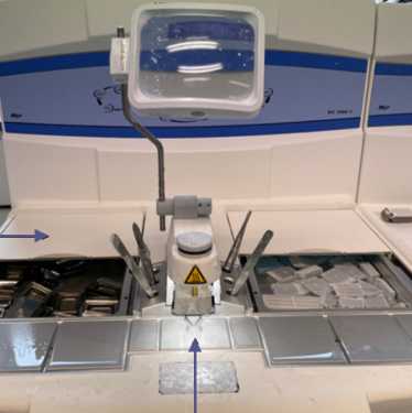
Tissue dehydration, clarification and impregnation baths
Automatic dehydrator
Tissue embedding
Embedding allows the specimen to be oriented and fixed with a paraffin embedding medium so that it can be sectioned later
Place the specimen in the bottom of a metal mould
Place the appropriate cassette on the mould to allow identification of the tissue embedded in the paraffin block
Once the mould is filled, position the tissue
Allow the block to solidify
Wait approximately 15 minutes in the refrigerated zone until the block has stiffened and can be removed from the mould
This step can be performed using an embedding station
Paraffin embedding station
Cut the blocks into trapezoidal shapes to make it easier to cut strips to prepare the slides
Histological sections
Surface of the paraffin blocks to reveal the tissue. Use a microtome set for a 15 micron cut
Slightly rehydrate the surfaced samples by placing them on damp paper Overnight at 4 °C . This will reduce the brittleness of the tissues and make them easier to cut
The samples are sectioned using an automatically rotating microtome (Histocore autocut). This is an important stage in the preparation of slides as it determines how well the tissue can be observed under the microscope
Make ribbons of cuts:
- Set the cut size to 5 microns
- Position the block, with the smaller side of the trapezoid facing up, as close as possible to the blade using the slider
- Unlock the blade guard
- Turn the crank clockwise
- Gently pull the ribbon with a brush to help it unwind and to avoid creasing the tissue
Example of ski cuts ribbon
The paraffin ribbons are placed in a water bath for a few seconds to allow the heat to unfold them
The ribbon is then ready to be placed on a glass slide
40°C water bath containing a ribbon
Preparation of histological slides
The use of adhesive slides is recommended
The slide is then dried on a slide holder in a laboratory at 40 °C for at least Overnight
Slides, that have not been stained at this stage, are called "white" slides
White slide
Before staining, the paraffin must be removed to allow the dyes to penetrate the tissue. This step is usually included in the automated steps of a staining robot
Slides staining
Haematoxylin, eosin (HE) is a morphological usual stain. Haematoxylin is a basic dye that stains acidic structures a purplish blue, such as DNA in nuclei. Eosin is anionic and acts as an acid stain. It will therefore stain basic structures such as cytoplasm pink
Scanning and digitisation of histological slides
The slides are then automatically scanned and digitised using the Pannoramic SCAN/3DHistech tool
Pannoramic SCAN/3DHistech scanner
They can then be viewed from any computer using the dedicated CaseViewer software (freeware). The quality of the scanner allows colour sections to be magnified up to 63 times
Protocol references
@BRIDGe (Animal Biological Resources for Integrated and Digital Genomics) platform of the UMR GABI, Université Paris Saclay, INRAE, AgroParisTech, GABI, 78350, Jouy-en-Josas, France.