Nov 19, 2020
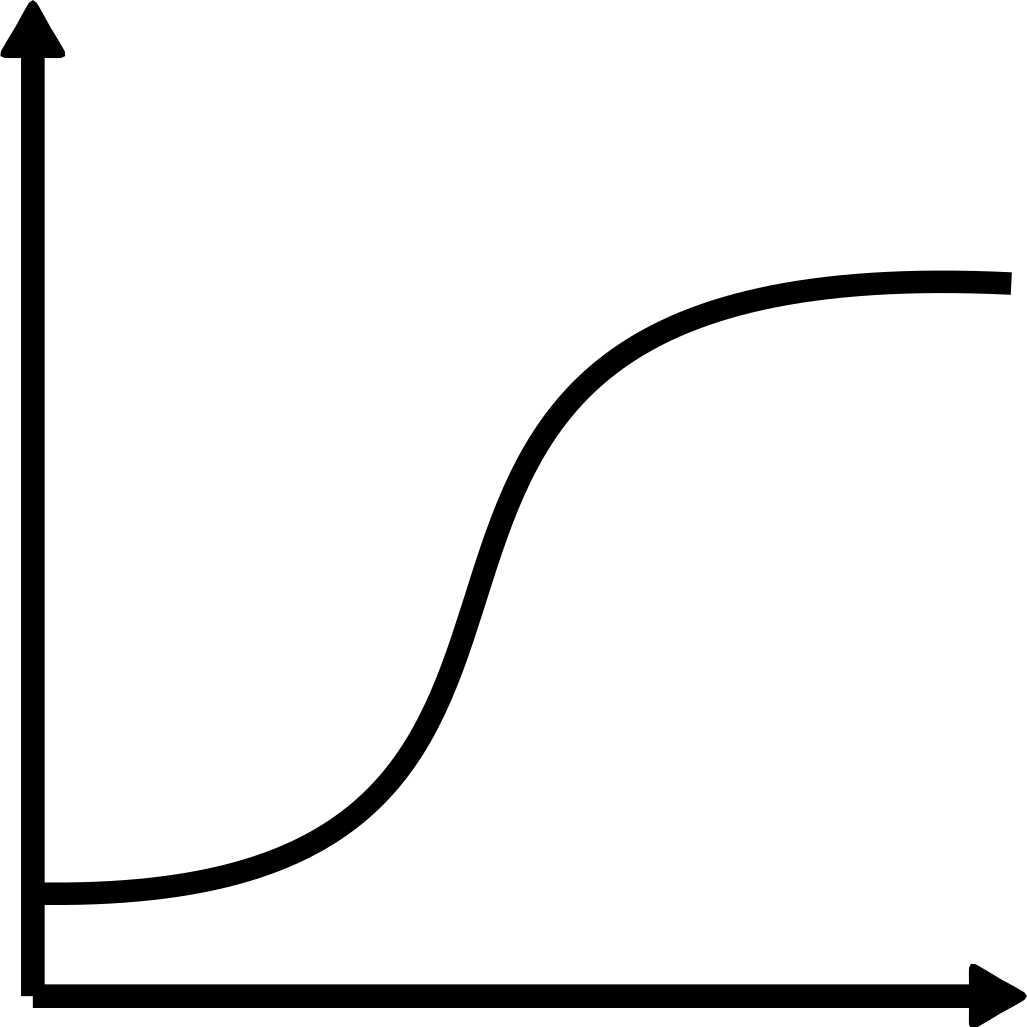
Growth curve for Chlamydomonas reinhardtii
- Joao Vitor Molino1
- 1Ronin Institute


Protocol Citation: Joao Vitor Molino 2020. Growth curve for Chlamydomonas reinhardtii. protocols.io https://dx.doi.org/10.17504/protocols.io.bpvbmn2n
License: This is an open access protocol distributed under the terms of the Creative Commons Attribution License, which permits unrestricted use, distribution, and reproduction in any medium, provided the original author and source are credited
Protocol status: Working
We use this protocol and it's working
Created: November 19, 2020
Last Modified: November 19, 2020
Protocol Integer ID: 44675
Keywords: Growth curve, Chlamydomonas, Absorbance, 96 well plate, growth curve of chlamydomonas reinhardtii, chlamydomonas reinhardtii, chlamydomona, fluorescent protein expression of mvenus, fluorescent protein expression, growth curve, fluorescent,
Disclaimer
DISCLAIMER – FOR INFORMATIONAL PURPOSES ONLY; USE AT YOUR OWN RISK
The protocol content here is for informational purposes only and does not constitute legal, medical, clinical, or safety advice, or otherwise; content added to protocols.io is not peer reviewed and may not have undergone a formal approval of any kind. Information presented in this protocol should not substitute for independent professional judgment, advice, diagnosis, or treatment. Any action you take or refrain from taking using or relying upon the information presented here is strictly at your own risk. You agree that neither the Company nor any of the authors, contributors, administrators, or anyone else associated with protocols.io, can be held responsible for your use of the information contained in or linked to this protocol or any of our Sites/Apps and Services.
Abstract
This protocols describe the steps required for obtain a growth curve of Chlamydomonas reinhardtii and fluorescent protein expression of mVenus and mCherry.
Protocol materials
IsoPlateTM - 96F (Black Frame & Clear well 96-well)Perkin ElmerCatalog #6005020
Material
- Erlenmeyer flask
- Orbital shaker
- Light source
- 96 well plate, Black Frame, Clear bottom (Ex: IsoPlateTM - 96F (Black Frame & Clear well 96-well)Perkin ElmerCatalog #6005020 )
Plate reader Settings
Reading are performed in a Black 96 well plate with clear bottom.
Absorbance set to 750 nm .
Fluorescence set as the Table below.
| Excitation (nm) | Emission (nm) | Gain | Optics position | ||
| Chlorophyll | 440 | 680 | 70 | bottom | |
| mVenus | 500 | 530 | 120 | bottom | |
| mCherry | 583 | 613 | 150 | bottom |
Inocullum
2w 5d
Innoculate 1 mL of the cells from a culture in stationary phase (120:00:00 to 168:00:00 )in an 100 mL erlenmeyer flask containing 50 mL of TAP media.
- Place the flask in a150 rpm, 25°C ,1cm of orbit with 80 μmol/m2s of incident white light ( 60 μmol/m2s to 120 μmol/m2s works)
- Take a initial sample 100 µL of culture and measure it in the plate reader according to the settings above.
- During 168:00:00 take samples at least once a day.
- The final culture can be used for further test, as dry cell weight (DCW) determination.
Note
Frequent sampling increase data quality, but it is advice to not remove more than 10% of culture in sampling during the entire procedure. Technical replicates are advice for each time point.
All culturing conditions are set initially, and can be change accordingly to the experiment goal.
Example of DCW protocol.
2w 5d
- Analytical balance with high precision (The higher the precision the better. For example a balance with a 0.1mg readability, could account to approximately 10% error alone in a measurement of 1mL sample of a culture at 1g/L)
- Microcentrifugal tubes
- Microcentrifuge
- Label microcentrifugal tubes
- Dry the tubes at 90 °C , Overnight
- Cool tubes at Room temperature for 00:30:00
- Record the weight of the tubes
- Harvest 2 mL of culture in a previously weighted tube
- Centrifuge the sample at 20000 rcf, 25°C, 00:01:00
- Carefully remove the supernatant by pipetting
- Wash the cells with ddH20, and centrifuge the sample at 20000 rcf, 25°C, 00:01:00
- Carefully remove the supernatant by pipetting
- Dry the tubes at 90 °C , Overnight
- Cool tubes at Room temperature for 00:30:00
- Record the weight of the tubes
- Subtract the initial tube weight to achieve the dry cell weight