Aug 15, 2025
GeoMx Digital Spatial Profiler Immuno-oncology (IO) Proteome Atlas (IPA) Assay for Human Pancreas
- Alexandra Cuaycal1,
- Dongtao Ann Fu1,
- Heather Kates1,
- Samuel Ewing1,
- Jing Chen1,
- Maigan Brusko1,
- Martha Campbell-Thompson1,
- Clayton E Mathews1
- 1University of Florida
- Alexandra Cuaycal: Postdoctoral Associate
- Dongtao Ann Fu: Molecular Pathology Core, RRID:SCR_016601
- Heather Kates: Data Management Analyst III
- Samuel Ewing: Biological Scientist
- Jing Chen: Associate Research Scientist
- Maigan Brusko: Assistant Professor
- Martha Campbell-Thompson: Professor
- Clayton E Mathews: Professor
- Human BioMolecular Atlas Program (HuBMAP) Method Development CommunityTech. support email: [email protected]



Protocol Citation: Alexandra Cuaycal, Dongtao Ann Fu, Heather Kates, Samuel Ewing, Jing Chen, Maigan Brusko, Martha Campbell-Thompson, Clayton E Mathews 2025. GeoMx Digital Spatial Profiler Immuno-oncology (IO) Proteome Atlas (IPA) Assay for Human Pancreas. protocols.io https://dx.doi.org/10.17504/protocols.io.5jyl8qyy8l2w/v1
License: This is an open access protocol distributed under the terms of the Creative Commons Attribution License, which permits unrestricted use, distribution, and reproduction in any medium, provided the original author and source are credited
Protocol status: Working
This protocol was used to spatially profile targeted proteins in human pancreas through the HuBMAP program.
Created: June 11, 2025
Last Modified: August 15, 2025
Protocol Integer ID: 219976
Keywords: pancreas, FFPE, GeoMx, Proteome atlas, IPA, NGS, Nanostring, morphology, insulin, cytokeratin, CD31, spatial proteomics, fluorescense, digital spatial profiler, HuBMAP, RNA sequencing, UF Diabetes Institute, cell morphology marker, tissue spatial information, geomx digital spatial profiler immuno, oncology proteome atlas, nanostring geomx digital spatial profiler, proteome atlas, labeled antibody, rna probe, florida molecular pathology core, defined antibody panel, morphology marker, hybridized rna probe, nanostring geomx, protein quantification via bulk rna, antibody, protein quantification, bioinformatics, antibody panel, antigen, geomx dsp instrument use, oncology, bulk rna, antibodies against antigen, following geomx ngs guideline, marker, paraffin sections from human pancrea, geomx ngs guideline, human pancreas sections within region
Funders Acknowledgements:
NIH
Grant ID: 1R01DK122160
NIH
Grant ID: P01AI042288
NIH
Grant ID: U54DK127823
NIH
Grant ID: 5R01DK123329
Disclaimer
For detailed information about this protocol, please visit Nanostring University: https://university.nanostring.com/geomx-dsp-spatial-proteogenomic-protocol
Abstract
Purpose: This protocol details processing fixed paraffin sections from human pancreas for the Nanostring GeoMX digital spatial profiler (DSP) immuno-oncology proteome atlas (IPA). This technology is based upon a defined antibody panel of 575 markers. Each antibody is conjugated to an RNA probe. The RNA probes are designed with UV cleavable linkers and DSP barcodes. Fluorescent-labeled antibodies against antigens that define specific cell types were used as morphology markers. Morphology markers are used to identify cell types of interest with nuclear counterstaining permitting tissue spatial information. Sample regions of interest (ROI) are defined based on the three morphology markers and each ROI is illuminated with UV light thereby cleaving hybridized RNA probes. Cleaved probes are aspirated via microcapillary and deposited into a unique well of a 96-well collection plate. The collection plate is used for next generation sequencing (NGS). Scope: This document was written following GeoMX NGS guidelines (university.nanostring.com) with minor modifications for the University of Florida Molecular Pathology Core. The entire workflow is described for manual slide staining and GeoMX DSP instrument use. Steps for library construction, sequencing, bioinformatics, and data analysis are in outline form only. Library preparation and sequencing are outsourced to the University of Florida ICBR facility. Expected outcome: Targeted-protein quantification via bulk RNA-sequencing will be obtained from fixed human pancreas sections within regions of interest and subareas defined by cell morphology markers.
Image Attribution
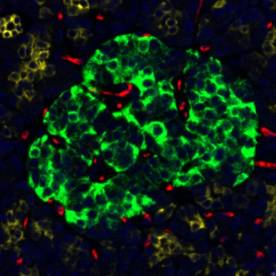
Human pancreas stained with panCytokeratin, Insulin, and CD31 with Syto83 nuclear marker.
Guidelines
Maintain the DSP instrument according to vendor recommendations including weekly wash buffer changes. Maintain an RNAse-free workspace. Validate morphology marker antibodies using assay conditions with antigen retrieval and assay incubation steps but use mock IPA antibody probe solution. Prepare all solutions using DEPC-water and RNAse-free supplies. Clean equipment with RNase AWAY, rinse with DEPC-treated water, and air-dry.
Materials
Chemicals (store at Room temperature except where noted):
Deparaffinization reagents (xylene or substitute, ethanols 100%, 95%)
10xPBS pH 7.4 (PBS, FisherScientific BP399-1 or comparable)
20x SSC (DNase RNase free, Sigma, S6639)
10x Citrate Buffer, pH 6.0 Antigen Retrieval Solution (Sigma, C9999-100ML)
10% neutral buffered formalin (NBF, FisherScientific, 50-980-502, EMS 1574004)
Formamide (deionized or molecular grade, FisherScientific, AM9342) store at 4 °C
DEPC-treated water (DEPC water, FisherScientific, BP561-1)
RNase AWAY (FisherScientific, 7003PK)
Tween 20 (FisherScientific, BP337-100)
TRIS-base (Sigma, 10708976001 or equivalent)
Glycine (Sigma G7126 or equivalent)
Paraformaldehyde, 4% in PBS (ThermoFisher, J61899.AK)
Solution preparations:
1xPBS (PBS, 1 liter) - add 100ml 10xPBS to 900ml DEPC water, store at 4 °C .
1xAntigen Retrieval Solution (100ml) - add 10ml 10xCitrate Buffer, pH 6.0 to 90ml DEPC water for final 1x Citrate Buffer, pH 6.0.
2xSSC (1 liter) - add 100ml 20x SSC and 900ml DEPC water.
2xSSC/T (100ml) - add 10ml 2x SSC, 1ml 10% Tween-20, and 89ml DEPC water.
4xSSC (1 liter) - add 200ml 20x SSC and 800 ml DEPC water.
2x SSC/50% formamide - add 100ml each 4x SSC and 100% formamide.
Morphology markers:
Primary antibodies are conjugated to fluorophores for cell types of interest based on fluorophores compatible with the DSP instrument (AF488, AF532, AF594, AF647 or similar). Use up to 3 conjugated primaries with suitable nuclear marker. Nuclear markers are SYTO 83 (AF532, S11364, FisherScientific, 5mM in DMSO). Antibodies used: Insulin, Mouse anti-all, AF488 (NICBTACLS) FisherScientific 53-9769-82. Cytokeratin (PanCK, Mouse anti-all, AF594 NAE/1/AE/3) Novus Biologicals NBP2-33200AF594. CD31, Mouse anti-hu, AF647 (NJC/70A) Abcam ab215912.
| A | B | C | D | |
| Antibody Name: | Insulin | PanCK | CD31 | |
| Fluorophore: | AF488 | AF594 | AF594 | |
| Clone: | (ICBTACLS) | (AE-1/AE3) | (JC/70A) | |
| Source: | mouse | mouse | mouse | |
| Dilutions: | 1:200 | 1:400 | 1:40 | |
| Company: | Invitrogen | Novus | Abcam | |
| Cat #: | 50-112-4641 | NBP2-33200AF594 | Ab215912 |
Morphological markers used for human pancreas.
Lab Supplies:
Superfrost Plus slides (FisherScientific, 22-035813)
Glass desiccator for slide drying and storage
Kimwipes
RNAse-free beakers and cylinders to prepare buffers
Coplin jars or slide trays
Pipettes (5-1000, 12-channel P20) and filter tips (DNase/RNase free)
Reagent reservoir, 20ml, sterile (FisherScientific, 8094)
Microcentrifuge tubes (DNase/RNase free)
Microcentrifuge rack
Adhesive PCR plate foils (ThermoFisher, AB0626).
Nanostring GeoMX DSP IPA reagents and kits:
GeoMX collection plate (4pk, 100473)
GeoMX instrument buffer kit (100474)
GeoMx Protein Slide Prep Kit (121300312) 4 °C
GeoMX Pro Code Pack Z and Y -20 °C (121400207)
GeoMx Human IO Proteome Atlas Kit (121300160) -80 °C .
Equipment:
Oven for drying slides
Leica XL slide autostainer or alternative
TintoRetriever pressure cooker (BioSB, MM 0023 - 090121 or alternative)
Waterbath (FisherScientific, FSGPD02)
Slide incubation chamber (black or opaque)
Nanostring GeoMX DSP instrument
PCR thermocycler
Centrifuge with rotor for 96-well PCR plates (at least 2000 x g)
Minicentrifuge for quick spins.
Safety warnings
Follow Environmental Health and Safety protocols for all hazardous chemicals (xylene, formalin, formamide).
Slide preparation
3h 50m
Cut paraffin block to fresh sample. Section at ~5 µm and place sections centrally on Superfrost Plus slides as shown in Fig. 1. Tissue samples should have maximum width and length of ~14 and 36 mm, respectively, for inclusion within the GeoMX instrument slide holder gasket. Tissue outside the gasket region should be carefully removed without creating folds or leaks to gasket seal. Air dry mounted slides overnight before use or storage. Recommended storage is for < 2 weeks in a desiccator at 4 °C .
Figure 1. Slide dimensions for GeoMx DSP. Tissue is placed in green scan area.
Bake slides at 60 °C for 00:30:00 to 03:00:00 immediately before use.
3h 30m
Dewax and rehydrate sections using slide tray stations or a Leica XL slide autostainer. Do not allow tissue sections to dry once rehydrated and avoid touching tissue when drying around edges.
Xylene 3 x 00:05:00 . CitriSolv or another xylene substitute can be used.
5m
100% Ethanol 2 x 00:05:00 .
5m
95% Ethanol 2 x 00:05:00 .
5m
ddH2O 2 x 00:05:00 .
5m
Antigen retrieval
1h 45m
Prepare Antigen Retrieval Solution (1x Citrate Buffer, pH 6) in Coplin jar.
Transfer slides to Antigen Retrieval Solution jar and put into BioSB pressure cooker.
Secure the pressure cooker lid and run on high pressure and high temperature for 00:15:00 .
15m
Allow pressure cooker to release pressure before opening the BioSB pressure cooker, and cool down for 00:25:00 without the lid.
25m
Immediately transfer slides to 1x TBS-T.
Wash slides in TBS-T 1x 00:05:00 .
5m
Slides can be stored in TBS-T up to 01:00:00 .
1h
Blocking and antibody incubations
17h
Draw a closed hydrophobic barrier around the tissue with hydrophobic pen (Vector).
Block with 200 µL of Buffer W per slide for 01:00:00 at Room temperature in a covered humidity chamber.
1h
During blocking, thaw the Protein Probe (antibody) mixes, mix by flicking and spin down. Do not vortex.
Prepare a working antibody solution by diluting the Protein Probe (antibody) mixes and Morphology Markers (MM, antibodies for protein markers) into Buffer W according to Table 1.
Keep working solution on ice until ready to use:
| A | B | C | D | E | F | G | H | |
| Pro Core Ab mix (IPA tube 1 of 2) | IPA Module (IPA tube 2 of 2) | MM 1: Ins | MM 2: PanCK | MM 3 CD31 | Buffer W | Total volume | ||
| Channel | - | - | AF488 | AF594 | AF647 | - | ||
| Dilution (v/v) | - | - | 1:200 | 1:400 | 1:40 | - | ||
| Volume per sample | 8.4 uL x n | 52.5 uL x n | 1.05 uL x n | 0.53 uL x n | 5.25 uL x n | 142.3 uL x n | 210 uL x n |
Table 1. Working antibody mix solutions for IPA slide prep (n = number of slides).
Remove Buffer W from slides (one at a time) by gently tapping each slide on a clean paper towel, then using a kimwipe to remove excess without touching the tissue.
Add 200 µL of working antibody solution to each slide. Cover the humidity chamber and incubate overnight at 4 °C .
16h
Post-fixation for pancreas
40m
Wash slides in 1x TBS-T 3 x 00:05:00 .
5m
Incubate in 4% PFA for 00:30:00 at room temperature.
30m
Wash slides in 1x TBS-T 2 x 00:05:00 .
5m
Nuclear staining
20m
Prepare enough of a 1:6000 solution of SYTO83 in 1x TBS-T.
Add 200 µL of the nuclear stain solution to each slide and incubate for 00:15:00 at Room temperature .
15m
Wash slides in 1x TBS-T 2 x 00:05:00 .
5m
Slides can be stored in 1x TBS-T wash solution up to 1 hour at Room temperature or up to 6 hrs at 4 °C
Scrape off hydrophobic pen barrier on a slide with a razor prior to GeoMs DSP run.
GeoMX DSP run
2h
Load slides in DSP within 6 hours of last wash for optimal results.
Slides must be stored in dark to avoid cleavage of DSP tags by UV light.
Start run by selecting New/Continue Run under Data Collection in the GeoMx DSP Control Center. Include a non-template control (NTC).
Clean the bottom of slide with 70% ethanol and place slide in slide holder with slide label towards front of instrument.
Complete loading slides then lower slide tray clamp. Cover sections with 6 ml Buffer S. Record slots for each slide.
Load a 96-well aspiration plate in DSP. Check solution containers are filled and waste container is empty.
Follow prompts in the GeoMx DSP Control Center to identify slides.
Name slides according to protocol https://dx.doi.org/10.17504/protocols.io.n2bvj6dnblk5/v1. Include UF HuBMAP sampleID and morphology marker abbreviations in order of channels followed by date (MMDDYY). Example: P1-21_DNA_PanCK_INS_CD31_121024.
Upload unzipped configuration files to DSP kits used for Morphology, Core, and Module kits used in slide preparations (nanostring/dsconfigfiles). Config files must match names of assays used on slides.
Define scan area for each sample using x- and y-sliders and scan. The minimum tissue slide filters particles and small tissue areas from scan. The sensitivity slider adjusts intensity of tissue identification.
Draw regions of interest (ROI) using geometric, segmented or other tools. Maximum ROI size is ~650 µm diameter circle.
Segment ROIs for AOI (area of illumination) for cell types of interest based on positive or negative selection by morphology markers. Minimum of ~ 50 nuclei/AOI is recommended for sufficient probe reads.
Fluorescently labeled antibodies were used as morphology markers to detect cell types of interest. Anti-Insulin is used to designate beta-cells in the endocrine compartment, anti-CD31 is used to define endothelium, and anti-pan cytokeratin (PanCK) is used to define ductal epithelium in the exocrine compartment. Acinar cells are defined by the lack of staining for CD31 and PanCK in the exocrine ROI.
Exit scan workspace and approve ROI to start ROI collection. 02:00:00 .
2h
After ROI/AOI collections are completed, follow prompts to remove slide holder and collection plate.
Remove buffer from each slide with pipette and dispose. Open clamps and unload slides. Store slides in 2x SSC at 4 °C up to 7 days.
Clean slide holder as directed and replace in instrument. Follow prompts to close instrument door.
Fill in the GeoMX Seq Code plate to be used with the plate barcode in the Readout Group Information grid. Click update button and finalize and download readout package buttons to USB drive.
Slide reuse
Protein assay slides can be restained following UV exposure for 3 minutes to strip tags from bound probes followed by washes using with 2xSSC/T. Proceed with additional staining.
NGS library and sequencing
Transfer collection plate to the University of Florida ICBR Gene Expression and Genotyping Core or other NGS core facility for library preparation and Illumina sequencing. Refer to Nanostring user manual for additional information.
Library will be prepared after indexing PCR with pooling and purification.
Library will be evaluated for quality and fragment size distribution using a BioAnalyzer 2100 (Agilent).
Core facility will perform Illumina sequencing.
Data analysis
Core facility will process Illumina FASTQ sequencing files and transfer back to laboratory.
Share FASTQ files and annotation file with data analyst who will analyze RNAseq data using R scripts for GeoMx DSP workflow (Bioconductor - GeoMxWorkflows). Data analysis may also be performed with GeoMX DSP software on auxiliary server.
Manage annotations (tags). Add columns to further define regions and cell types from morphology markers.
Run various testing including QC, data scaling, normalization, background, statistical tests, and ratios.
Analyze in spatial context using spatial deconvolution software and scRNAseq data.
Complete analysis and upload data to HuBMAP HIVE.