Dec 12, 2024
Genomic DNA Extraction and Genotyping PCR on Mouse Tissues
- Jayshen Arudkumar1,2,
- Yu Chinn Joshua Chey1,2,
- Sandra Piltz1,2,
- Paul Quinton Thomas1,2,
- Fatwa Adikusuma1,2
- 1University of Adelaide;
- 2SAHMRI


External link: https://doi.org/10.1186/s44330-024-00019-y
Protocol Citation: Jayshen Arudkumar, Yu Chinn Joshua Chey, Sandra Piltz, Paul Quinton Thomas, Fatwa Adikusuma 2024. Genomic DNA Extraction and Genotyping PCR on Mouse Tissues. protocols.io https://dx.doi.org/10.17504/protocols.io.ewov1qw6ygr2/v1
Manuscript citation:
Arudkumar, J., Chey, Y.C.J., Piltz, S.G. et al. CRISPR-mediated generation and comprehensive phenotyping of Duchenne Muscular Dystrophy mouse models. BMC Methods 1, 19 (2024). https://doi.org/10.1186/s44330-024-00019-y
License: This is an open access protocol distributed under the terms of the Creative Commons Attribution License, which permits unrestricted use, distribution, and reproduction in any medium, provided the original author and source are credited
Protocol status: Working
We use this protocol and it's working
Created: January 30, 2024
Last Modified: December 12, 2024
Protocol Integer ID: 94369
Keywords: Genomic, CRISPR, Mouse, DNA, Therapy, DMD, murine models of duchenne muscular dystrophy, crispr microinjection, crispr microinjection of embryo, duchenne muscular dystrophy, crispr, broader landscape of genetic disorder investigation, molecular testing at the dna, cas9 gene, pcr on mouse tissues paper, genetic disorder investigation, molecular testing, genotyping pcr, mouse tissues paper, using mouse model, generation of diverse animal model, mouse model, genomic dna, permanent modifications to dna, serum creatine kinase, murine model, diverse animal model, dna, embryo
Disclaimer
These protocols are for research purposes only.
Abstract
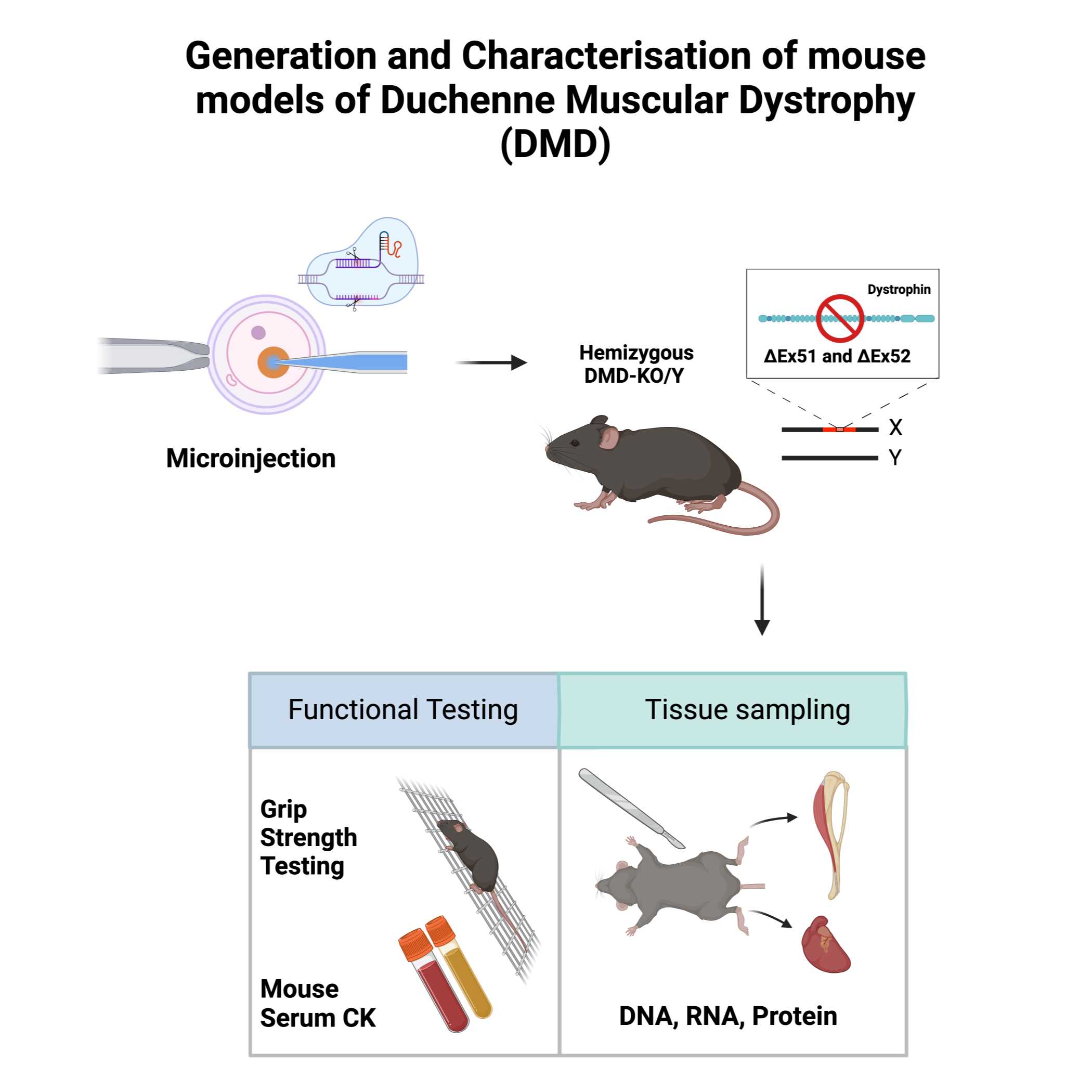
Paper abstract: CRISPR-Cas9 gene-editing technology has revolutionised the creation of precise and permanent modifications to DNA, enabling the generation of diverse animal models for investigating potential treatments. Here, we provide a protocol for the use of CRISPR-Cas9 to create murine models of Duchenne Muscular Dystrophy (DMD) along with a step-by-step guide for their phenotypic and molecular characterisation. The experimental procedures include CRISPR microinjection of embryos, molecular testing at the DNA, RNA, and protein levels, forelimb grip strength testing, immunostaining and serum creatine kinase (CK) testing. We further provide suggestions for analysis and interpretation of the generated data, as well as the limitations of our approach. These protocols are designed for researchers who intend on generating and using mouse models to study DMD as well as those seeking a detailed framework of phenotyping to contribute to the broader landscape of genetic disorder investigations.
Image Attribution
Image was generated using Biorender.
Guidelines
This protocol is designed for use with 25-50 mg of ear or tail tissue. The High Pure PCR Template Preparation kit (Roche) was followed for DNA purification. PCR conditions may vary based on considerations such as primer design and the type of polymerase employed in amplification.
Materials
• PPE (Personal Protective Equipment)
• High Pure PCR Template Preparation Kit (Roche)
Troubleshooting
Safety warnings
Wear proper PPE (gloves, safety goggles, enclosed shoes and lab coat) and prepare solvents in a chemical fume hood. Dispose used solvents or waste material in an appropriate biohazard waste containers.
Ethics statement
Animal work described in this manuscript has been approved and conducted under the oversight of the Animal Ethics Committee of South Australian Health and Medical Research Institute (SAHMRI) and The University of Adelaide.
Lysis and Protein Digestion
Add 200 μL of Roche Tissue Lysis Buffer to a 1.5 mL tube containing the tissue sample.
Note
Ensure the buffer maintains a pH of 7.4, as Tris-HCl buffers the pH
Note: EDTA chelates Mg2+/Ca2+ (essential for DNases), and urea lyses cells while disrupting hydrophobic interactions to destabilise proteins, including nucleases.
Note
For a higher DNA yield, it's advisable to chop up the tissue sample.
Add 40 μL of approximately 413 μg/μL Roche Proteinase K solution.
Vortex the tube to mix the contents.
Incubate the tube at 55 °C for 3 hours.
Note
Longer incubation times, including overnight or multiple days, can increase DNA yield as Proteinase K degrades proteins, solubilising them (including DNases).
Incubate a sufficient amount of Roche Elution Buffer in a 1.5/10 mL tube at 75 °C until ready for use.
Vortex the tube
DNA Column Binding
Add 200 μL of Roche Binding Buffer to the lysed sample.
Note
Ensure a pH of 4.4 is maintained with Tris-HCl
Note
Guanidinium chloride and urea disrupt hydrophobic interactions, destabilizing proteins and disrupting hydrogen bonds between DNA and water, allowing stronger binding to the silica filter. Triton-X 100 solubilizes lipids, allowing them to flow through the filter tube.
Add 100 μL of isopropanol.
Vortex the tube
Centrifuge the tube at 13,000 RCF for 5 minutes.
Decant the solution into a Roche High Pure Filter Tube in a collection tube.
Centrifuge the tube at 8,000 RCF for 1 minute.
Note
Soluble contaminants, including proteins, lipids, and polysaccharides, are washed through the filter, while protein and salt residues remain with the DNA.
Discard the flow-through and place the filter tube in a new collection tube.
Add 500 μL of Roche Inhibitor Removal Buffer to the filter tube.
Note
Guanidium chloride removes residual proteins and pigments. Ethanol removes the salts.
Centrifuge the tube at 8,000 RCF for 1 minute.
Discard the flow-through and place the filter tube in a new collection tube.
Additional Washing
Add 500 μL of Roche Washing Buffer to the filter tube.
Discard the flow-through and place the filter tube in the same collection tube.
Add 500 μL of Roche Washing Buffer to the filter tube.
Discard the flow-through and place the filter tube in the same collection tube.
Note
Ethanol removes remaining salts including leftover guanidium chloride.
Centrifuge the tube at 8,000 RCF for 1 minute.
Discard the flow-through and place the filter tube in the same collection tube.
Centrifuge the tube for 10 seconds at maximum speed to remove any residual ethanol, drying the filter column.
Elution of DNA
Transfer the filter tube to a 1.5 mL tube.
Add 200 μL of pre-warmed 55°C Roche Elution Buffer (pH 8.5 Tris-HCl).
Note
Re-hydrates the soluble DNA to allow it to flow through the filter. DNA is more stable and dissolves faster at this slightly basic pH than in water
Centrifuge the tube at 8,000 RCF for 1 minute to elute the DNA.
Note
The quantification of DNA concentrations (ng/µl) can be done using a Nanodrop spectrophotometer.
Genotyping PCR
The New England Biolabs (NEB) website can be consulted for the finer details of reaction volumes and components when amplifying using a standard NEB Taq DNA Polymerase and the 10X Standard Taq Reaction Buffer. The primers we used are listed in Supplementary Figure S2. Note that the annealing temperature is subject to variability according to the primer pair sequence. We recommend using 50-100 ng of genomic DNA as the template, as well as assembling all reaction components on ice prior to transferring to the preheated thermocycler. After the PCR, confirm the expected band sizes by carefully visualising the amplified products on a 1% agarose gel.
As a guide, the PCR program on the thermal cycler for a 1KB amplicon is as follows:
| A | B | C | |
| 1 cycle | 95°C | 2 minutes | |
| 25 cycles | 95°C | 30 seconds | |
| 60°C | 30 seconds | ||
| 72°C | 60 seconds | ||
| 1 cycle | 72°C | 5 minutes (to finish replication on all templates) | |
| 1 cycle | 4-10°C | indefinite period (storing the sample prior to further analysis) |