Apr 28, 2026
Gametocyte enrichment methods: MACS & Percoll
- Wouter Graumans1
- 1RadboudUMC


Protocol Citation: Wouter Graumans 2026. Gametocyte enrichment methods: MACS & Percoll . protocols.io https://dx.doi.org/10.17504/protocols.io.ewov1rbkklr2/v1
License: This is an open access protocol distributed under the terms of the Creative Commons Attribution License, which permits unrestricted use, distribution, and reproduction in any medium, provided the original author and source are credited
Protocol status: Working
We use this protocol and it's working
Created: April 13, 2026
Last Modified: April 28, 2026
Protocol Integer ID: 314921
Keywords: plasmodium, gametocyte enrichment, membrane feeding, serum replacement, magnetic cell sorting (MACS), Percoll density gradient., plasmodium vivax gametocytes through magnetic cell, plasmodium vivax gametocyte, gametocyte enrichment method, step by step ex vivo enrichment, step ex vivo enrichment, percoll density gradient, percoll, magnetic cell
Abstract
This protocol describes the step by step ex vivo enrichment of plasmodium vivax gametocytes through magnetic cell sorting and Percoll density gradient prior to membrane feeding.
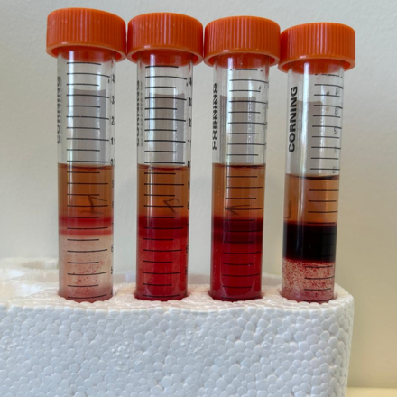
Image Attribution
The image shows the results of a Percoll 70% density enrichment experiment where blood failed to penetrate the gradient after centrifugation. See warnings for further information.
Safety warnings
Percoll enrichment can be performed with multiple densities. In our optimized protocol we used Percoll 65%. During testing we also tried Percoll 70%, but during several experiments blood failed to penetrate the gradient after centrifugation. Because this was observed repeatedly, independent of centrifugation speed, this condition was dropped for further evaluation. For further information see the protocol image under Description.
Ethics statement
Working with blood from human participants requires prior approval by the users relevant ethics committee. This protocol was formulated during a Plasmodium vivax controlled human infection study; the study was approved by the Dutch Central Committee on Research Involving Human Subjects (NL-005553).
Before start
Prewarm the centrifuge and all material at 37°C. Keep tubes at all times warm at 37°C.
Serum replacement with universal donor
- Transfer 1.2mL blood from the Li-hep tube to an Eppendorf tube in the pre-warmed heat-block
- Centrifuge tubes for 20 sec at 10,000 rpm
- Remove two times 200µL of plasma
- Add 400µL of AB serum to the pellet
- Mix with vortex, half speed
- Fill a 2mL syringe with blunt needle
- Feed to mosquitoes
MACS gametocyte enrichment
- Place a bottle of incomplete medium and the MACS stand in the incubator (at 37°C). Attach needles to LS columns.
- Use a syringe to take up the blood, subsequently deplete white blood cells from RBCs using a Plasmodipur filter (max 10mL per filter). Filtrate by applying gentle pressure and collect the filtered blood in a new 15mL tube. Flush the Plasmodipur filter with 2mL of RPMI, followed by air to retain all the liquid, stop when bubbles are seen
- Transfer 1000µL of blood from each volunteer to an Eppendorf tube, spin down using a benchtop centrifuge and remove the plasma with a 1mL syringe with blunt needle. Keep tube warm in the heating block to use later on
- Equilibrate LS columns with 1mL incomplete culture medium at 37°C
- Observe dripping to note when flow through ceases
- Invert blood tube a few times to ensure a homogenous blood solution. Add 3mL of fresh warm whole blood of volunteer from the heparin tube to column
- Wait until it passes through the column, pipette a few times up and down, and add another 3mL
- Wait until it passed through the column, pipette a few times up and down, and add the remaining volume of blood or repeat the sequence
- When flow through ceases, add 1mL culture medium to wash the column
- Add again 1mL culture medium to wash the column
- Remove column from magnet. Place column with needle in a 15mL tube
- Add 1mL culture medium, allow to flow into tube
- Add another 1mL culture medium, use plunger (gently) directly to collect retained liquid
- Repeat plunger procedure with air
- Spin down tubes at 37°C: r 0 min, 10 min 760×g (NOT RPM), 1 min
- Remove the supernatant, 1mL of media is left behind on top of the pellet. Vortex and transfer the volume to an Eppendorf tube and keep warm in the heating block
- Add 180µL of kept whole blood from the same volunteer to the Eppendorf tube (see bullet 3 of this section).
- Spin down tubes using a benchtop centrifuge, full speed for 20 seconds. Very carefully remove supernatant and leave a little bit on top of pellet (approximately 30µL)
- Add 150µL AB serum to the Eppendorf tube and vortex briefly
- Feed the entire volume to one cup containing 50 mosquitoes
Percoll gametocyte enrichment
- Use a syringe to take up the blood, subsequently deplete white blood cells from RBCs using a Plasmodipur filter (max 10mL per filter). Filtrate by applying gentle pressure and collect the filtered blood in a new 15mL tube. Flush the Plasmodipur filter with 2mL of RPMI, followed by air to retain all the liquid, stop when bubbles are seen
- Transfer 1000µL of blood from each volunteer to an Eppendorf tube, spin down using a benchtop centrifuge and remove the plasma with a 1mL syringe with blunt needle. Keep tube warm in the heating block to use later on
- Spin down tubes at 37°C: r 0 min, 5 min 760×g (NOT RPM) or 1500×g (NOT RPM), 1 min
- Remove the plasma, but leave enough to not disturb the top layer of the RBCs. Resuspend pellet with prewarmed incomplete RPMI medium to a HCT of 50%. Mix well
- Prepare 15mL tubes with 6mL aliquots of 65% Percoll working solution
- Collect all the blood by dividing it evenly over three 5mL syringes with blunt needles, with a maximum of 3mL per syringe
- Gently layer the blood from the syringe via the wall of the tube - on top of the Percoll. Hold the pipette with the end against the inner wall of the 15mL tube. Be careful that the interface is not disturbed and temperature is maintained at 37°C
- Once all the blood is layered transfer tubes to the prewarmed centrifuge, select slow centrifuge speed up (~3 min) and down (~3min), turn off the break. Spin tubes at 760×g (NOT RPM) and/or 1500×g (NOT RPM) for 15 mins at 37°C
- After spinning keep tubes warm and use a syringe with blunt needle to remove directly the gametocyte containing band from the gradient. Transfer the collected liquid (<1,0mL) to a new 15mL tube
- Top up the samples to 10mL with pre-warmed incomplete RPMI and spin at 760×g for 5 minutes at 37°C in a table top centrifuge (for 15mL tubes)
- Remove 7mL of the supernatant, be careful not to disturb the pellet, and add again 7mL of RPMI. Mix well by vortex
- Repeat washing procedure another two times
- After the final wash remove 9mL supernatant, 0.5mL of media is left behind on top of the pellet. Vortex and transfer the volume to an Eppendorf tube and keep warm in the heating block
- Add 180µL of kept whole blood from the same volunteer to the Eppendorf tube (see bullet 2 of this section).
- Spin down tubes using a benchtop centrifuge, full speed for 20 seconds. Very carefully remove supernatant and leave a little bit on top of pellet (approximately 30µL)
- Add 150µL AB serum to the Eppendorf tube and vortex briefly
- Feed the entire volume to one cup containing 50 mosquitoes