Nov 20, 2020
Fungal extraction from Xylosandrus compactus
- James Skelton1,
- Craig Bateman1,
- Jiri Hulcr1
- 1University of Florida
- Protocols Bark Beetle Mycobiome


Document Citation: James Skelton, Craig Bateman, Jiri Hulcr 2020. Fungal extraction from Xylosandrus compactus. protocols.io https://dx.doi.org/10.17504/protocols.io.bnuumeww
License: This is an open access document distributed under the terms of the Creative Commons Attribution License, which permits unrestricted use, distribution, and reproduction in any medium, provided the original author and source are credited
Created: October 23, 2020
Last Modified: November 20, 2020
Document Integer ID: 43636
Keywords: fungal extraction from xylosandrus compactus, bark beetle mycobiome, part of the bark beetle mycobiome, ambrosia fungi from xylosandrus compactus, fungal extraction, ambrosia fungi, xylosandrus compactus, fungus symbiosis, fungi, fungus, widespread insect, symbiosis, more information on the bbm, extraction, black twig borer
Abstract
This protocol describes how to extract ambrosia fungi from Xylosandrus compactus, the black twig borer.
This protocol is part of the Bark Beetle Mycobiome (BBM) Research Coordination Network. For more information on the BBM international network: Hulcr J, Barnes I, De Beer ZW, Duong TA, Gazis R, Johnson AJ, Jusino MA, Kasson MT, Li Y, Lynch S, Mayers C, Musvuugwa T, Roets F, Seltmann KC, Six D, Vanderpool D, & Villari C. 2020. Bark beetle mycobiome: collaboratively defined research priorities on a widespread insect-fungus symbiosis. Symbiosis 81: 101–113 https://doi.org/10.1007/s13199-020-00686-9.
Troubleshooting
For one beetle, assemble thirteen 1.5 mL micro centrifuge tubes containing .5 mL of sterile PBS and label them as follows:
(W)
(SW.1), (SW.01), (SW.001)
(MY.1), (MY.01), (MY.001) OR (G.1), (G.01), (G.001
(HP.1), (HP.01), (HP.001)
(A.1), (A.01), (A.001)
W=Water, SW=Surface Wash, MY=Mycangium, G=Gallery, HP=Head+ pronotum, A=Meso+metathorax+abdomen
- Add one drop of tween oil only to (SW.1).
- Wipe down microscope area with ethanol. Put the following items under UV sterilization for at least 10 minutes:
- Weak forceps, strong forceps, paraffin, minuten pins, 000 pins, and centrifuge tubes (see below). Open the cap of the tube with tween oil.
- Add the beetle to the tube with tween oil (SW.1). Vortex for 20 seconds at 2100 rpm.
- Remove the beetle from the tube with tween and add to a tube with sterile water or PBS (W). Vortex for 20 seconds at 2100 rpm.
- Transfer the beetle with sterile weak forceps to a dry kimwipe, and then onto the paraffin.
- Secure beetle with dorsal side up using minuten pins. Place four pins total in between the mesonotum and pronotum. Two of these pins should almost form a \/ shape in the parafilm, making a cradle for the beetle. The other two pins should form a /\ shape, holding the beetle down and preventing it from rotating.
- Insert a minuten pin into the anterior portion of the pronotum. Once inserted, decrease the angle the pin so that it is parallel to the paraffin and so that the pronotum of the beetle slides forward, revealing the mycangium. Secure this pin in the pronotum down with more pins to maintain its position.
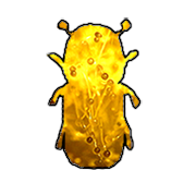
- Using a sterile 000 pin, work the edge of the mycangial membrane to lift up the entire mycangium as a cohesive mass. The color is typically orangish or beige in compactus, and the texture looks like very fine sand or eggs. Transfer the entire mycangium into a centrifuge tube. This may take a few transfers.
- Using sterile scalpel, sever the beetle just anterior to the location of the mycangium. Transfer the head and pronotum into one tube, and the rest of the beetle into another tube.