Oct 24, 2020
Fluorescent labeling Bacillus mycoides
- Andreea S1
- 1University of Groningen
- iGEM Groningen 2020


Protocol Citation: Andreea S 2020. Fluorescent labeling Bacillus mycoides. protocols.io https://dx.doi.org/10.17504/protocols.io.bkuukwww
License: This is an open access protocol distributed under the terms of the Creative Commons Attribution License, which permits unrestricted use, distribution, and reproduction in any medium, provided the original author and source are credited
Protocol status: Other
The protocol is developed based on literature and has not been tested yet.
Created: September 04, 2020
Last Modified: October 24, 2020
Protocol Integer ID: 41588
Keywords: fluorescent labeling bacillus mycoides gfp method, superfolder green fluorescent protein, peptide in the bacteria, bacillus mycoide, tagged bacterial strain, red fluorescent protein, bacterial strain, bacteria, desired peptide, protein, plant during colonization, chinese cabbage plant
Abstract
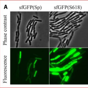
GFP method is performed in bacteria to localize a desired peptide in the bacteria. In this experiment, we are focusing on the Nlp-14a. In the paper written by (Yi,2017) GFP was mutated on a B. mycoides E18 strain to study the plant-interaction studies. In their paper, they constructed a superfolder green fluorescent protein (sfGFP) and red fluorescent protein (mKate2) in the B. mycoides E18 strain. They tracked the GFP- tagged bacterial strain after inoculating in Chinese cabbage plants in a hydroponic system. They noticed that the bacterial strain rapidly attached to the plant during colonization and formed a matrix.
Citation
LINK
Strains and Growth conditions of bacteria
Routinely culture Bacillus strains in Luria-Bertani (LB) medium at 30 °C with aeration at 200 rpm.
Preparation of B. mycoides cells for GFP
Prepare the B. mycoides strain aliquotes for electroporation.
Cultivate the bacterial strain overnight in LB broth at 30 °C and at 180 rpm.
Transfer 1 mL of the overnight culture into 100 mL of LB medium (with 2% [wt/wt] glycine) and incubate it at 30 °C and 180rpm until optical density at 600 nm is 0.4 to 0.7.
Centrifuge the cells and wash the pellets with increasing concentrations of ice-cold glycerol (2.5%, 5%, and 10%). Resuspend this pellet in precooled electroporation buffer (10% glycerol) and shock freeze in liquid nitrogen.
Add the library vector DNA in an amount of 2 µg to the cells, and perform electroporation. The settings for electroporation are 2.0 kV, 25 lF and 200 X in a 2-mm cuvette using a Bio Rad Gen Pulser II electroporation system (Bio-Rad).
Add 1 mL of LB medium and grow the cells for 02:00:00 at 30 °C and 150 rpm for recovery and then plate on LB-Cm4 agar.
After 24:00:00 of growth at 30 °C , harvest the colonies from the plates and pool in LB medium
Store the libraries at -80 °C as 15% glycerol stocks.
FACS
Inoculate the B. mycoides strain mKate2mut library in 50 mL of LB-Cm4 and grow at 7 or 6 to an OD600nm of 0.3-0.6.
B. mycoides has been seen to show extensive cell-chaining and hence a mild sonication step of 4 rounds of 3 X 10 pulses of 1s with an amplitude of 30% can be applied to disassemble the aggregated cells.
Sort the cells on a flow cytometer at 20 psi using a 70 micromolar (µM) nozzle at a flow rate of 1.0 with the highest sort precision mode (0– 32-0 sort purity mask).
Using a sequential gating strategy with FCS height versus widths, followed by SCC height versus width, cellular debris, and chained cells can be excluded.
To separate the brightest variants choose a cutoff of 3% of the brightest event in the first round of cell sorting and 0.3% of the brightest events in the second round of sorting with the light scatter parameters.
Screening of FP variants and flow cytometry measurements
After FACS sorting, plate the final fluids containing bright cells on LB-Cm4 plates and grown them overnight at 30 °C .
Observe the colonies using a fluorescence microscope. Keep the filter setting for GFP as excitation at 460/480nm and emission at 495/540 nm with a 485 nm dichromatic mirror; for RFP, the filter setting can be kept as excitation at 545/580 nm and emission at 610 nm with a 600 nm dichromatic mirror.
Capture the images on a camera and calculate the intensity of single-cell with Image J software.
Calculate the total cell fluorescence the formula is: Corrected total cell fluorescence (CTCF) = Integrated Density – (Area of selected cell x Mean fluorescence of background readings)
Citations
Yi, Y., Frenzel, E., Spoelder, J., Elzenga, J. T. M., van Elsas, J. D., & Kuipers, O. P.. Optimized fluorescent proteins for the rhizosphere-associated bacterium Bacillus mycoides with endophytic and biocontrol agent potential
10.1111/1758-2229.12607