Jun 30, 2026
Flow Cytometry Protocol for CD19 Expression Detection Using CD19 Recombinant Monoclonal Antibody (CSB-RA004888MA4HU)
- Rosie Liu1
- 1CUSABIO
- CUSABIO TECHNOLOGY LLC


Protocol Citation: Rosie Liu 2026. Flow Cytometry Protocol for CD19 Expression Detection Using CD19 Recombinant Monoclonal Antibody (CSB-RA004888MA4HU). protocols.io https://dx.doi.org/10.17504/protocols.io.6qpvr1d9pgmk/v1
License: This is an open access protocol distributed under the terms of the Creative Commons Attribution License, which permits unrestricted use, distribution, and reproduction in any medium, provided the original author and source are credited
Protocol status: Working
We use this protocol and it's working
Created: June 26, 2026
Last Modified: June 30, 2026
Protocol Integer ID: 319882
Keywords: CD19 protein; CD19 Recombinant Monoclonal Antibody; CD19 detection; flow cytometry (FC); flow cytometry (FC) validation, flow cytometry protocol for cd19 expression detection, using cd19 recombinant monoclonal antibody, cd19 recombinant monoclonal antibody, performance of the cusabio cd19 recombinant monoclonal antibody, cusabio cd19 recombinant monoclonal antibody, surface expression of cd19 protein, cd19 expression detection, human raji cells via flow cytometry, cd19 protein, flow cytometry protocol, flow cytometry application, flow cytometry, human raji cell, human igg1 isotype control
Abstract
This protocol aims to detect the surface expression of CD19 protein on human Raji cells via flow cytometry (FC), verify the performance of the CUSABIO CD19 Recombinant Monoclonal Antibody (CSB-RA004888MA4HU) in a flow cytometry application, and eliminate non-specific binding signal with a Human IgG1 Isotype Control (CSB-RA011156MA1HU) for accurate result interpretation.
Image Attribution
Flow Cytometry for CD19 Expression Detection Using CD19 Recombinant Monoclonal Antibody
Guidelines
Principle
Flow cytometry is a high-throughput single-cell analysis technology that enables simultaneous multiparameter quantitation of thousands of cells per second. In this assay, indirect immunofluorescence staining is adopted: the non-conjugated anti-CD19 primary antibody specifically recognizes and binds to the CD19 antigen on the surface of Raji cells; subsequently, the FITC-labeled secondary antibody targeting the human IgG Fc region binds to the primary antibody. When cells pass through the laser detection zone of the flow cytometer, the FITC fluorophore is excited to emit fluorescence, and the signal intensity is proportional to the expression level of the CD19 antigen. The isotype control antibody with the same IgG1 isotype is used to distinguish specific antigen binding from non-specific background binding.
Key Notes for Operation
1. Since CD19 is a membrane protein, fixation and permeabilization are not required for surface staining. Fixation is only needed when samples need to be stored for a short period.
2. Maintain cell viability above 90% throughout the experiment, as dead cells tend to bind antibodies non-specifically and increase background fluorescence.
3. Strictly follow the recommended antibody dilution range of 1:50 to 1:200, and optimize the optimal dilution according to the actual cell antigen expression level.
4. Fully resuspend cells during each washing step to ensure complete removal of unbound free antibodies.
5. Keep samples at 4°C during staining (antibody incubation) to prevent endocytosis of antibodies. Protect from light to avoid fluorophore degradation.
6. If samples cannot be tested immediately, fix cells with 4% formaldehyde for 15 min at room temperature, and store at 4°C in the dark for up to 1 week.
7. Accurately gate on live cells to exclude debris/dead cells (which may show artifactual fluorescence).
Materials
Cell Line
· Raji cells (Human Burkitt’s lymphoma suspension cell line)
Antibodies
· CD19 Recombinant Monoclonal Antibody (CUSABIO; Catalog No.: CSB-RA004888MA4HU; Isotype: hIgG1; Non-conjugated)
· Human IgG1&Igk Isotype Recombinant Monoclonal Antibody (CUSABIO; Catalog No.: CSB-RA011156MA1HU; Isotype: hIgG1; Non-conjugated)
· FITC-conjugated anti-Human IgG Fc secondary antibody
Reagents
· Sterile Phosphate Buffered Saline (PBS, 0.01M, pH 7.4)
· Normal Human Serum
· Trypan blue (for cell counting)
Equipment and Consumables
· Flow cytometer
· Benchtop centrifuge
· Cell counting device (hemocytometer or automated cell counter)
· 1.5 mL microcentrifuge tubes, 15 mL centrifuge tubes, flow cytometry tubes
· Pipettes and disposable pipette tips
· Ice bath, aluminum foil for light protection
Troubleshooting
Problem
No significant fluorescence shift between sample and isotype control (Posssible causes: 1. Antibody concentration is too low due to excessive dilution 2. CD19 antigen is not expressed or expressed at extremely low level in target cells 3. Secondary antibody does not match the primary antibody isotype 4. Fluorescence quenching caused by light exposure)
Solution
1. Reduce the dilution ratio and increase antibody concentration within the recommended range 2. Verify the CD19 expression profile of the target cell line 3. Confirm the secondary antibody is specific for human IgG Fc region 4. Protect all fluorescent reagents and stained samples from light throughout the experiment.
Problem
High non-specific background fluorescence (Posssible causes: 1. Insufficient blocking procedure 2. Antibody concentration is too high 3. High proportion of dead cells in the sample 4. Inadequate washing steps)
Solution
1. Extend blocking time or increase serum concentration in blocking buffer 2. Increase the antibody dilution ratio to reduce non-specific binding 3. Use freshly cultured cells with high viability; add a dead cell exclusion dye if necessary 4. Increase the number of washes and resuspend cells fully during each wash.
Problem
Low cell recovery rate and excessive debris (Posssible causes: 1. Overly high centrifugation speed or long centrifugation duration 2. Vigorous pipetting that damages cell integrity 3. Cell apoptosis during long-term incubation)
Solution
1. Maintain centrifugation parameters at 1000 rpm for 5 min 2. Resuspend cells gently to avoid mechanical damage 3. Perform all incubation steps at 4℃ to reduce cell apoptosis.
Problem
Broad or poorly resolved fluorescence peaks (Posssible causes: 1. Large amount of cell aggregates in the sample.2. Flow cytometer is not properly calibrated)
Solution
1. Filter cell suspension through a 40 μm cell strainer before detection; resuspend cells thoroughly 2. Perform instrument calibration and quality control before sample acquisition.
Problem
Weak signal (Possible causes: 1. Low antibody concentration; 2. poor cell viability)
Solution
1. Increase primary antibody concentration; 2. ensure cells are >90% viable.
Problem
Cell clumping (Possible causes: 1. Inadequate mixing; 2. overcentrifugation)
Solution
1. Mix cells gently before staining; 2. reduce centrifugation force/time.
Safety warnings
1. Biosafety Risk: Raji cells are human tumor cell lines. All live cell operations must be carried out in a biosafety cabinet in accordance with BSL-2 biosafety specifications. All contaminated consumables and waste must be sterilized before disposal.
2. Antibody Handling: Avoid repeated freeze-thaw cycles (aliquot antibodies and store at -20°C).
3. Cell Viability: Use only viable cells (viability 3e90%) to ensure accurate results. Dead cells may bind antibodies non-specifically.
4. Fluorescence Quenching: Fluorescent secondary antibodies and stained cell samples are extremely sensitive to light. Prolonged light exposure will cause fluorescence quenching and reduce detection sensitivity.
Before start
Prior to sample processing, all antibodies and reagents shall be handled per storage specifications. Fluorescent reagents must be protected from light at all times. Prepare antibody working solutions fresh on the experiment day following recommended dilutions, avoid repeated freeze-thaw of antibody stocks, and pre-chill sterile PBS to preserve cell viability and prevent antigen internalization.
Reagent Preparation
Blocking Buffer: 10% (v/v) normal human serum diluted in PBS, prepared fresh before use.
Anti-CD19 Primary Antibody Working Solution: Reconstitute the CD19 Recombinant Monoclonal Antibody (CSB-RA004888MA4HU) to a concentration of 2 µg per 1×10^6 cells.
Isotype Control Working Solution: Reconstitute the Human IgG1 Isotype Recombinant Monoclonal Antibody (CSB-RA011156MA1HU) at the identical concentration as the primary antibody.
Secondary Detection Antibody Working Solution: Dilute FITC-conjugated anti-Human IgG Fc secondary antibody according to the manufacturer’s recommended dilution, prepared under dark conditions.
Cell Resuspension Buffer: Ice-cold sterile PBS for final sample preparation.
Assay Procedures
Cell Harvesting
For Raji suspension cells, transfer the cell culture to a 15 mL centrifuge tube and centrifuge at 1000 rpm for 5 min to pellet cells.
Discard the culture supernatant, resuspend the cell pellet with ice-cold sterile PBS, and centrifuge again at 1000 rpm for 5 min. Repeat this wash procedure for 3 times.
Count the viable cells (trypan blue exclusion) and adjust cell density to ensure each reaction contains 1×10^6 cells per staining reaction.
Blocking
Aliquot 1×10^6 cells per tube into 1.5 mL microcentrifuge tubes. Add 100 µL blocking buffer to each tube, mix gently, and incubate at room temperature for 30 min to block non-specific Fc receptor binding.
Centrifuge at 1000 rpm for 5 min and discard the supernatant.
Primary Antibody Incubation
Aliquot 1×10⁶ cells into separate tubes (one for CD19 antibody, one for isotype control).
Add 100 μL anti-CD19 primary antibody working solution to the experimental tube. Add 100 μL isotype control working solution to the negative control tube.
Incubate at 4℃ overnight with gentle shaking, protected from light.
Washing After Primary Incubation
Add 1 mL ice-cold PBS to each tube, resuspend the cell pellet thoroughly, and centrifuge at 1000 rpm for 5 min. Discard the supernatant. Repeat this wash step 3 times to remove unbound primary antibody.
Secondary Antibody Incubation
Resuspend the cell pellet in 100 μL FITC-conjugated anti-Human IgG Fc secondary antibody working solution.
Incubate at 4℃ for 30 min in the dark.
Post-secondary Incubation Washing
Wash cells with ice-cold PBS for 3 times following the same centrifugation protocol to remove residual unbound secondary antibody.
Sample Preparation for Detection
Resuspend the final cell pellet in 500 μL ice-cold PBS, and transfer the cell suspension to a flow cytometry analysis tube. Keep samples on ice and in the dark.
Flow Cytometry Acquisition
Detect the FITC fluorescence signal on a flow cytometer as soon as possible. Collect at least 10,000 cellular events per sample for statistical analysis.
Result Analysis
Population Gating: Perform forward scatter (FSC) and side scatter (SSC) gating to gate the intact Raji cell population, excluding cell debris, aggregates, and impurities.
Fluorescence Histogram Analysis: Generate a histogram of FITC-A fluorescence intensity for the gated cell population.
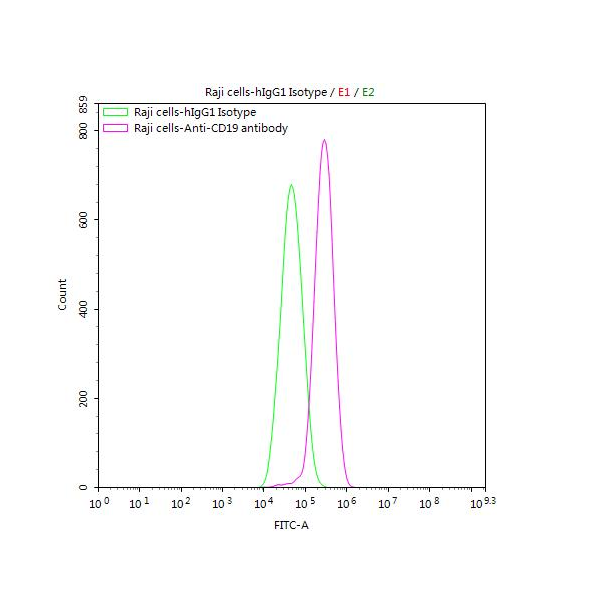
Raji cells were stained with Human IgG1 Isotype Control (CSB-RA011156MA1HU)(green line) and anti-CD19 antibody (CSB-RA004888MA4HU) (2µg/1*106cells) (red line), washed and then followed by FITC-conjugated anti-Human IgG Fc antibody and analyzed with flow cytometry.
The peak from the isotype control group (green line) represents the background fluorescence caused by non-specific antibody binding, which is used to set the positive gate threshold.
A significant rightward shift of the fluorescence peak in the anti-CD19 antibody group (red line) compared to the isotype control indicates positive CD19 expression on Raji cells.
Quantitative Assessment: Calculate the percentage of CD19-positive cells and the mean fluorescence intensity (MFI) of the positive population to quantitatively evaluate CD19 expression level.
Protocol references
○ CD19 Recombinant Monoclonal Antibody (CSB-RA004888MA4HU)
○ Human IgG1&Igκ Isotype Recombinant Monoclonal Antibody (CSB-RA011156MA1HU)
○ Target Species: Homo sapiens (Human)
○ Target Analyte: CD19