May 21, 2026
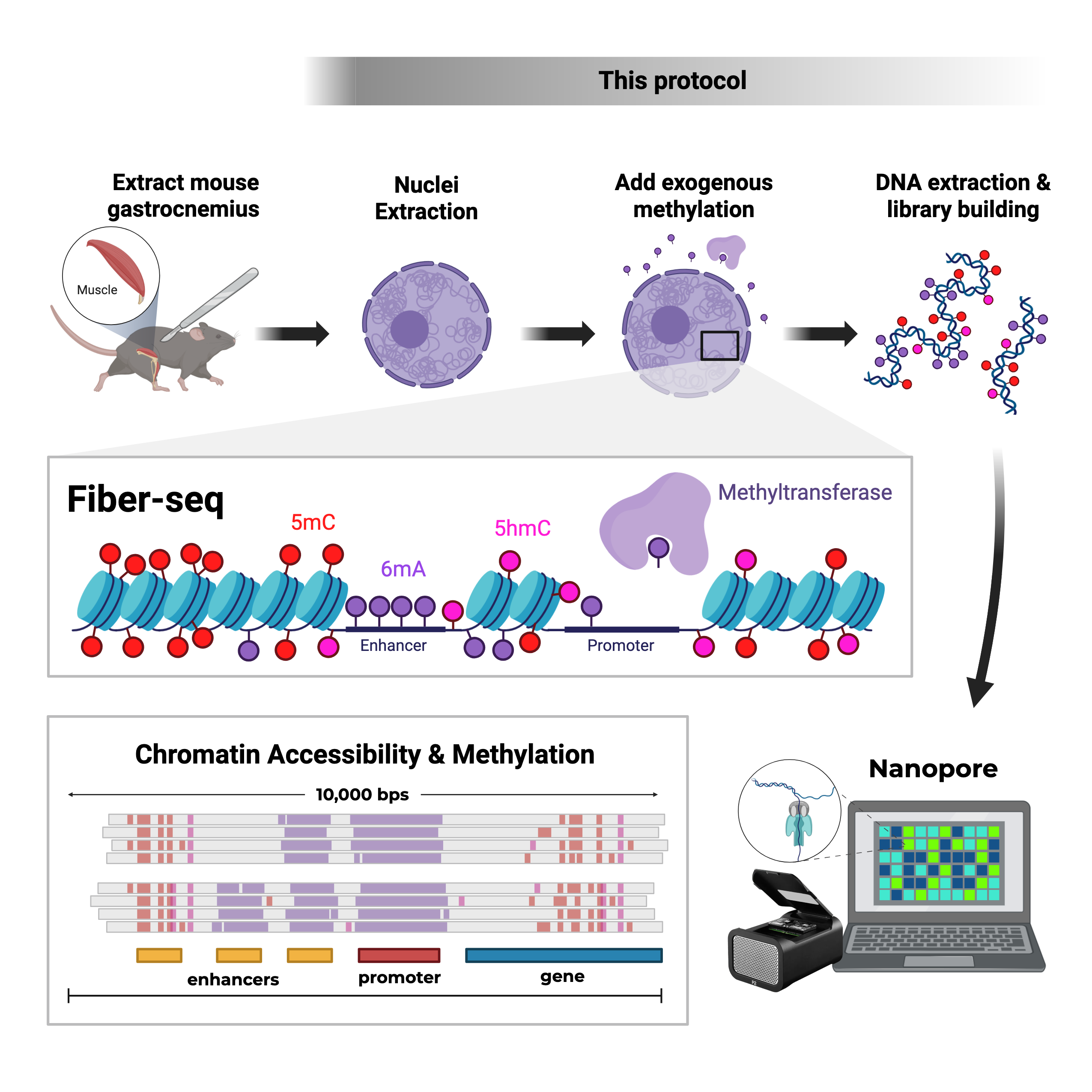
Fiber-seq for flash frozen mouse gastroc tissue for IGVF
Forked from Fiber-seq for flash frozen mouse tissue for IGVF
- Jasmine Sakr1,
- Negar Fattahi1,
- Jessica Valladolid1,
- Ali Mortazavi1
- 1University of California, Irvine


Protocol Citation: Jasmine Sakr, Negar Fattahi, Jessica Valladolid, Ali Mortazavi 2026. Fiber-seq for flash frozen mouse gastroc tissue for IGVF. protocols.io https://dx.doi.org/10.17504/protocols.io.x54v95dk4l3e/v1
License: This is an open access protocol distributed under the terms of the Creative Commons Attribution License, which permits unrestricted use, distribution, and reproduction in any medium, provided the original author and source are credited
Protocol status: Working
We use this protocol and it's working
Created: August 09, 2025
Last Modified: May 21, 2026
Protocol Integer ID: 224370
Keywords: Fiber-seq, open chromatin, methylation, m6A, methyltransferase, mouse, IGVF, UCI, whole genome sequencing, DNA, genome, Mortazavi, nuclei isolation, seq for flash frozen mouse tissue, seq experiment for frozen mouse tissue, hmw dna extraction kit for tissue, nuclei from flash frozen mouse, flash frozen mouse tissue, frozen mouse tissue, hmw dna extraction kit, nuclei extraction, flash frozen mouse, hprc fiber, fiber, seq experiment, seq protocol, tissue, gastroc, sequencing, NEB, Miltenyi Biotec, Miltenyi, EcoGII Methyltransferase, EcoGII, New England Biolabs, 5mC, 6mA, DNA extraction, 5hmC, seq for flash frozen mouse gastroc tissue, seq experiment for frozen mouse gastroc tissue, flash frozen mouse gastroc tissue, frozen mouse gastroc tissue, hmw dna extraction kit for tissue, gastrocnemius for igvf cc line, dna extraction, hmw dna extraction kit, nuclei extraction, frozen lcl cell, igvf this protocol, hprc fiber, igvf cc line, nuclei from flash, frozen mouse, fiber, igvf, seq experiment, seq protocol, seq protocol on dmso, dna, tissue
Funders Acknowledgements:
NHGRI
Grant ID: HG012077
Abstract
This protocol describes Fiber-seq experiment for frozen mouse gastroc tissue and has been optimized from the following protocols:
Nuclei extraction: Protocol to isolate and fix nuclei from flash-frozen mouse left gastrocnemius for IGVF CC Lines set, Version 2.1 (February 2025)
Labeling: HPRC Fiber-seq protocol on DMSO frozen LCL cells, (March 2024)
DNA extraction: Monarch‱ HMW DNA Extraction Kit for Tissue (NEB #T3060S/L), Version: 2.1_4/21
Image Attribution
Graphical abstract made using BioRender and other graphical images were taken from NEB DNA extraction protocol.
Guidelines
Try working quickly to help maintain nuclei integrity and prevent over labelling.
Materials
Reagents
| Name | Manufacturer | Cat # | |
| Nuclei Isolation/Extraction Buffer | Miltenyi Biotec | 130-128-024 | |
| Phosphate Buffered Saline (PBS) | HyClone | SH30256.02 | |
| 7.5% Bovine Serum Albumin (BSA) | Life Technologies | 15260037 | |
| Debris Removal Solution | Miltenyi Biotec | 130-109-398 | |
| NucBlue Fixed Cell ReadyProbes | ThermoFisher | R37606 | |
| 1 M Tris-Cl, pH 8.0 | ThermoFisher | AM9856 | |
| NaCl | Sigma-Aldrich | S9888-500G | |
| KCl | Sigma-Aldrich | P9541-500G | |
| 0.5 M EDTA, pH 8.0 | ThermoFisher | AM9260G | |
| 0.5 M EGTA, pH 8.0 | Calbiochem | LC4100 | |
| Spermidine | ThermoFisher | A19096 | |
| 20% Sodium Dodecyl Sulfate (SDS) | ThermoFisher | AM9820 | |
| EcoGII Methyltransferase (200 U) | New England Biolabs | M0603S | |
| Monarch® HMW DNA Extraction Kit for Tissue | New England Biolabs | T3060 |
Supplies and Equipment
| Name | Manufacturer | Cat # | |
| gentleMACS™ C Tube | Miltenyi Biotec | 130-093-237 | |
| gentleMACS™ Octo Dissociator | Miltenyi Biotec | 130-095-937 | |
| gentleMACS™ Octo Coolers | Miltenyi Biotec | 130-130-533 | |
| Centrifuge 5810 R | Eppendorf | 022-625-004 | |
| MACS SmartStrainers (70 um) | Miltenyi Biotec | 130-110-916 | |
| MACS SmartStrainers (30 um) | Miltenyi Biotec | 130-098-458 | |
| DNA LoBind® 2.0 mL Tubes | Eppendorf | 022-431-048 | |
| DNA LoBind® 5.0 mL Tubes | Eppendorf | 030-108-310 | |
| DNA LoBind® 15.0 mL Tubes | Eppendorf | 030-122-208 | |
| Disposable Hemacytometer | Sigma-Aldrich | MDH-2N1-50PK | |
| Eppendorf ThermoMixer® C | Eppendorf | - | |
| Eppendorf SmartBlock 2.0 mL | Eppendorf | 5362000035 | |
| Thermo Scientific™ HulaMixer™ Sample Mixer | Invitrogen | 50-272-2983 | |
| Qubit 4 Fluorometer | ThermoFisher | Q33226 | |
| Invitrogen™ Qubit™ Assay Tubes | ThermoFisher | Q32856 |
Nuclei Extraction Set up
Make enough Buffers for the total number of samples and keep on ice.
| Name | Reagent | Final conc. | 1 | 2 | |
| Lysis buffer (LB) | Nuclei Isolation/Extraction Buffer | NA | 4.5 mL | 9 mL | |
| Resuspension Buffer (RSB) | Phosphate-buffered saline (PBS) | NA | 3.45 mL | 6.41 mL | |
| 7.5% BSA | 0.1% | 46.67 µL | 86.67 µL | ||
| Phosphate-buffered saline (PBS) | Phosphate-buffered saline (PBS) | NA | 5 mL | 10 mL | |
| Debris Removal Solution (DRS) | Debris Removal Solution (Miltenyi) | NA | 1 mL | 2 mL |
Or a large stock can be stored at 4ºC for a month.
Prepare and label the following tubes (for each sample) for nuclei extraction:
- Pre-chilled gentleMACS C Tube
- 5 mL tube
- 15 mL tube
- MACS SmartStrainers (70 µm)
- MACS SmartStrainers (30 µm)
- Disposable Hemacytometer
Keep flash frozen tissue samples on dry ice until lysis.
Add 2.5 mL Nuclei Extraction Buffer to a chilled gentleMACS C Tube.
Nuclei Extraction
55m 10s
Drop whole frozen tissue into the chilled gentleMACS C Tube with 2.5 mL Nuclei Extraction Buffer.
Invert immediately, ensuring tissue is not stuck to the bottom or sides. Proceed immediately to load onto the Octo Dissociator and add an Octo Cooler over each gentleMACS C Tube.
Run the gentleMACS Program 4C_nuclei_1 on the Octo Dissociator (00:05:00 ).
5m
Remove the gentleMACS C Tube and ensure tissue did not get stuck on the sides by tapping the tube.
* If the tissue sample fails to dissociate, try rerunning the program. After the third unsuccessful attempt, remove the tissue from LB and place it in a new 1.5 mL tube. Place the tube in -80 freezer for 5-10 mins to refreeze tissue, and then try again.
Spin down in4 °C centrifuge for 00:00:10 .
10s
Mix the nuclei suspension gently and thoroughly by pipetting with a 5 mL pipette.
Filter the nuclei suspension through 70 µm MACS SmartStrainer into a 5 mL tube.
- Hold your 5 mL pipette perpendicular to the filter. Press your tip against its center and slowly pass the suspension through the filter while moving your tip in a circular motion. Try to minimize the area of the circle around the center point. Follow these instructions for any filtering unless stated otherwise.
Wash the empty, used gentleMACS C Tube with an additional 2 mL Nuclei Extraction Buffer. Close the cap and swish to recover any nuclei stuck to the sides.
Filter the 2 mL additional Nuclei Extraction Buffer through the 70 µm MACS SmartStrainer into the same 5 mL tube (total volume now 4.5 mL).
5m
Centrifuge the 4.5 mL nuclei suspension at 700 x g, 4°C, 00:05:00 .
5m
Remove the supernatant without disrupting the pellet.
Resuspend nuclei pellet in 3.1 mL RSB.
- Mix by pipetting up and down 7-10 times using a 5 mL pipette. Performing this step poorly might influence the accuracy of the nuclei count and, consequently, the input into the next step.
Filter nuclei suspension through 30 µm MACS SmartStrainer into a 15 mL tube.
Add 900 µL DRS and mix by pipetting 10 times slowly up and down using a 5 mL pipette.
Overlay with 4 mL PBS using a P1000 or P5000 pipette, whichever you are more comfortable with.
- Place the tube on a flat surface, tilt it at a 45-degree angle, and slowly add the first 1 mL. You can increase speed gradually after the initial addition of PBS.
10m
Centrifuge at 3000 x g, 4°C, 00:10:00 with no brake.
- Three phases will be formed:
- A top transparent buffer layer,
- A middle cloudy debris band/ring,
- A bottom transparent layer containing nuclei. The pellet is usually visible.
10m
Aspirate the two top phases (the buffer layer and the cloudy debris band, as shown within the red box in the picture) and discard them into a designated waste container.
- Aspirate the first phase, then the second phase. Stay above the third layer of nuclei to prevent loss. Usually, it will take ~1000 µL to take out the debris band, and the nuclei will be suspended in the remaining ~2000 µL.
5m
Add cold RSB to the remaining suspension until a final volume of 5 mL is reached. Gently invert the tube three times. DO NOT vortex.
Centrifuge at 1000 x g, 4°C, 00:10:00 with full break.
10m
Discard the supernatant into a designated waste container.
Fully resuspend the nuclei pellet in 187.5 µL RSB.
Count nuclei:
- Use 1:11 dilution factor: 2 µL sample + 20 µL dye.
- Mix by pipetting up and down 7-10 times.
- Load a 10 µL sample onto a disposable hemocytometer.
* If the counts taken from two opposing squares are vastly different (i.e., one square has around half the amount of nuclei of the other square), then take a count from a third square to decide which count is the more accurate one.
5m
Make aliquots of 3 million nuclei in 1.5 mL tubes and proceed to labeling and DNA extraction.
Fiber-seq & DNA Extraction Set Up
Make buffers and solutions.
- Buffer A
| Final Concentration | Stock Concentration | Volume stock soln | |
| Nuclease-free H2O | 100% | 95.8 mL | |
| 15 mM Tris-Cl, pH 8.0 | 1 M Tris-Cl, pH 8.0 | 1.5 mL | |
| 15 mM NaCl | 5 M NaCl | 300 μL | |
| 60 mM KCl | 3 M KCl | 2 mL | |
| 1 mM EDTA, pH 8.0 | 0.5 M EDTA, pH 8.0 | 200 μL | |
| 0.5 mM EGTA, pH 8.0 | 0.5 M EGTA, pH 8.0 | 100 μL |
This buffer is used in the labeling reaction and is table at room temperature.
- 0.5 Molarity (M) Spermidine and store at 4 °C .
- 20 Mass / % volume Sodium Dodecyl Sulfate (SDS) and store at Room temperature .
Pre-chill centrifuge to 4 °C ..
Set two thermomixers at 56 °C and 25 °C .
Prepare and label the following tubes for DNA extraction (for each labeling reaction):
- Bead strainer in a collection tube
- 2 mL tube for wash step
- 2 mL tube for elution step
- 1.5 tube for final elution
Add 100 µL of gDNA Elution Buffer II to the 2 mL elution tubes.
Prepare an ice bucket and keep the following on ice:
- Proteinase k and RNAse A
- 0.5 Molarity (M) Spermidine
Labeling
15m
Spin 3 million nuclei at 500 x g, 4°C, 00:05:00 .
5m
During the 5 min spin, mix the following and keep the Reaction Master Mix On ice .
Buffer A + Spermidine Room temperature
| Number of tubes | 1 | 2 | 3 | 4 | |
| Buffer A | 200 μL | 400 μL | 600 mL | 800 mL | |
| 0.5 M Spermidine | 0.2 μL | 0.4 μL | 0.6 μL | 0.8 μL |
This is made fresh on the day of the experiment.
Reaction Master Mix (75 U) On ice
| Number of tubes (+0.2 extra) | 1 | 2 | 3 | 4 | |
| Buffer A + Spermidine | 157.5 µL | 315 µL | 472.5 µL | 630 µL | |
| EcoGII Methyltransferase (200 U) | 15 µL | 33 µL | 48 µL | 63 µL | |
| 32 mM SAM | 7.5 µL | 16.5 µL | 24 µL | 31.5 µL |
This reaction uses 75 units of enzyme and final 160 µM SAM concentration with a total reaction volume of 180 µL.
Remove the supernatant and resuspend the pellet in 180 µL of Reaction Master Mix.
Incubate on 25 °C heat block for 00:10:00 .
10m
Add20 µL Proteinase k and 10 µL RNAse A to each tube. Mix using wide-bore pipette tip.
* Skip this step if you are going to freeze the lysate and extract DNA later.
Add 20% SDS to each sample for a final concentration of 1%.
- Calculate the amount of 20% SDS you need.
- Insert the pipette tip into the solution.
- Add one small drop of SDS.
- Gently move the tip to a different location in the tube and add another drop.
- Repeat until the total calculated volume of 20% SDS is added.
- Avoid creating bubbles — the dropwise method ensures even distribution.
Thoroughly mix by gently inverting tube. Do not vortex or pipette to mix.
* If you are going to extract DNA later, flash freeze and store at -80 °C .
DNA extraction
1h 41m 30s
Incubate in a thermal mixer at 2000 rpm, 56°C, 00:10:00 .
* If you are working from thawed reaction, first add20 µL Proteinase k and 10 µL RNAse A to each tube. Mix using wide-bore pipette tip.
10m
Add 180 µL Protein Separation solution.
Mix on vertical rotating mixer for 20 rpm, 00:01:00 .
1m
Centrifuge for 16000 x g, 25°C, 00:10:00 .
10m
Using a wide-bore tip, transfer as much of the upper phase containing the DNA to a labeled, empty 2 mL tube without disrupting the pellet at the bottom. Note the total volume for later steps.
Add 2 DNA capture beads to each sample.
Calculate and add 0.7x volume isopropanol to each sample.
Mix on a vertical rotating mixer at 10 rpm, 00:10:00 to attach DNA to the beads.
10m
During the wash steps, work quickly to prevent DNA from drying out on beads!
Discard liquid by keeping tube upright, insert pipette tip and gently push beads aside to remove liquid.
Add 500 µL Wash Buffer.
Mix by gently inverting the tube 3 times.
Discard liquid by keeping tube upright, insert pipette tip and gently push beads aside to remove liquid.
Add 500 µL Wash Buffer.
Mix by gently inverting the tube 3 times.
Discard liquid by keeping tube upright, insert pipette tip and gently push beads aside to remove liquid.
Pour the beads into the bead retainer that is in a collection tube.
Pulse spin (≤ 1 second) the tubes to remove any residual wash buffer from the beads.
Separate the bead retainer from the collection tube and pour the beads into the labeled tube with 100 μL of elution buffer and save the bead retainer for a future step.
Incubate on thermal mixer at 56 °C for 00:10:00 and during incubation, make sure all sides of the beads are exposed to elution buffer by tilting the tube almost horizontally and gently shaking after the first 5 min.
10m
Incubate for 01:00:00 at room temperature. During incubation, make sure all sides of the beads are exposed to elution buffer by tilting the tube almost horizontally and gently shaking every 5 min.
1h
Pour the eluate and the glass beads into the bead retainer that is placed in the final labeled tube.
Centrifuge for 12000 rpm, 25°C, 00:00:30 to separate the eluate from the glass beads.
30s
Incubate DNA by one of the following methods to make sure it is homogeneously dissolved:
- at 37 °C for 01:00:00
- at 4 °C Overnight
Mix gently by pipetting using wide-bore tip.
Qubit each tubes and keep on ice until library prep or store in -20°C
Library building
Follow "ONT Library Prep for Whole Genome Sequencing" protocol to build libraries for whole genome sequencing using Oxford Nanopore platform.